online | memorias.ioc.fiocruz.br
Taenia solium is a parasite whose larvae (metaces-todes) can be found in the central nervous system of hu-mans, causing neurocysticercosis (NC). This parasitosis is a zoonotic disease that is endemic in the developing countries of Latin America, Asia and Africa (Sciutto et al. 2000, Pawlowski 2008, Raccurt et al. 2009, Flores Castro 2010, Praet et al. 2010, Ito et al. 2011). Cysti- cercosis has emerged as a cause of severe neurologic diseases in the United States that primarily affects im-migrants from Latin America. Moreover, the relevance of cysticercosis as a public health problem is highlight-ed by local transmission (Sorvillo et al. 2011). Human cysticercosis is acquired accidentally by the ingestion of T. solium eggs and its transmission is related to a lack of sanitation, bad personal hygiene and the inadequate disposal of human faeces (Vianna et al. 1986, Huerta et al. 2008, Flisser & Correa 2010).
NC is classified as active or inactive and inflam-matory or non-inflaminflam-matory, according to the stage of metacestode development when viewed by neuroimag-ing tests associated with the presence or absence of a host immune response (Sotelo et al. 1985, Barcelos et al. 2007). The diagnosis of human NC is determined after the analysis of clinical, epidemiological, neuroimaging
and immunological data (Del Brutto et al. 2001). The combination of enzyme linked immunosorbent assay (ELISA) and western blotting (WB) to detect specific antibodies against metacestodes extracts of T. solium in the serum samples or in the cerebrospinal fluid (CSF) of patients with NC represents a viable alternative for the diagnosis of the disease in developing countries (Shiguekawa et al. 2000, Barcelos et al. 2001, Sahu et al. 2009, Nunes et al. 2010).
Differences in the infectivity and the pathogenicity of T. solium metacestodes may be related to the genetic variability of the parasite (Campbell et al. 2006). Vari-ance in T. solium metacestode DNA can generate anti-genic variability according to clonal lineage behaviour (Maravilla et al. 2008).
The aim of the present study is to investigate the genetic polymorphisms in T. solium metacestodes from different Brazilian geographical areas and to relate them to each other using antibody recognition in the serum samples of NC patients.
SUBJECTS, MATERIALS AND METHODS
Parasites - T. solium metacestodes obtained from the skeletal muscles of naturally infected pigs were washed in saline solution (0.15 M NaCl) four times and were stored at -70ºC for subsequent analysis. The parasites were collected from one individual pig in each of the fol-lowing Brazilian geographical areas: (i) Distrito Federal (DF), Central West Region, (ii) Bahia (BA), Northeast Region, (iii) Minas Gerais (MG) and (iv) São Paulo (SP), Southeast Region. The samples were collected when a pig with a massive infection was found in each area. Financial support: CNPq, CAPES, FAPEMIG
+ Corresponding author: [email protected] Received 5 April 2011
Accepted 23 August 2011
Genetic polymorphism in
Taenia solium
metacestodes
from different Brazilian geographic areas
Ivanildes Solange da Costa Barcelos1,2, Maria Aparecida Souza3, Janethe Deolinda de Oliveira Pena3,
Gleyce Alves Machado1,4, Lísia Gomes Martins de Moura1, Julia Maria Costa-Cruz1/+
1Laboratório de Diagnóstico de Parasitoses 3Laboratório de Biologia Molecular, Instituto de Ciências Biomédicas, Universidade Federal de Uberlândia, Av. Pará 1720, 38400-902 Uberlândia, MG, Brasil 2Laboratório de Ciências Biomédicas - Imunologia, Campus Jataí 4Departamento de Ciências Biológicas, Campus Catalão, Universidade Federal de Goiás, Goiânia, GO, Brasil
The aim of the present study is to investigate genetic polymorphisms in Taenia solium metacestodes from dif-ferent Brazilian geographical areas and to relate them to antibody recognition in serum samples of neurocysticer-cosis (NC) patients. Metacestodes were obtained from the Distrito Federal (DF), Bahia, Minas Gerais (MG) and São Paulo (SP) regions of Brazil. Samples of human sera from 49 individuals with NC, 68 individuals with other helminthiasis and 40 healthy volunteers were analysed (157 individuals in total). Antigens were prepared and used in enzyme-linked immunosorbent assay and western blotting assays to detect specific immunoglobulin G antibod-ies. Genetic distances between metacestode populations were analysed using random amplified polymorphic DNA (RAPD) analysis. Our results show that there was a higher frequency of reactivity in the DF region in the sera from NC patients (p < 0.05), while discrimination between active and inactive NC was seen only in extracts from the MG and SP regions (p < 0.05). Using RAPD, the sample from the DF region presented a greater increase compared to the other regions. A relationship between genetic polymorphisms among T. solium metacestodes from different areas in Brazil and the differences in antibody detection in patients with NC were established.
Preparation of antigens - The saline extracts of T. so-lium metacestodes from different geographic areas were prepared as described by Costa et al. (1982) with some modifications. Briefly, 50 metacestodes from each area were disrupted in 5 mL distilled water in an ice bath for 5 min, were homogenised for 4 min and were subse-quently submitted to ultrasonic treatment (Thornton, In- pec Electronics, Brazil) at 40 kHz for four periods of 30 s each in an ice bath. After adding 5 mL of 0.3 M NaCl, the ultrasonic treatment was repeated; the mixture was stirred at 4ºC for 2 h and was centrifuged at 12,400 g at 4ºC for 30 min (Du Pont SORVALL Products New-town, Connecticut, USA). The supernatants were analy-sed for protein content using the Lowry method (Lowry et al. 1951) and were stored in aliquots at -70ºC until use in the ELISA and WB analyses.
Patients - With the exception of those individuals with Echinococcus granulosus, who came from South-ern Brazil and the normal volunteers, who were recruited among students and their relatives, serum samples were collected from 157 individuals at the university hospital in Uberlândia. All individuals were from similar back-grounds and socio-economic conditions. Group 1 (G1) consisted of 49 patients (30 females and 19 males) with NC (19 active and 30 inactive) who were between 14-82 years of age (average age was 42 years) with a diagnosis based on clinical and epidemiological data, computerised tomography and/or magnetic resonance imaging and a positive immunological test (Del Brutto et al. 1996). Group 2 (G2) consisted of 68 individuals (40 females and 28 males) between two-71 years of age (average age was 36 years) who were monoinfected with the follow-ing helminths: Taenia sp. (20), Strongyloides stercoralis (20), Schistosoma mansoni (10), Hymenolepis nana (8) from the southeast area and E. granulosus (10) from the southern area. Group 3 (G3) (control) consisted of 40 vol-unteers (20 females and 20 males) between 21-80 years of age (average age was 32 years) who were apparently healthy based on clinical features. Although they came from an area with endemic cysticercosis (southeastern Brazil), none had household contact with T. solium in-fection or a previous history of taeniasis or cysticerco-sis. This group also underwent three faecal sample tests (Baermann 1917, Lutz 1919), which were negative.
ELISA - All reagents were tested previously and ELISA was carried out according to Shiguekawa et al. (2000). Polystyrene microplates (Corning, USA) were coated with the saline extracts at a concentration of 5 µg/ mL (50 µL/well). Serum samples were diluted 1:200 and peroxidase-conjugated goat anti-human immunoglobulin G (IgG) (Fc chain specific) (Sigma, St. Louis, USA) was used at a titre of 3,000 for all antigens. The assay was developed by adding the enzyme substrate consisting of 0.03% H2O2 (Merck, Germany) in 0.1 M citrate-Na2PO4 buffer (pH 5.0) containing 0.4 mg/mL of o-phenylenedi-amine (Merck). After 15 min of incubation at room tem-perature (RT), the reaction was stopped by adding 2N H2SO4 (25 mL/well) and the absorbance was determined at 492 nm using a microplate reader (Titertek Multiskan, Flow Laboratories, USA). The cut-off was established using the mean of three non-reactive samples plus two
standard deviations. The results were expressed as the reactivity index (RI), calculated by dividing the read-ing values of the test (absorbance = optical density) by the cut-off (Pardini et al. 2002). All samples showing an RI > 1 were considered positive.
WB - The WB assay was performed according to Shiguekawa et al. (2000) with some modifications. All saline extracts were subjected to electrophoresis and were transferred to nitrocellulose membranes (0.45 µm) (Sigma) as described by Towbin et al. (1979), using a semi-dry transfer apparatus (Multiphor II, Pharmacia-LKB). Serum samples from 98 patients (49, 39 and 10 in-dividuals from G1, G2 and G3, respectively) were diluted 1:50 before analysis. Peroxidase-conjugated anti-human IgG (whole molecule) (Sigma) was used at a titre of 200. The results were considered positive when at least two or more immunodominant proteins (18, 24, 28-32, 39-42, 47-52, 64-68 and 70 kDa) (Barcelos et al. 2007) were recognised by the serum samples.
Genomic DNA and random amplified polymorphic DNA (RAPD) - Genomic DNA extraction was performed in metacestodes from each region using methods that were described by Casas et al. (1995) and modified by Gonza-lez et al. (2002). The negative control consisted of DNA that was extracted from 10 mg skeletal muscle derived from an uninfected pig. Briefly, the samples were ho-mogenised in liquid nitrogen and were extracted in 50 µL of lysis buffer (4 M guanidine isothiocyanate, 25 mM so-dium citrate, pH 7, 0.5% sarcosyl and 1 mM dithiothre-itol) for 10 min at RT and were centrifuged at 4,000 g. DNA was precipitated from the supernatant using one volume of isopropanol followed by another centrifugation at 11,750 g for 10 min. DNA pellets were washed in 70% ethanol and were resuspended in water. The RAPD assay was carried out using 30 ng of DNA template, 400 mM of dNTPs (Amersham Biosciences, USA), 3 mM of MgCl2 (Lab Trade, Brazil), 1.5 U of Taq DNA polymerase (Lab Trade, Brazil) and 1 nmol of primers (35 decamer prim-ers) (Gene Link, USA) (Table I). Sterile water was used as a non-DNA control for the reaction. Polymerase chain reaction amplification was performed using the following parameters: an initial denaturation of 5 min at 94ºC, 35 cycles of 94ºC for 30 s, 36ºC for 1 min and 72ºC for 2 min, and a final extension at 72ºC for 10 min (Perkin Elmer, New Jersey, USA). Amplified products were analysed by electrophoresis in 2% agarose gels (Acros Organics, Belgium) that were stained with ethidium bromide and visualised under ultraviolet light. RAPD band patterns were recorded and analysed giving each band a value of 1 (if present) or 0 (if absent). The bands were also compared against RAPD profiles of pork muscle as a control.
The genetic distances among the samples of T. solium metacestodes were analysed using STAT 4.5 software using percentage of disagreement and unweighted pair-group method using arithmetic average.
Ethics - This study received approval from the Re-search Ethics Committee of the Federal University of Uberlândia, MG, Brazil.
RESULTS
All serum samples were analysed using ELISA and the saline extracts of T. solium metacestodes that were collected from different geographic areas (Fig. 1). The data shows that the RI was significantly higher in the NC patient sera (G1) (p < 0.05) when compared with the con-trol group (G3) or the other helminths group (G2) with the exception of patients with an E. granulosus infection.
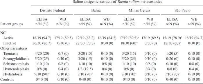
Table II shows the reactivity frequency that was ob-tained using ELISA and WB assays with regard to the four antigenic extracts in the serum samples of G1, G2 and G3. ELISA showed reactivity differences between the patients with active and inactive NC (G1) only in those antigenic extracts from the MG and SP geographic areas (p < 0.05). In G2, there was higher frequency (90% in the extracts from the DF region and 70% in the other antigens) of reactivity in the ELISA assays for samples from E. granulosus patients (p < 0.05). The active NC serum samples reacted in the range of 84.2-94.7% in the WB assays when the different antigens were used. In contrast, all serum samples from individuals with in-active NC, other parasitosis and the apparently healthy volunteers had no reaction with any of the antigens that were analysed (Table II).
The frequency of the different antigens that were rec-ognised by the IgG antibodies in WB from patients with active NC is shown in Fig. 2. In patients with active NC, there was a variation of reactivity among the different an-tigens (DF, BA, MG and SP). The 24 kDa peptide was rec-ognised with the DF, BA and MG antigens; peptides from the 28-32 kDa range were recognised with the DF, MG and SP antigens, whereas peptides from the 39-42 kDa range were recognised with the MG and SP antigens. The peptides from the 47-52 kDa and 64-68 kDa range were recognised with all of the antigens that were analysed.
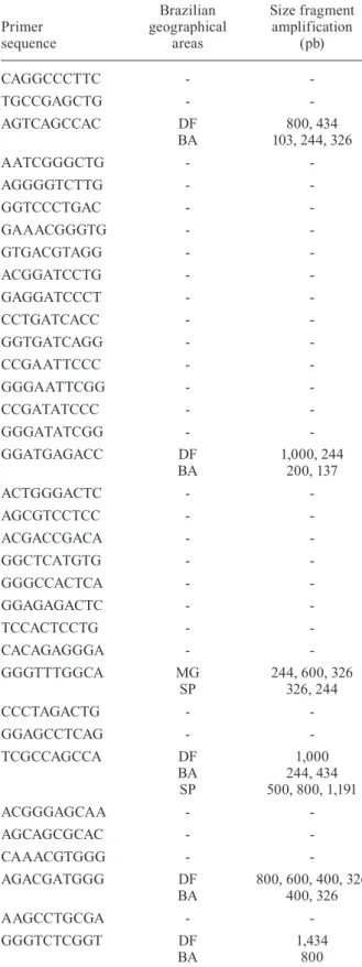
The analysis of RAPD profiles indicated the existence of a genetic polymorphism between different Brazilian geographical areas. In the banding patterns generated by RAPD, only amplicons 103, 137, 200, 244, 326, 400, 434, 500, 600, 800, 1,000, 1,191 and 1,434 were polymorphic and as seen in Table I, only cysticerci from DF generated bands of 800 and 434 bp, whereas cysticerci from BA pro-duced amplicons of 103, 244 and 326 bp and those from MG and SP did not generate these amplicons. The repro-ducibility of the RAPD analysis was confirmed. Genetic relationships among metacestodes that were collected from different Brazilian geographical areas using the 13 RAPD markers are shown in a dendrogram (Fig. 3). The sample from the DF region exhibits a greater difference in relation to the other samples and is followed by the sample from the BA region, whereas the samples from the SP and MG regions exhibit a smaller difference.
TABLE I
Primers and polymorphic fragments by random amplified polymorphic DNA of Taenia solium metacestodes
from different Brazilian geographical areas
Primer sequence
Brazilian geographical
areas
Size fragment amplification
(pb)
CAGGCCCTTC -
-TGCCGAGCTG -
-AGTCAGCCAC DF
BA
800, 434 103, 244, 326
AATCGGGCTG -
-AGGGGTCTTG -
-GGTCCCTGAC -
-GAAACGGGTG -
-GTGACGTAGG -
-ACGGATCCTG -
-GAGGATCCCT -
-CCTGATCACC -
-GGTGATCAGG -
-CCGAATTCCC -
-GGGAATTCGG -
-CCGATATCCC -
-GGGATATCGG -
-GGATGAGACC DF
BA
1,000, 244 200, 137
ACTGGGACTC -
-AGCGTCCTCC -
-ACGACCGACA -
-GGCTCATGTG -
-GGGCCACTCA -
-GGAGAGACTC -
-TCCACTCCTG -
-CACAGAGGGA -
-GGGTTTGGCA MG
SP
244, 600, 326 326, 244
CCCTAGACTG -
-GGAGCCTCAG -
-TCGCCAGCCA DF
BA SP
1,000 244, 434 500, 800, 1,191
ACGGGAGCAA -
-AGCAGCGCAC -
-CAAACGTGGG -
-AGACGATGGG DF
BA
800, 600, 400, 326 400, 326
AAGCCTGCGA -
-GGGTCTCGGT DF
BA
1,434 800
DISCUSSION
Differences in the reactivity of IgG antibodies in se-rum samples from patients with NC were demonstrated using ELISA and WB for antigenic extracts from the four populations of T. solium metacestodes that were collect-ed from different geographical areas of Brazil. A higher frequency of positive samples from patients with NC was observed in the antigen extracts from the DF region in the ELISA assay. Subsequently, discrimination between serum samples of patients with active and inactive forms of NC was possible in extracts from MG and SP regions and there was a low reactivity of samples from patients with inactive NC. Espinoza et al. (1986) and Barcelos et al. (2005) have shown that in paired samples of CSF and in serum from patients with active and inactive NC, analysed by ELISA, there are higher IgG antibody levels in samples from the patients with active NC.
The results of serologic tests for the diagnosis of NC are directly influenced by differences in biologi-cal samples, if total or purified antigenic extracts are used, the patient’s origin and the clinical phases of NC. This could explain why the efficiency of these tests var-ies according to the published reports (Del Brutto et al. 1996, Schantz 2006). In this study, ELISA and WB as-says using the saline extract of T. solium metacestodes showed a high sensitivity. ELISA had a low specific-ity and WB demonstrated specificspecific-ity for the
detec-tion of specific bands (≤ 70 kDa) in the serum samples
from patients with active NC. Using the saline extract of T. solium metacestodes in our study, WB identified four of the seven antigenic markers (18, 24, 39-42 and 50 kDa) that are similar to the glycoproteins, described by Tsang et al. (1989), for the diagnosis of NC. In this study, the highest reactivity was found with 64-68 kDa antigens. Shiguekawa et al. (2000) and Barcelos et al. (2007) showed that the 64-68 kDa antigens were immu-nodominant in serum samples. Analysing paired CSF and serum samples from NC patients, Simac et al. (1995) showed an additional 60-75 kDa band in CSF samples and explained this finding by referring to a Miller et al. (1985) publication, which proposed that the intrathecal secretion of specific antibodies was responsible for re-sults such as these. The low sensitivity of the WB assay of serum samples from patients with inactive NC that was observed in this study was previously reported by our group (Barcelos et al. 2007).
The cross-reactivity observed in serum samples from patients infected by other helminth species using ELISA varied according to the four T. solium metacestodes an-tigens used. The group diagnosed with other parasitosis is representative of the general population, particularly in Brazil, where parasitic diseases are highly prevalent. Therefore, cross-reactions may occur when screening for active NC using tests, such as ELISA and/or WB, which require further confirmation using antigens with more specificity, e.g., purified antigens (Machado et al. 2007). The presence of common antigenic epitopes in different species of parasites has been described (Shiguekawa et al. 2000, Ishida et al. 2003). The higher cross-reactivity with E. granulosus can be explained by its phylogenetic closeness with Taenia parasites (Hoberg 2006).
The present study demonstrates DNA polymor-phism in T. solium metacestode samples from different geographical regions in Brazil. Martinez-Hernandez et al. (2009) analysed the genetic structure of the T. solium population based on previously published molecular data of mitochondrial and nuclear ribosomal DNA se-quences that were deposited in the GenBank from Af-rica, Asia, Latin America and Oceania and the report demonstrated polymorphisms in cysticerci sequences from Brazil and other countries and provided gene flow values between them. RAPD employing T. solium cysticerci that was recovered from naturally infected
pigs from Mexico, Honduras, Madagascar and Tanza-nia showed several fixed alleles with linkage disequi-librium in some parasites, suggesting the existence of local lineages (Maravilla et al. 2003, Vega et al. 2003). Genetic variability of the 45W gene family has been demonstrated between Chinese and Mexican T. solium (Zheng et al. 2008). In the present study, the parasite material originated from only one pig and it is possible that antigenic variation exists within the parasites of one state. Moreover, even though the same protocol has been used for collecting and isolating the antigens, dif-ferences in antigenic properties can also be due to other factors, such as the age of the cysts and viability. TABLE II
Reactivity frequency by enzyme linked immunosorbent assay (ELISA) and Western Blotting (WB)
Patient groups
Saline antigenic extracts of Taenia solium metacestodes
Distrito Federal Bahia Minas Gerais São Paulo
ELISA n/N (%)
WB n/N (%)
ELISA n/N (%)
WB n/N (%)
ELISA n/N (%)
WB n/N (%)
ELISA n/N (%)
WB n/N (%)
NC
Active 18/19 (94.7) 17/19 (89.5) 12/19 (63.2) 16/19 (84.2) 17/19 (89.5)a 17/19 (89.5) 15/19 (78.9)b 18/19 (94.7)
Inactive 26/30 (86.7) 0/30 (0) 22/30 (73.3) 0/30 (0) 18/30 (60)a 0/30 (0) 18/30 (60)b 0/30 (0)
Other parasitosis
Taeniasis 4/20 (20) 0/7 (0) 3/20 (15) 0/10 (0) 3/20 (15) 0/10 (0) 1/20 (5) 0/10 (0)
Strongyloidiasis 5/20 (25) 0/10 (0) 3/20 (15) 0/10 (0) 5/20 (25) 0/10 (0) 0/20 (0) 0/10 (0)
Schistosomiasis 1/10 (10) 0/8 (0) 1/10 (10) 0/8 (0) 1/10 (10) 0/8 (0) 0/10 (0) 0/8 (0)
Hymenolepiasis 0/8 (0) 0/4 (0) 1/8 (12.5) 0/4 (0) 0/8 (0) 0/4 (0) 0/8 (0) 0/4 (0)
Hydatidosis 9/10 (90)c 0/10 (0) 7/10 (70)c 0/10 (0) 7/10 (70)c 0/10 (0) 7/10 (70)c 0/10 (0)
Controls 0/40 (0) 0/10 (0) 0/40 (0) 0/10 (0) 0/40 (0) 0/10 (0) 0/40 (0) 0/10 (0)
a, b: difference by two proportions (p < 0.05) between active neurocysticercosis (NC) and inactive NC with Minas Gerais and São Paulo extracts of T. solium metacestodes, respectively; c: difference by two proportions (p < 0.05) between Distrito Federal extracts of T. solium metacestodes and others extracts; n/N: serum sample reagents/total serum sample analyzed.
Fig. 2: percentage of reactivity frequency of serum samples from pa-tients with active neurocysticercosis by western blotting among dif-ferent antigens. BA: Bahia; DF: Distrito Federal; IgG: immunoglobu-BA: Bahia; DF: Distrito Federal; IgG: immunoglobu-lin G; MG: Minas Gerais; SP: São Paulo.
Because a large number of potential nuclear and mito-chondrial markers can be generated by RAPD using read-ily available primers (Williams et al. 1990), it is possible that some polymorphic loci found using this technique are translated to a certain phenotypic variability (i.e., some antigenic variants). Therefore, in the present study, although genetically divergent from other populations that were analysed, cysticerci from the DF region were strongly recognised by patients with NC. Furthermore, knowledge of the genetic structure of T. solium may be applied to understanding its epidemiology and transmis-sion because differences in the infectivity and pathoge-nicity of T. solium metacestodes, which are manifested by different antigen recognition by host antibodies, may imply genetic variability of the parasite (Campbell et al. 2006, Hinojosa-Juarez et al. 2008) and a more complex disease presentation in areas where NC predominates and in regions where there is a similar prevalence of both clinical forms (Ito et al. 2003). Because of its genetic variability, T. solium could be a significant source of the clinical heterogeneity observed in this disease and could contribute to the difficulties encountered in the design of reliable immunodiagnostic tools and vaccines (Vega et al. 2003, Sciutto et al. 2008). RAPD technology has been an effective tool for the study of intra-species genetic variability. Interestingly, cysticerci from the DF region, which presented more variability, as shown in the den-drogram (Fig. 3), exhibited more reactivity with serum samples from patients with active and inactive NC.
REFERENCES
Baermann G 1917. Eine einfache methods zur Auffindung von Anky-lostomum (Nematoden) larven in endproben, Mededeel mit. h. Geneesk, Lab Weltevreden Feestbundel, Batavia, p. 41-47.
Barcelos ISC, Mineo JR, de Oliveira Silva DA, Ferreira MS, de Moura LP, Biondi GF, Costa-Cruz JM 2001. Detection of IgG in cerebrospinal fluid for diagnosis of neurocysticercosis: evalua-tion of saline and SDS extracts from Taenia solium and Taenia crassiceps metacestodes by ELISA and immunoblot assay. Trop Med Int Health 6: 219-226.
Barcelos ISC, Ferreira MS, Moura LP, Biondi GF, Costa-Cruz JM 2005. Use of the paired samples (cerebrospinal fluid and serum) in immunodiagnostic of active and inactive human neurocys- ticercosis. Mem Inst Oswaldo Cruz 100: 427-429.
Barcelos ISC, Moura LP, Costa VP, Ferreira MS, Costa-Cruz JM 2007. Taenia solium metacestode immunodominant peptides recognized by IgG antibodies in cerebrospinal fluid and serum paired samples from patients with active and inactive neurocys- ticercosis. Mem Inst Oswaldo Cruz 102: 713-717.
Campbell G, Garcia HH, Nakao M, Ito A, Craig PS 2006. Genetic variation in Taenia solium. Parasitol Int 55 (Suppl.): S121-S126.
Casas I, Powell L, Klapper PE, Cleator GM 1995. New method for the extraction of viral RNA and DNA from cerebrospinal fluid for use in the polymerase chain reaction assay. J Virol Methods 53: 25-36.
Costa JM, Ferreira AW, Makino MM, Camargo ME 1982. Spinal flu-id immunoenzymatic assay (ELISA) for neurocysticercosis. Rev Inst Med Trop Sao Paulo 24:337-341.
Del Brutto OH, Wadia NH, Dumas M, Cruz M, Tsang VCW, Schantz PM 1996. Proposal of diagnostic criteria for human cysticercosis and neurocysticercosis. J Neurol Sci 142: 1-6.
Del Brutto OH, Rajshekhar V, White AC Jr, Tsang VC, Nash TE, Takayanagui OM, Schantz PM, Evans CA, Flisser A, Correa D,
Botero D, Allan JC, Sarti E, Gonzalez AE, Gilman RH, Garcia HH 2001. Proposed diagnostic criteria for neurocysticercosis.
Neurology 57: 177-183.
Espinoza B, Ruiz-Palacios G, Tovar A, Sandoval MA, Plancarte A, Flisser A 1986. Characterization by enzyme-linked immuno-sorbent assay of the humoral immune response in patients with neurocysticercosis and its application in immunodiagnosis. J Clin Microbiol 24: 536-541.
Flisser A, Correa D 2010. Neurocysticercosis may no longer be a pub-lic health problem in Mexico. PLoS Negl Trop Dis 4: e831.
Flores Castro R 2010. Current situation of the most frequent zoonosis in the world. Gac Med Mex 146: 423-429.
Gonzalez LM, Montero E, Puente S, López-Velez R, Hernández M, Sciutto E, Harrison LJS, Parkhouse RME, Gárate T 2002. PCR tools for the differential diagnosis of Taenia saginata and Taenia solium taeniasis/cysticercosis from different geographical loca-tions. Diagn Microbiol Infect Dis 42: 243-249.
Hinojosa-Juarez AC, Sandoval-Balanzario M, McManus DP, Mon-roy-Ostria A 2008. Genetic similarity between cysticerci of Tae-nia solium isolated from human brain and from pigs. Infect Genet Evol 8: 653-656.
Hoberg EP 2006. Phylogeny of Taenia: species definitions and origins of human parasites. Parasitol Int 55 (Suppl): S23-S30.
Huerta M, Avila R, Jiménez HI, Díaz R, Díaz J, Díaz Huerta ME, Hernández M, Martinez JJ, Garate T, Gómez E, Abad T, Fragoso G, Fleury A, Sciutto E 2008. Parasite contamination of soil in households of a Mexican rural community endemic for neuro-cysticercosis. Trans R Soc Trop Med Hyg 102: 374-379.
Ishida MMI, Rubinsky-Elefant G, Ferreira AW, Hoshino-Shimizu S, Vaz AJ 2003. Helminth antigens (Taenia solium, Taenia crassi-ceps, Toxocara canis, Schistosoma mansoni and Echinococcus granulosus) and cross-reactivities in human infections and im-munized animals. Acta Trop 89: 73-84.
Ito A, Yamasaki H, Nakao M, Sako Y, Okamoto M, Sato MO, Nakaya K, Margono SS, Ikejima T, Kassuku AA, Afonso SM, Ortiz WB, Plancarte A, Zoli A, Geerts S, Craig PS 2003. Multiple genotypes of Taenia solium ramifications for diagnosis, treatment and con-trol. Acta Trop 87: 95-101.
Ito A, Okamoto M, Li T, Wandra T, Dharmawan NS, Swastika KI, Dekumyoy P, Kusolsuk T, Davvajav A, Davaasuren A, Dorjsuren T, Mekonnen SM, Negasi ZH, Yanagida T, Sako Y, Nakao M, Na-kaya K, Lavikainen AJ, Nkouawa A, Mohammadzadeh T 2011. The first workshop towards the control of cestode zoonoses in Asia and Africa. Parasit Vectors 4: 114.
Lowry VH, Rosebrouch NJ, Farr AL, Randal RJ 1951. Protein meas-urement with the Folin phenol reagent. J Biol Chem 193: 265-275.
Lutz AV 1919. O Schistosomum mansoni e a schistosomose segundo observações feitas no Brasil pelo Dr. Adolpho Lutz. Mem Inst Oswaldo Cruz 11: 121-155.
Machado GA, Santiago FM, Mineo JR, Costa-Cruz JM 2007. As-sessment of antigenic fractions of varying hydrophobicity from
Taenia solium metacestodes for the diagnosis of human neuro-cysticercosis. Trop Med Int Health 12: 1369-1376.
Maravilla P, Souza V, Valera A, Romero-Valdonivos M, Lopez-Vi-dal Y, Dominguez-Alpizar JL, Ambrosio J, Kawa S, Flisser A 2003. Detection of genetic variation in Taenia solium. J Para-sitol 89: 1250-1254.
Martinez-Hernandez F, Jimenez-Gonzalez DE, Chenillo P, Alonso-Fernandez C, Maravilla P, Flisser A 2009. Geographical wide-spread of two lineages of Taenia solium due to human migrations: can population genetic analysis strengthen this hypothesis? Infect Genet Evol 9: 1108-1114.
Miller BL, Staugaitis SM, Tourtellotte WW, Shapshak P, Goldberg M, Heiner D, Weil M 1985. Intra-blood-brain barrier IgG synthesis in cerebral cysticercosis. Arch Neurol 42: 782-784.
Nunes DS, da Silva Ribeiro V, Manhani MN, Costa-Cruz JM 2010. Jacalin-unbound fraction of Taenia saginata in immunodiagno-sis of neurocysticercoimmunodiagno-sis in human cerebrospinal fluid. Diagn Microbiol Infect Dis 68: 259-264.
Pardini AX, Peralta RH, Vaz AJ, Machado LD, Peralta JM 2002. Use of Taenia crassiceps cysticercus antigen preparations for detection of antibodies in cerebrospinal fluid samples from pa-tients with neurocysticercosis (Taenia solium). Clin Diag Lab Immunol 9: 190-193.
Pawlowski ZS 2008. Control of neurocysticercosis by routine medical and veterinary services. Trans R Soc Trop Med Hyg 102: 228-232.
Praet N, Kanobana K, Kabwe C, Maketa V, Lukanu P, Lutumba P, Polman K, Matondo P, Speybroeck N, Dorny P, Sumbu J 2010.
Taenia solium cysticercosis in the Democratic Republic of Con-go: how does pork trade affect the transmission of the parasite?
PLoS Negl Trop Dis 4: e817.
Raccurt CP, Agnamey P, Boncy J, Henrys JH, Totet A 2009. Se-roprevalence of human Taenia solium cysticercosis in Haiti.
J Helminthol 83: 113-116.
Sahu PS, Parija SC, Narayan SK, Kumar D 2009. Evaluation of an IgG-ELISA strategy using Taenia solium metacestode somatic and excretory-secretory antigens for diagnosis of neurocysticercosis revealing biological stage of the larvae. Acta Trop110: 38-45.
Schantz PM 2006. Progress in diagnosis, treatment and elimination of echinococcosis and cysticercosis. Parasitol Int (Suppl.) 55: S7-S13.
Sciutto E, Fragoso G, Fleury A, Laclette JP, Sotelo J, Aluja A, Vargas L, Larralde C 2000. Taenia solium disease in humans and pigs: an ancient parasitosis disease rooted in developing countries and emerging as a major health problem of global dimensions. Mi-crobes Infect 2: 1875-1890.
Sciutto E, Fragoso G, de Aluja AS, Hernández M, Rosas G, Larralde C 2008. Vaccines against cysticercosis. Curr Top Med Chem 8: 415-423.
Shiguekawa KYM, Mineo JR, Moura LP, Costa-Cruz JM 2000. ELI-SA and western blotting tests in the detection of IgG antibodies to Taenia solium metacestodes in serum samples in human neu-rocysticercosis. Trop Med Int Health 5: 443-449.
Simac C, Michel P, Andriantsimahavandy A, Esterre P, Michault A 1995. Use of linked immunosorbent assay and enzyme-linked immunoelectrotransfer blot for the diagnosis and monitor-ing of neurocysticercosis. Parasitol Res 81: 132-136.
Sorvillo F, Wilkins P, Shafir S, Eberhard M 2011. Public health im-plications of cysticercosis acquired in the United States. Emerg Infect Dis 17: 1-6.
Sotelo J, Guerrero V, Rubio F 1985. Neurocysticercosis: a new classi-fication based on active and inactive forms. A study of 753 cases.
Arch Intern Med 145: 442-445.
Towbin H, Staehelin T, Gordon J 1979. Electrophoretic transfer of pro-teins from polyacrylamide gels to nitrocellulose sheets: procedure and some applications. Proc Natl Acad Sci USA 76: 4350-4354.
Tsang VC, Brand JA, Boyer AE 1989. An enzyme-linked immunoe-lectrotransfer blot assay and glycoprotein antigens for diagnosing human cysticercosis (Taenia solium). J Infect Dis 159: 50-59.
Vega R, Piñero D, Ramanankandrasana B, Dumas M, Bouteille B, Fleury A, Sciutto E, Larralde C, Fragoso G 2003. Population ge-netic structure of Taenia solium from Madagascar and Mexico: implications for clinical profile diversity and immunological technology. Int J Parasitol 33: 1479-1485.
Vianna LG, Macêdo V, Costa JM, Mello P, Souza D 1986. Estudo soroepidemiológico da cisticercose humana em Brasília, Distrito Federal. Rev Soc Bras Med Trop 19: 149-156.
Williams JGK, Kubelik AR, Livak KJ, Rafalski JA, Tingey SV 1990. DNA polymorphisms amplified by arbitrary primers are useful as genetic markers. Nucleic Acids Res 18: 6531-6535.