Se ro diagno sis o f
Helicob acter p ylori
infe ctio n by Co bas Co re ELISA in
adults fro m Minas Ge rais, Brazil
1Laboratório de Pesquisa em Bacteriologia and
2Departamento de Clínica Médica, Faculdade de Medicina, and 3Departamento de Microbiologia, Instituto de Ciências Biológicas,
Universidade Federal de Minas Gerais, Belo Horizonte, MG, Brasil G.A. Rocha1,
A.M.R. O liveira1, D.M.M. Q ueiroz1, E.N. Mendes1, S.B. Moura3, C.A. O liveira2 and T.C.A. Ferrari2
Abstract
We evaluated the accuracy of a 2nd generation ELISA to detect
Helicobacter pylori infection in adults from a developing country in view of variations in sensitivity and specificity reported for different populations. We studied 97 non-consecutive patients who underwent endoscopy for evaluation of dispeptic symptoms. The presence of H. pylori was determined in antral biopsy specimens by culture, by the preformed urease test and in carbolfuchsin-stained smears. Patients were considered to be H. pylori positive if at least two of the three tests presented a positive result or if the culture was positive, and negative if the three tests were negative. Sixty-five adults (31 with peptic ulcer) were H. pylori positive and 32 adults were H. pylori negative. Anti-bodies were detected by Cobas Core anti-H. pylori EIA in 62 of 65 H. pylori-positive adults and in none of the negative adults. The sensitiv-ity, specificity and positive and negative predictive values of the test were 95.4, 100, 100 and 91.4%, respectively. The Cobas Core anti-H. pylori EIA presented high sensitivity and specificity when employed for a population in Brazil, permitting the use of the test both to confirm the clinical diagnosis and to perform epidemiologic surveys.
Co rre spo nde nce D.M.M. Q ueiroz Laboratório de Pesquisa em Bacteriologia, FM, UFMG Av. Alfredo Balena, 190, S. 4026 30130-100 Belo Horizonte, MG Brasil
Fax: + 55-31-274-2767
E-mail: dqueiroz@ medicina.ufmg.br
Research supported by CNPq, FAPEMIG, PRPq/UFMG and FINEP.
Received January 8, 1998 Accepted August 3, 1998
Ke y wo rds
•Helicobacter pylori •Serodiagnosis •ELISA
Intro ductio n
Helicobacter pylori is now recognized as the major etiologic agent of gastritis, as an essential factor in the pathogenesis of peptic ulcer (1) and also as a factor involved in the etiopathogenesis of gastric carcinoma (2,3). Several methods have been employed for the diagnosis of H. pylori infection. Methods such as culture (4), determination of the presence of the microorganism in stained smears (5) and in histological sections (6),
the preformed urease test (7) and PCR (8) require esophagogastroduodenoscopy to ob-tain fragments of gastric mucosa and, al-though sensitive and specific, are not indi-cated for epidemiological surveys. The meth-ods most often used for this purpose are the detection of serum antibodies (9,10) and the respiratory test with 13C-labelled urea (11).
specific-ity of the ELISA depend on the antigen preparation employed (14,15) and the gold standard used to compare tests (16,17). Tests that employ partially or highly purified anti-gens free of flagellar proteins (considered as 2nd generation) have improved the accuracy of ELISA (14,18). With respect to the gold standard, the use of 2 or more tests has been recommended for the diagnosis of infection. Also, it is recommended that a result should be considered truly positive only when at least 2 tests are positive, and truly negative when all the tests are negative. The result should also be considered truly positive when only the culture is positive because, in this case, no false-positive results are expected (17).
In addition to these considerations the literature has reported variations in the sen-sitivity and specificity of serologic tests when different populations are studied. Goossens et al. (12), using the same commercial ELISA kit in populations of different origins, dem-onstrated high specificity (95%) of the test for the population of Belgium and only 70% specificity for the population of Mediterra-nean origin. For these reasons, it is highly advisable to evaluate an ELISA on the popu-lation to be studied by testing a well-defined serum panel before its use as a diagnostic tool for H. pylori (12,16,19).
Therefore, the present study was designed to evaluate the accuracy of a 2nd generation ELISA to detect H. pylori infection in adults from Minas Gerais, Brazil, employing 3 methods (culture, urease test and stained smears) as the gold standard and adopting
specific and rigorous criteria to establish the diagnosis of infection.
Patie nts and Me tho ds
This project was approved by the Ethics Committee of Hospital das Clínicas, Univer-sidade Federal de Minas Gerais, Brazil, and informed consent was obtained from all pa-tients.
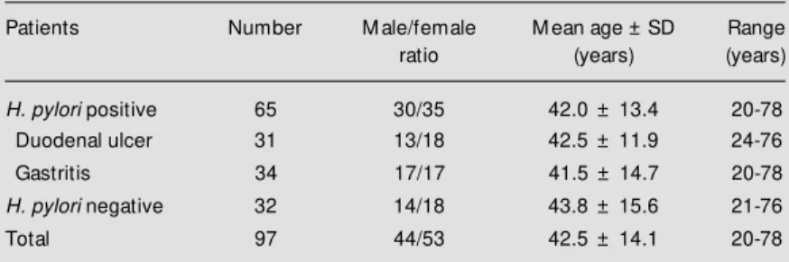
From January to December 1995 we stud-ied prospectively 97 non-consecutive pa-tients (44 men and 53 women; mean age, 42.5 years; range, 20 to 78 years) who under-went upper gastrointestinal endoscopy for evaluation of symptoms related to the upper gastrointestinal tract. Patients taking antimi-crobial drugs during the last 3 months before endoscopy were not included in the study. Among the H. pylori-positive patients 31 (13 males and 18 females) presented peptic ul-cer. The characteristics of the studied popu-lation are listed in Table 1.
At endoscopy, 3 biopsy specimens were obtained from the antral gastric mucosa for
H. pylori identification. One fragment was placed in a tube containing Christensen’s 2% urea agar. Another specimen was smeared on a glass slide, heat fixed, stained with 40% carbolfuchsin (20) and examined under an immersion lens. The specimen for culture was kept in thioglycolate broth (Difco Labo-ratories, Detroit, MI) at 4oC for no longer
than 5 h before processing. The tissue speci-mens were homogenized in tissue grinders and plated onto Belo Horizonte medium (BHM) (4). The plates were incubated at 37oC in a microaerophilic environment for
up to 7 days. Colonies of H. pylori were identified by Gram-negative appearance, positive oxidase and catalase tests, and a rapidly positive urease test.
Patients were considered to be H. pylori
positive if at least two of the three tests were positive or if the culture alone was positive, and negative, if the three tests were negative. Blood samples were drawn from each
Table 1 - Characteristics of the population studied.
Patients Number M ale/female M ean age ± SD Range
ratio (years) (years)
H. pylori positive 65 30/35 42.0 ± 13.4 20-78
Duodenal ulcer 31 13/18 42.5 ± 11.9 24-76
Gastritis 34 17/17 41.5 ± 14.7 20-78
H. pylori negative 32 14/18 43.8 ± 15.6 21-76
patient for serology at the time of endoscopy and the sera were stored at -20oC before
testing.
H. pylori antibodies were assayed by the Cobas Core anti-H. pylori IgG EIA (Roche Diagnostic Systems, Basel, Switzerland), a second generation EIA that employs a well-characterized and highly immunogenic puri-fied H. pylori-specific multi-component an-tigen preparation containing urease and free of cross-reacting flagellar proteins (18,21). The test was performed according to manu-facturer’s instructions. Briefly, patient and control samples were diluted and allowed to react for 15 min at 37oC with H. pylori
antigen-coated beads. After a washing step, the peroxidase-conjugated goat anti-human IgG antibody was added and incubated for 15 min at 37oC. After removal of the
un-bound conjugate by a washing step, the beads were incubated with a substrate solution con-taining tetramethylbenzidine and hydrogen peroxide. The enzymatic reaction was stopped by the addition of acid, and absorb-ance at 450 nm was determined. The con-centrations of IgG antibody in the serum samples were determined by interpolation from a standard curve constructed by dilu-tion of the positive control and by plotting absorbance values obtained for each stan-dard against the corresponding anti-H. py-lori concentrations (in U/ml). The values within ± 10% of the cutoff were considered to be equivocal.
The performance of the test was evalu-ated by determining the sensitivity, specific-ity and positive and negative predictive val-ues with a 95% confidence interval (CI).
The interassay reproducibility of the test was evaluated by testing 20 positive serum samples (10 from patients with duodenal ulcer and 10 from patients with gastritis) and 10 negative serum samples (selected at ran-dom) on different days and the intra-assay reproducibility by testing twice the same sera on the same day.
The χ2 test was used to compare
propor-tions between groups (sex distribution and sensitivity). Analysis of variance for vari-ables of normal distribution and the Kruskal-Wallis test for variables of asymmetrical distribution were employed to evaluate dif-ferences in IgG concentration and differ-ences in age among groups. Spearman’s cor-relation was used to compare IgG concentra-tion and patient age. The level of signifi-cance was set at P<0.05.
Re sults
H. pylori was identified by microbiologi-cal methods in 65 of the 97 (67.0%) patients studied. The H. pylori-positive (with and without peptic ulcer) and -negative groups were similar with respect to age (P = 0.81) and gender (P = 0.78).
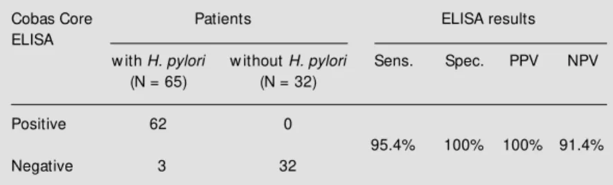
Anti-H. pylori antibodies were detected in 62 of 65 (95.4%) H. pylori-positive sub-jects and in none of the negative ones (Table 2). There were no equivocal results. Among the 3 patients who were H. pylori-positive by microbiological methods but who were nega-tive by serology, 2 had gastritis alone and 1 had peptic ulcer. The sensitivity, specificity and positive and negative predictive values of ELISA, with their 95% CI, for adults were 95.4% (88.5-98.4%), 100% (96.4-100%), 100% (96.4-100%) and 91.4% (83.5-95.9%), respectively (Table 2). There was no signifi-cant difference in sensitivity between pa-tients with (96.7%) and without peptic ulcer
Table 2 - Sensitivity, specificity, and predictive values of Cobas Core ELISA in 97 patients undergoing endoscopy.
Sens., Sensitivity; Spec., specificity; PPV, positive predictive value; NPV, negative predictive value.
Cobas Core Patients ELISA results
ELISA
w ith H. pylori w ithout H. pylori Sens. Spec. PPV NPV
(N = 65) (N = 32)
Positive 62 0
95.4% 100% 100% 91.4%
(94.1%) (P = 1.0). No significant differences (P = 0.71) in IgG concentration were ob-served in these groups (median = 56.2 U/ml, range 5.0 to 597.9 U/ml for patients with peptic ulcer, and median = 37.1 U/ml, range 2.1 to 1041.2 U/ml for patients without pep-tic ulcer).
In regard to inter- and intra-assay repro-ducibility, 29 of 30 serum samples showed identical qualitative results on repeated test-ing on different days and on the same day.
There was no significant difference in IgG concentration between the sera from males (mean = 98.5 U/ml, SD = 120.0) and females (mean = 120.9 U/ml, SD = 169.8) with peptic ulcer (P = 0.68). In contrast, the IgG concentration in the sera from females with gastritis (median = 53.3 U/ml, range 15.8 to 1041.2 U/ml) was significantly higher than in males with gastritis (median 14.2 U/ ml, range 2.1 to 436.4 U/ml) (P = 0.008). No differences in IgG concentration were ob-served among adults of different ages (P = 0.12).
D iscussio n
Several ELISA tests are commercially available to diagnose H. pylori infection in adults. Their sensitivity and specificity de-pend on the gold standards used to compare the tests, the nature of the antigens employed and the value chosen for the cutoff. Employ-ing 3 methods as gold standard - culture on BHM whose sensitivity is comparable to that of PCR (21), carbolfuchsin-stained smears and preformed urease test - and con-sidering H. pylori positivity to be present when at least 2 tests were positive or if the culture was positive, and H. pylori negativ-ity to be present when all 3 tests were nega-tive, we observed high sensitivity (95.4%) and specificity (100%) of Cobas Core for the diagnosis of H. pylori infection in adults.
As reported by others (12,15), the results of the present study demonstrated that the intra- and the interassay reproducibility of
this ELISA was excellent.
Although the number of patients in this study was not very large, the size of the sample was sufficient, as demonstrated by the less than 10% amplitude of the 95% CI for all indicators of the test.
Thus, the high sensitivity (95%) observed permits the safe use of the test in epidemio-logic surveys. However, this value is not sufficiently high to exclude the diagnosis of infection in the presence of a negative test at the individual level, although the probability of the existence of disease in this situation is low. On the other hand, the 100% specificity we observed permits us to confirm the diag-nosis of infection when the result is positive. Employing the same ELISA kit and con-sidering a truly positive result when culture and/or histology was positive, a similar re-sult was demonstrated by Goossens et al. (12) for Belgian patients (93 and 96% sensi-tivity and 95 and 91% specificity for adults 18-40 years old and >40 years old, respec-tively). Debongnie et al. (22), also in Bel-gium, using culture, and Trautmann et al. (18) in Germany, using bacterial detection in histological sections, culture, urease test and Western blot as gold standard (with the pa-tient considered to be positive when one of the tests was positive), demonstrated 98.6 and 96% sensitivity and 94.4 and 97% speci-ficity for the Cobas Core, respectively.
diagnosing infection used and the rigid crite-ria for establishing negativity contributed to the fact that no patient was erroneously clas-sified as H. pylori negative. However, in some populations the specificity of the test is very low as observed by Goossens et al. (12) for Mediterranean patients and by Antoine et al. (13) for the French patients, indicating that the ELISA must be validated for differ-ent populations.
The serologic test was negative in three patients found to be H. pylori positive by microbiological methods (two with gastritis and one with duodenal ulcer). Similar false-negatives have been reported by several in-vestigators (12,13). The most likely explana-tion is that in some patients no systemic IgG antibodies will occur although H. pylori can be detected from the gastric biopsy speci-mens, i.e., they failed to seroconvert. An-other explanation is that, although the kit employed contained several antigenic com-ponents including urease, which is highly immunogenic, some patients may harbor bacterial strains with few antigenic determi-nants in common with the strains used to obtain the antigen. In this case, the use of another ELISA employing a different anti-gen or of another method for antibody detec-tion such as immunoblotting may
demon-strate the presence of anti-H. pylori IgG. Furthermore, since in Brazil the infection occurs at an early age, the patients with gastritis may develop atrophy of the gastric mucosa with a consequent decrease in num-ber of bacteria, a fact that may alter the production of antibodies. However, histo-logical examination of the false-negative cases observed here did not reveal atrophy but only the presence of chronic gastritis of moderate intensity.
As reported by others (23,24), in the present study we also observed that IgG concentration in adults does not permit the discrimination between gastritis and duode-nal ulcer. We have no explanation for the observation that the IgG concentration in the sera from females with gastritis was signifi-cantly higher than in males with gastritis.
In conclusion, the ELISA for H. pylori
diagnosis presented high sensitivity and specificity when employed for a population in one state in Brazil, permitting the use of the test both to confirm the clinical diagnosis and to perform epidemiologic surveys. How-ever, in order to rule out H. pylori infection with safety in the presence of a negative test, the use of a more sensitive method or of a combination of methods becomes necessary for clinical diagnosis.
Re fe re nce s
1. M égraud F & Lamouliatte H (1992). Heli-cobacter pylori and duodenal ulcer. Diges-tive Diseases andSciences, 37: 769-772. 2. Parsonnet J, Friedman GD, Vandersteen DP, Chang Y, Vogelman JH, Orentreich N & Sibley RK (1991). Helicobacter pylori infection and the risk of gastric carcino-ma. New England Journal of M edicine, 325: 1127-1131.
3. Blaser M J & Parsonnet J (1994). Parasit-ism by the “ slow ” bacterium Helicobacter pylori leads to altered gastric homeosta-sis and neoplasia. Journal of Clinical In-vestigation, 94: 4-8.
4. Queiroz DM M , M endes EN & Rocha GA (1987). Indicator medium for isolation of Campylobacter pylori. Journal of Clinical
M icrobiology, 25: 2378-2379.
5. Jones DM , Lessels AM & Eldridge J (1984). Campylobacter-like organisms on the gastric mucosa: culture, histological and serological studies. Journal of Clinical Pathology, 37: 1002-1006.
6. Gray SF, Wyatt J & Rathbone BJ (1986). Simplified techniques for identifying C. pylori. Journal of Clinical Pathology, 39: 1279-1280.
7. M arshall BJ, Warren JR, Francis GJ, Langton SR, Goodw in CS & Blicow ED (1987). Rapid urease test in the manage-ment of Campylobacter pyloridis -associ-ated gastritis. American Journal of Gas-troenterology, 82: 200-210.
8. Hammar M , Tyszkiew icz T, Wadstrom T
& O’Toole PW (1992). Rapid detection of Helicobacter pylori in gastric biopsy mate-rial by polymerase chain reaction. Journal of Clinical M icrobiology, 30: 54-58. 9. M itchell HM , Lee A, Berkow ickz J &
Borody T (1988). The use of serology to diagnose active Campylobacter pylori in-fection. M edical Journal of Australia, 149: 604-609.
AR & Boutton TW (1987). Campylobacter pylori detected non invasively by the car-bon13-urea breath test. Lancet, 1:
1174-1177.
12. Goossens H, Glupczynski Y, Burette A, van den Borre C & Butzler JP (1992). Evaluation of a commercially available sec-ond-generation immunoglobulin G en-zyme immunoassay for detection of Heli-cobacter pylori infection. Journal of Clini-cal M icrobiology, 30: 176-180.
13. Antoine C, Lozniew ski A, De Korw in JD, Conroy M C, Feldmann L, Duprez A & Weber M (1995). Étude comparative de quatre méthodes sérologiques commer-cialisées pour le diagnostic de l’infection gastrique à Helicobacter pylori. Gastroent-érologie CliniqueetBiologique, 19: 182-188.
14. Hirschl AM , Rathbone BJ, Wyatt JI, Berger J & Rotter M L (1990). Comparison of ELISA antigen preparations alone or in combination for serodiagnosing Helico-bacter pylori infections. Journal of Clinical Pathology, 43: 511-513.
15. Lelw ala-Guruge J, Nilsson I, Ljungh A & Wadstrom T (1992). Cell surface proteins of Helicobacter pylori as antigens in an ELISA and a comparison w ith three com-mercial ELISA. Scandinavian Journal of
Infectious Diseases, 24: 457-465. 16. Fauchère JL (1996). Evaluation of the
anti-Helicobacter pylori serum antibody re-sponse. In: Lee A & M égraud F (Editors), Helicobacter pylori: Techniques for Clini-cal Diagnosis and Basic Research. W.B. Saunders Company Ltd., London. 17. M égraud F (1996). Advantages and
disad-vantages of current diagnostic tests for the detection of Helicobacter pylori. Scan-dinavian Journal of Gastroenterology, 215: 57-62.
18. Trautmann M , M oldrzyk M , Vogt K, Korber J, Held T & M arre R (1994). Use of a receiver operating characteristics in the evaluation of tw o commercial enzyme im-munoassays for detection of Helicobacter pylori infection. European Journal of Clini-cal M icrobiology and Infectious Diseases, 13: 812-819.
19. Jensen AKV, Andersen LP & Wachmann CH (1993). Evaluation of eight commer-cial kits for Helicobacter pylori IgG anti-body detection. Acta Pathologica M icro-biologica et Immunologica Scandinavica, 101: 795-801.
20. Queiroz DM M , Rocha GA, M endes EN, Lage AP, Carvalho ACT & Barbosa AJA (1990). A spiral microorganism in the stomach of pigs. Veterinary M icrobiology,
24: 199-204.
21. van Zw et AA, Thijs JC, Kooistra-Smid AM D, Schirm J & Snijder JAM (1993). Sensitivity of culture compared w ith that of polymerase chain reaction for detec-tion of Helicobacter pylori from antral bi-opsy samples. Journal of Clinical M icrobi-ology, 31: 1918-1920.
22. Debongnie JC, Durez P & Luyasu V (1993). Validation et usage clinique et épidémiologique d’un test sérologique commercialisé dans le diagnostic de l’infection à Helicobacter pylori. Gastroen-térologie Clinique et Biologique, 17: 98-102.
23. Andersen LF & Gaarslev K (1992). IgG subclass antibodies against Helicobacter pylori heat-stabile antigens in normal per-sons and in dyspeptic patients. Acta Pa-thologica M icrobiologica et Immunologica Scandinavica, 100: 747-751.