5 1 5 1 5 1 5 1 5 1 Mem Inst Oswaldo Cruz, Rio de Janeiro, Vol. 94, Suppl. I: 51-63, 1999
A Role for Extracellular Amastigotes in the
Immunopathology of Chagas Disease
Julio Scharfstein
+, Alexandre Morrot
Laboratório de Imunologia Molecular, Instituto de Biofisica Carlos Chagas Filho, Universidade Federal do Rio de Janeiro, 21944-970 Rio de Janeiro, RJ, Brasil
In spite of the knowledge obtained about immune control of Trypanosoma cruzi (T. cruzi) infection, the mechanisms responsible for the variable clinico-pathological expression of Chagas disease remain unknown. In a twist from previous concepts, recent studies indicated that tissue parasitism is a pre-requisite for the development of chronic myocarditis. This fundamental concept, together with the real-ization that T. cruzi organisms consist of genetically heterogeneous clones, offers a new framework for studies of molecular pathogenesis. In the present article, we will discuss in general terms the possible implications of genetic variability of T. cruzi antigens and proteases to immunopathology. Peptide epitopes from a highly polymorphic subfamily of trans-sialidase (TS) antigens were recently identified as targets of killer T cell (CTL) responses, both in mice and humans. While some class I MHC restricted CTL recognize epitopes derived from amastigote-specific TS-related antigens (TSRA), others are tar-geted to peptide epitopes originating from trypomastigote-specific TSRA. A mechanistic hypothesis is proposed to explain how the functional activity and specificity of class I MHC restricted killer T cells may control the extent to which tissues are exposed to prematurely released amastigotes. Chronic im-munopathology may be exacerbated due the progressive accumulation of amastigote-derived antigens and pro-inflammatory molecules (eg. GPI-mucins and kinin-releasing proteases) in dead macrophage bodies.
Key words: cysteine proteinases - cruzipain - kinins - amastigotes - Trypanosoma cruzi - Chagas disease
DYNAMICS OF IMMUNE RESPONSE EARLY IN
T. CRUZI INFECTION
Host resistance to microbial infection integrates two major and overlapping defense systems, in-nate and adaptive immunity. Intracellular patho-gens can quickly relay activation signals that stimu-late non-specific humoral and cellular effector re-sponses in the infected host. Assisted by these in-nate defense responses, the rate of microbial growth is delayed for several days, while the adaptive branch of immunity gets prepared to confront the pathogen on the long term. The infection can only persist when the organisms succeed to counteract the selective pressure imparted by immune effec-tor cells and/or antibodies. In the case of chronic viral infections, immune subversion is often tar-geted against intracellular pathways involved in antigen processing and/or presentation by class I MHC molecules.
In the case of T. cruzi, the mechanisms which enable their persistent growth in mammalian tis-sues were not characterized. As discussed later in this text, there are reasons to think that the mo-lecular diversity of T. cruzi organisms may affect the dynamics of tissue and organ involvement. This is supported by recent evidences showing that acute infection with parasite stocks pertaining to different genotypic groups induce distinct histo-pathology patterns in acutely infected mice (de Diego et al. 1998) Recently confirmed by research in genetic epidemiology (Souto et al. 1996, Brisse et al. 1998), the concept that T. cruzi has a multi-clonal descent was earlier proposed on the basis of isoenzyme (Miles et al. 1978, Romanha et al. 1979, Ready et al. 1980, Tibayrenc et al. 1986) and fin-gerprint analysis of k-DNA (Morel et al. 1980). While not excluding the importance of host genet-ics as a determinant of host susceptibility in vivo (Trischman et al. 1978), studies performed with laboratory strains of T. cruzi (Brener 1965, Andrade & Andrade 1966, Mello & Brener 1978) and also with parasite clones (Postan et al. 1983, de Diego et al. 1998, Macedo & Pena, 1998) suggested that the variable expression of Chagas disease may be influenced, at least to some extent, by the genetic and biological diversity of the T. cruzi clones which circulate in sylvatic and domestic reservoirs. Supported by FUJB, Faperj and Pronex II.
+Corresponding author. Fax: +55-21-280.8193. E-mail:
5 2 5 2 5 2 5 2
5 2 Amastigotes in Immunophatology of Chagas Disease J Scharfstein, A Morrot
In spite of the poor knowledge about the in-nate responses which metacyclic trypomastigotes stimulate in wound mucosal tissues, this defense system should be fully operative by the time the first cycles of intracellular infection are completed. Once released from disrupted cells, the trypo-mastigotes spread the infection via the bloodstream and/or lymphatics. At this early stage of infection, a wide range of non-phagocytic host cells can be invaded by the trypomastigotes, but host cell tar-get preference can differ markedly from one para-site clone to another due to the variable composi-tion and expression levels of their cell surface ad-hesion molecules, some of which are highly poly-morphic (Affranchino et al. 1989, Tackle & Cross 1991, Colli 1993, Schenckman et al. 1994, Giordanno et al. 1994, Pereira et al. 1996, Salazar et al. 1996) or due to differential signaling ability of the invading parasite clones (Ming et al. 1995, Burleigh & Andrews 1998).
The survival strategies of the sub-populations of T. cruzi clones which preferentially invade mononuclear phagocytic cells (reticulotropic para-sites) are not sufficiently well characterized. These parasite sub-populations must either inhibit mac-rophage activation or somehow protect themselves from their microbicidal machinary in order to sur-vive in genetically resistent host. As true for other intracellular pathogens, innate immunity against T. cruzi depends on the release of γ-IFN by NK cells (Aliberti et al. 1996, Cardillo et al. 1996). In ge-netically resistant strains, the onset of this T cell independent pathway depends on IL-12 produc-tion by activated macrophages and appears to be stimulated by tGPI-mucins, a potent class of pro-inflammatory molecules expressed by trypo-mastigotes and by atrypo-mastigotes (Camargo et al. 1997). Synergized by TNF-α (Munoz-Fernandez et al. 1992), γ-IFN induces a heightened state of microbicidal activation of macrophages in geneti-cally resistant animals, at least so during the first days of infection. The mechanisms by which acti-vated macrophages ultimately exert their anti-para-site activity is somewhat controversial, but there are indications the production of nitric oxide (NO) metabolites is critically involved (Gazzinelli et al. 1992). As for the innately susceptible inbred mice strains, their response to infection is dominated by the macrophage down-regulatory cytokines IL-10 or TGF-β (Silva et al. 1992, Gazzinelli et al. 1992). Interestingly, recent evidences suggest that factors leading to the accumulation of cyclic AMP by macrophages may down-regulate the pro-inflam-matory response which tGPI-mucins otherwise stimulate in such cells (Procopio et al. 1999); un-der these conditions, tGPI-mucins can upregulate IL-10 expression by macrophages, thereby
inhib-iting the synthesis of both IL-12(p40) and
TNF-α. In short, parasite factors which induce the accu-mulation of c-AMP may convert a potentially resistant macrophage into a highly susceptible tar-get cell.
In contrast to parasite clones that preferentially invade macrophages, the sub-populations that vade non-phagocytic cells in the first days of in-fection will grow undisturbed because CTL effec-tors are not as yet prepared to act. The necrosis caused by host cell death is extensive and may in-volve multiple tissues and organs (Lenzi et al. 1996, Cotta de Almeida et al. 1977). Inflamed tis-sues are exposed to high amounts of parasite anti-gens, either released by extracellular parasites or leaked from killed organisms. Once captured by immature dendritic cells, these antigens are trans-ported to the proximal draining lymph nodes. Af-ter upregulating their MHC molecules, the matured dendritic cells present the MHC-bound peptide antigens to naive CD4+ and CD8+ T cells. Depend-ing on the genetic background of the individual, on the antigen load and on the cytokine balance, the functional characteristics of CD4+ T cells primed by dendritic cells can be rapidly polarized under the influence of type 1 or type 2 stimulating cytokines (IL-12 and IL-4, respectively). Driven by inflammatory chemokines, these circulating CD4+ Th1 and CD8+ T (Tc1) attach to the vascu-lar adhesins expressed by activated endothelial cells (Kumar & Tarleton 1998) and are recruited into the inflamed tissues. Upon antigen-stimulation, these effectors T cells secrete γ-IFN and TNF-α (Russo et al. 1988) and/or directly kill the class I MHC infected targets by apoptosis (Kumar & Tarleton 1998, Andrade et al. 1999).
RELATIVE ROLES OF T CELLS AND ANTIBODIES IN ADAPTIVE IMMUNITY
mac-5 3 5 3 5 3 5 3 5 3 Mem Inst Oswaldo Cruz, Rio de Janeiro, Vol. 94,Suppl. I, 1999
rophage activation is transiently blocked by Th2-type of cytokines (refer to the abortive cycles of amastigote replication, later in this text), opsonization may increase rather than diminish the parasite load. In contrast to B cell knockouts, mice lacking class I or class II MHC-restricted T cells died during the acute phase of the infection with the Brazil strain. Not surprisingly, the infection was even worsened in animals that were deficient in both class I and class II MHC expression. Interest-ingly, the tissue inflammatory responses were ab-sent in mice with the MHC-II class deficiency and the animals succumbed to infection due to high parasite load. Taken together, these studies have demonstrated that effector CD4+ and CD8+ T cells are critically involved in the acute control of T. cruzi infection. Similar processes may occur in humans, given the evidence that HLA-A2+ chagasic patients (indeterminate form) often display antigen-specific CD8+ CTL in their peripheral blood (Wizel et al. 1998). The molecular mechanisms of target cell killing was recently investigated in infected mice (Kumar & Tarleton 1998). These authors showed, somewhat unexpectedly, that mice with targeted deletion of genes encoding for perfurin or granzyme B could control T. cruzi (Brazil strain) infection as efficiently as wild type animals. Their data suggest that class I MHC restricted CD8+ T cells may kill T. cruzi infected targets using the Fas/Fas L interaction system, or alternatively en-gage TNF-α and IFN-γ cytokines in this process.
T. CRUZI PROTEASES AS FACTORS OF VIRULENCE AND PATHOGENICITY
In spite of the evidence indicating that tissue parasite burden correlates with the intensity of fo-cal immunopathology, non-infected cells are some-times also damaged in the proximity of the pri-mary site of infection (Andrade ZA, this volume). Ribeiro dos Santos and Hudson (1980) were the first to propose that the passive adsorption of T. cruzi antigens onto certain types of non-infected host cells may render them susceptible to by-stander antibody-dependent cellular cytotoxicity (ADCC). In addition to being possibly involved in the peripheral neuropathy which occurs in the acute phase (Koberle et al. 1968), ADCC may be in-volved in the microangiopathy which develops in acutely infected dogs (Andrade et al. 1994). Al-though the molecular basis of these lesions is still unknown, recent studies on the cysteine-protein-ases of the cruzipain family (Cazzulo et al. 1989, Murta et al. 1990, Eakin et al. 1992, Meirelles et al. 1992, McKerrow et al. 1995) may offer new clues to investigate the underlying mechanisms. Originally identified because of their antigenic properties in chagasic patients (Scharfstein et al.
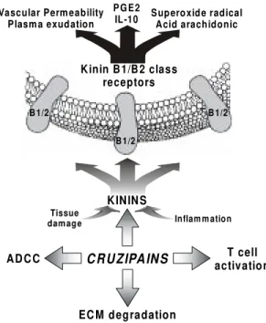
1983, 1986, Murta et al. 1990, Gazzinelli et al. 1990, Arnholdt et al. 1993, Morrot et al. 1997), the members of the cruzipain family consist of a heterogeneous group of closely related proteinase isoforms (Lima et al. 1994) encoded by about 130 genes (Campetella et al. 1992). The expression of the major isoform (cruzipain) is upregulated when the invading trypomastigotes transform into intra-cellular amastigotes (Tomas & Kelly 1996) and this process is accompanied by increased cell surface expression. It is thus conceivable that the amastigotes, once released into interstitial spaces, may use the cell surface cruzipain isoenzymes to degrade the adjacent extracellular matrix (ECM). By doing so, the parasite may prevent or disrupt ECM-interactions with TNF-α, chemokines and other inflammatory mediators involved in the re-cruitment and/or activation of immune cells T cells (Gillat et al. 1996). Cruzipain is worth studying in this context (Fig. 1), because antigenic deposits of this antigen were recently identified in sites of myocardial inflammation, in autopsies from pa-tients with severe chronic cardiomyopathy (Morrot et al. 1997). Given that some of the cruzipain isoen-zymes are stable and enzymatically active in neu-tral-acid pH, we reasoned that their half-life in ex-tracellular tissues would possibly depend on the levels of host proteinase inhibitors which perme-ate inflammatory sites. We thus sought to investi-gate the immunological implications of cruzipain interactions with α2-Macroglobulin, a non-specific plasma proteinase inhibitor (Morrot et al. 1997). In view of previous evidences indicating that CD4+ T cells from chagasic patients responded to cruzipain (Arnholdt et al. 1993), we verified if the functional inactivation of the proteinase by α2M could influence the efficiency of antigen presen-tation and processing by human monocytes. Our studies revealed that monocytes engage a highly endocytic scavenger receptor expressed by mono-cytes (CD91 or LRP/α2MR) to rapidly internalize
5 4 5 4 5 4 5 4
5 4 Amastigotes in Immunophatology of Chagas Disease J Scharfstein, A Morrot
integrate elements of innate defense systems with those in charge of acquired immunity.
As a follow-up from the studies involving
α2M, we turned our attention to kininogen (HK or LK), both of which are members of the cystatin superfamily of cysteine proteinase inhibitors (Barret et al. 1986). In addition to the cystatin-like domains, these multifunctional plasma glycopro-teins (HK) participates in the activation of the in-trinsic pathway of coagulation and modulate plate-let activation by thrombin. Important to our dis-cussion, HK or LK are the parent substrate mol-ecules from which bradykinin or lysil-bradykinin are released, upon proteolytic cleavage respectively by plasma or tissue kallikrein (Bhoola et al. 1992).
Kinins are short-lived peptides which engage dis-tinct subtypes of G-protein coupled (GPC) kinin receptors in a wide spectrum of biological pro-cesses, such as modulation of neuronal activity (Higashida et al. 1990), cell proliferation and vas-cular permeability (Bhoola et al. 1992), smooth muscle contraction or relaxation (Monbouli & Vanhoutte 1995). Depending on the host cell, ki-nin signaling is transduced by GPC-receptors that are either expressed constitutively (B2) or are in-duced (B1) during anoxia or noxious stimuli (Regoli 1980, Burch & Kyle 1992).
The initiative to investigate the relationship of T. cruzi with the kinin system was initially moti-vated by enzymatic specificity studies carried out with cruzipain 2 (Lima et al. 1994), a minor isoform whose recombinant form was only recently ex-pressed in S. cerevisae. After noting that its sub-strate specificity somewhat resembled that of tis-sue kallikrein, we were able to demonstrate that these isoforms could act as kinin-releasing en-zymes. (Del Nery et al. 1997), although with dif-ferent efficiency. On a first look, the finding that cruzipain was able to proteolyze HK/LK seemed paradoxical because these plasma glycoproteins were previously reported to inactivate cruzipain by means of their tight-binding cystatin domains (Stoka et al. 1995, Scharfstein et al. 1995). Clues to understand the mechanism of kinin-release emerged when the Ki’s obtained for HK were de-termined for different isoforms of cruzipain: inter-estingly, we observed that r-cruzipain 2 was mark-edly less sensitive to inhibition by HK as compared to cruzain (Lima et al. in preparation) the arche-type from the cruzipain family (McKerrow et al. 1995). Our data suggest that the kininogenase ac-tivity of T. cruzi cysteine-proteinases might have evolved due to structural diversification of the cata-lytic site of some isoforms, such as cruzipain 2. Notably, in the same study (Del Nery et al. 1997) we also showed that cruzipain could generate bradykinin indirectly, that is, by converting the plama prekallikrein zymogen in active kallikrein. In a parallel study, we used intravital microscopy to verify if the topical application of purified cruzipain on the hamster cheek pouch could stimu-late increases in vascular permeability (Svensjo et. 1997). Potent responses were indeed observed, suggesting that T. cruzi may use cruzipain isoezymes to generate kinins upon cellular contact with endothelial cells (Fig. 1). Ongoing studies in mice with targeted deletion of kinin-receptors should confirm if this hemoflagellate actively en-gages the kallikrein-kinin system to migrate across non-fenestrated blood capillaries.
The role of T. cruzi-induced kinin release in the pathogenesis of chronic heart disease, previ-KININ S
CRUZIPAINS
Inflam m ation
AD C C
EC M degradation
Tissue damage
B 1/2 B 1/2
B 1/2
K inin B 1/B 2 class receptors
Vascular Perm eability Plasm a exudation
PGE2
IL-10 Superoxide radicalAcid arachidonic
T cell activation
5 5 5 5 5 5 5 5 5 5 Mem Inst Oswaldo Cruz, Rio de Janeiro, Vol. 94,Suppl. I, 1999
ously suggested on the basis of animal studies (Morris et al. 1990, Rossi 1990) is worth explor-ing. In a detailed three-dimensional confocal mi-croscopy analysis of post-necropsy heart specimens derived from chronic chagasics autopsies, Higuchi et al. (1999) have noted extensive abnormalities of cardiac microcirculation as well as in the inter-stitial matrix patterns of myocardial tissues. Pecu-liar lesions, often manifested as arteriolar dilata-tion and capillary vessel tortuosity, were described, being thus far vaguely attributed to fibrosis and/or to as yet uncharacterized lesions induced by the parasites. Apart from initial studies looking at ef-fects of desialilation by TS enzymes (Libby et al. 1986), little is known about the biochemical basis of T. cruzi interaction with endothelial cells (Mor-ris et al. 1990). As discussed earlier in this section, the realization that cruzipain-isoenzymes are ca-pable of releasing pro-inflammatory kinins offers a new experimental framework to investigate the pathophysiological consequences of long-term stimulation of myocardial capillaries by vasoac-tive kinins. Our description of cruzipain effects on the microcirculation of the hamster cheek pouch (Svensjo et al. 1977) suggest that merely a few parasites may suffice to generate vasoactive kinins, thereby provoking plasma exudation from vicinal capillaries (Fig. 2, bottom panel). Similar processes, perhaps involving NO released by endothelial or smooth muscle cells may underlie the neuronal le-sions observed in the acute stage of infection.
SUBVERSION OF THE CLASS I MHC PATHWAY OF ANTIGEN PRESENTATION IN T. CRUZI INFECTED CELLS
During microbial infection, host class I or class II molecules encoded by polymorphic MHC genes bind to antigenic peptides (T epitopes) produced by the invasor. Once associated with the appropri-ate MHC allele product, the peptide:MHC com-plex is targeted to the cell surface of the infected cell, where it is “presented” to naive or effector T cells. The intracellular pathways that generate small antigenic peptides by proteolytic processing and then enable MHC loading are collectively referred as antigen processing and presentation system. In the past years, the multiple checkpoints that are sensitive to attack by virus and other intracellular parasites were characterized. In general terms, the viral products may target elements controlling (i) antigen-proteolysis by proteosomes, (ii) peptide transport to the ER (iii) MHC assembly and/or sort-ing and (iv) surface expression of class I MHC:peptide complexes.
As shown in mice, the analysis of HLA-A2-restricted CTL specificities in the peripheral blood
of chagasic patients has identified different mem-bers of a subfamily of TS-related antigens as killer cell targets (Wizel et al. 1997, Low et al. 1997): two originating from amastigote-derived antigens, ASP-1 and ASP-2, and one from trypomastigote-specific antigen, TSA-1. Teleologically, the usage of a vast array of polymorhic T. cruzi proteins as parent substrates for the proteosomal proteases makes sense because this should increase oppor-tunities for epitope loading of the highly diverse MHC-class I products that exist in genetically out-bred populations. In other words, had the antigen variability involved exclusively single genes, the parasite sub-populations which display variant se-quences should have increased chances to escape from detection. Thus, structural variability im-ported by multi-copy polymorphic genes may have been advantageous to host-parasite equilibrium be-cause it should reduce the excessive risk associ-ated with the growing diversification of parasite sub-populations.
The maneuvers which some T. cruzi clones use to subvert the class I presentation pathway are not clarified as yet. In a recent study focusing on class II MHC restricted CD4+ responses of inbred mice, Kahn and Wlekinski (1997) offered insight into this problem. They showed that the simultaneous expression of individual antigens from polymor-phic TS subfamily may limit the availability of processed epitopes from each antigen below the threshold level required to stimulate a protective IFN-γ response against the parasite. It will be in-teresting to know if similar mechanisms may al-low for parasite escape from detection by class I-MHC restricted killer T cells. Admittedly, how-ever, antigen variation per se may not ensure pro-tection to parasites that are multiplying in host cells from genetically outbred individuals (e.g. dogs and humans), because in such cases antigen-presenta-tion can involve a more diversified array of MHC allelic products. As discussed in the next section, there are reasons to think that amastigotes may be equipped with the molecular machinery required to subvert the class I MHC presentation pathway.
T. CRUZI CLONAL DIVERSITY AND AMASTIGOTE EVASION MECHANISMS
5 6 5 6 5 6 5 6
5 6 Amastigotes in Immunophatology of Chagas Disease J Scharfstein, A Morrot
the rates of intracellular division and transforming efficiency of amastigotes vary significantly among different clones (Dvorak & Hyde 1973, Revollo et al. 1998), the host cell ability to generate antigenic peptides to class I MHC molecules may be jected to similar constraints. Perhaps a given sub-population of amastigote clones may be more apt to subvert the class I MHC presentation pathway if they multiply slowlier. By reducing their me-tabolism, the availability of amastigote antigens (e.g., ASP) in the host cell cytoplasm may be also decreased; under these conditions, antigenic pep-tides from the slowly growing amastigotes (latent forms) may not reach the threshold required to load class I MHC molecules. Conversely, sub-popula-tions of amastigote clones which exhibit high rates of amastigote division should be able to release high contents of polymorphic antigens into the cytosol, thus allowing for efficient presentation of antigenic peptides at the target cell surface. It is thus conceivable that tissues from chronically in-fected outbred individuals may harbor a small num-ber of slowly dividing clones that tend to escape from amastigote-specific killer T cells. As dis-cussed later on, we propose that these parasite sub-populations may contribute to the exacerbation of immunopathology. Of course, parasite clones that sustain a high levels of proliferation may also suc-ceed at subverting the class I MHC pathway, but in this case they must actively block antigen pro-teolysis, peptide transport or cell surface exposure of the class I MHC:peptide complex at the target cell surface, by analogy to mechanisms described for virus.
DUAL ROLE OF TRYPOMASTIGOTE-SPECIFIC KILLER T CELLS
On a first impression, the ability of CTL to kill non-phagocytic targets which harbor intracellular trypomastigotes would seem to be a redundant pro-cess, because most of the infective parasites should be anyway released into the extracellular fluids upon host cell burst. However, target cell death by apoptosis should be advantageous to the host (Andrade et al. 1999), inasmuch as this mecha-nism would spare host tissues from the detrimen-tal effects of necrosis that would otherwise occur if the target cells collapse due to excessive num-bers of parasites. Since mutual destruction is not a fruitful evolutionary strategy, it is unlikely that host cell apoptosis also destroys all the intracellular parasites. During stages of chronic infection, the large majority of infected cells should harbor para-sites derived from a single parasite clone. Since amastigote division proceeds asynchronously (Dvorak & Hyde 1973), the clones which succeed at subverting the class I antigen presentation
path-way should gradually transform into trypo-mastigotes. The trypomastigote specific CTL may then attack the target cells as soon as the antigenic peptide epitopes (originating from TS-related or other dominant antigens) reach the threshold re-quired for class I MHC presentation. Although the rates of MHC-loading and surface expression may vary from one type of host cell to another, it is rea-sonable to predict that the most infected target cells should still harbor some intracellular amastigotes by the time the trypomastigote-specific CTL in-duced death by apoptosis. Killing at early stages of trypomastigote-transformation makes sense be-cause the antigen presentation machinery may not be functional at later stage of the intracellular cycle. Therefore, an early attack by trypomastigote-spe-cific killers is the best possible compromise be-tween the host and the parasite, because the re-lease of just a few intracellular trypomastigotes should suffice to ensure long term survival of both. According to this model, the extent to which tis-sues are exposed to prematurely released amastigotes should be critically influenced by the time course of target cell recognition by trypomastigote specific killer T cells. In short, we predict that high numbers of prematurely released amastigotes should be exposed to tissues when (i) the class I MHC presentation of amastigote-an-tigens is subverted or, alternatively, when amastigote-specific CTL are either deleted or down-regulated, (ii) trypomastigote-specific kill-ers attack host cells as soon as the trypomastigotes are transformed. Our model sustains that patho-genic sub-populations of trypomastigote clones should maximize (rather than minimize, as postu-lated for amastigotes) antigen presentation by the class I MHC presentation pathway.
ABORTIVE-CYCLES OF AMASTIGOTE REPLI-CATION: ROLE OF CLASS I MHC-KILLER T CELLS
5 7 5 7 5 7 5 7 5 7 Mem Inst Oswaldo Cruz, Rio de Janeiro, Vol. 94,Suppl. I, 1999
amastigote clones may succeed in subverting the class I MHC presentation pathway. Hence, the kill-ing of target cells by CTL (secretkill-ing type 2 cytokines, Tc2) specific for trypomastigote pep-tides may release a substantial number of amastigotes into the interstitium. New cycles of abortive replication may ensue, as the extracellu-lar amastigotes infect macrophages that are re-cruited into the primary site of infection. Our model (Fig. 2) predicts that the ultimate fate (death or replication) of the intracellular amastigotes lodged in the macrophages should be determined by the local balance between cytokines released by acti-vated Th1 or Tc1 (eg. IFN-γ, TNF-α) and mac-rophage down-regulatory cytokines (IL-10 and TGF-β) released by Tc2/Th1cells or by equivalent cytokines released by Tc2 CD8+ lymphocytes. As discussed earlier in this text, macrophage stimula-tion by parasite-factors which induce PGE2 may down-regulate the pro-inflammatory response oth-erwise elicited by by t-GPI-mucins (Procopio et al. in press).
In the acute infection, the tissue parasite load in lymphoid organs, such as the spleen, is initially high but it tends to decrease as cellular immune effectors intervene. It is possible that the extracel-lular amastigotes which are prematurely released from host cells, due to attack by trypomastigote-specific CTL, may somehow contribute to the massive polyclonal activation of lymphocytes (D’Imperio-Lima et al. 1985) and/or to the tran-sient state of non-specific immunosuppression (Teixeira et al. 1978). Interestingly, there is a close temporal relationship between the drop in tissue parasite load and the onset of the immunoregulatory changes which occur in peripheral lymphoid tis-sues (Dos Reis 1997). It is thus possible that the pool of spleen CD4+ T cells which undergo acti-vation-induced cell death (AICD) upon in vitro stimulation with anti-CD3 (Dos Reis et al. 1995) is enriched by amastigote-specific CD4+ T cells. The recent demonstration that macrophages ex-posed to live amastigotes activate CD4+ and CD8+ T cells from lymph nodes and spleen (Caulada-Benedetti et al. 1998) is consistent with the above mentioned concept. As pointed out by Dos Reis et al. (1997), the polarization of Th2-type responses at late stages of the acute infection may occur due to the elimination of activated Th1-type cells by apoptosis. It will be interesting to determine if the accumulation of amastigote-derived antigens is a pre-requisite for the induction of anergy, clonal de-letion and/or Th2-dependent down-regulation of amastigote-specific CD4+ Th1 lymphocytes (Fig. 2, upper panel). If true, animals or patients which exhibit deficit in trypomastigote-specific CTL
(Tc2) would not be able to release substantial num-bers of intracellular amastigotes to lymphoid tis-sues. Under these hypothetical conditions, amastigote-specific CD4+ Th1 cells would not be as efficiently eliminated or down-regulated and the Th1 inflammatory subset would expand, thereby aggravating tissue damage. These general predictions are consistent with recent data sug-gesting that early immunological events can influ-ence the development of inflammation at chronic stages of infection (Mariano et al. 1999). Perhaps the hypothesis outlined here (Fig. 2 upper panel) may offer clues to understand the mechanisms un-derlying the contrasting behavior of the Colombiana-Balb/C model of chronic carditis (Ribeiro dos Santos et al. 1992). Instead of the CD8+ T cells (anti-parasite) which dominate the chronic myocardial infiltrates in the majority of mice models (Tarleton et al. 1997) as well as in humans (Reis et al. 1997), the infection of Balb/C mice with Colombiana favors the induction of autoreactive CD4+ T cell effectors which attack cardiac tissues. According to our model, the Balb/ C animals infected by the Colombiana strain may have failed to efficiently eliminate or down-regu-late cross-reactive amastigote-specific Th1 CD4+ cells in lymphoid tissues, perhaps because trypomastigote-specific CD8+ T cells (Tc2) are not functionally active in this model, at least so in the chronic stages of infection. Our hypothesis also predicts that the self-epitopes recognized by these CD4+ Th1 cell clones should cross react with amastigote antigens from the Colombiana strain.
5 8 5 8 5 8 5 8
5 8 Amastigotes in Immunophatology of Chagas Disease J Scharfstein, A Morrot
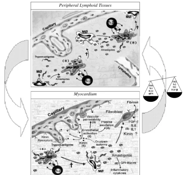
Fig. 2: role of abortive cycles of amastigote replication in the immunopathology of Chagas disease. The model predicts that the blockade of macrophage activation by anti-inflammatory cytokines (IL10 and/or TGF-β) allows for the replication of amastigotes in the cytosol (S). Some amastigote clones can subvert the class I MHC antigen-presentation pathway, thus avoiding immune detection by amastigote-specific killer T cells. As soon as the first trypomastigotes are transformed, peptide-antigens from these forms are generated in the host cell cytoplasm. The peptide:MHC complex is displayed at the cell surface of the macrophages, propitiating the attack by trypomastigote-specific Tc2 killer cells (=). Because amastigote division is asynchronous, the macroph-ages still harbor several intracellular amastigotes at the time of attack by trypomastigote-specific killer T cells. Extracellular amastigotes are then released to the interstitium, together with a few trypomastigotes. While the trypomastigotes move away from the site of infection, the nonmotile amastigotes stay in the periphery. The extracellular amastigotes are then opsonized and rapidly internalized by tissue macrophages, which are kept suppressed by IL-10 and/or TGF-β released by Th2-type of cells, or by Tc2 effectors. These cytokines again prevents macrophage activation, allowing for amastigote escape into the cytosol, where they start to multiply. Once again, the amastigotes subvert the class I MHC presentation pathway, and consequently, are not recognized by amastigote-specific killer cells. Several rounds of abortive-infection are induced by trypomastigote-specific killers (Tc2) [(SÕ=)]. This feedback loop may persist for some time, allowing for the build up of amastigote-antigens and pro-inflamatory products inside dead macrophage bodies. It is proposed that the onset of the class I MHC-restricted abortive cycles of amastigote replication in lymphoid tissues (upper panel) may indirectly play a role in immunoregulation (refer to text). In the myocardium (right panel), this pathway may be involved in the exacerbation of the focal inflammatory responses, precipitating the transition from the indeter-minate form to the chronic form of the disease. Immunopathology is exacerbated due to the local deposition of antigens and/or pro-inflammatory products from pathogenic amastigotes. Living amastigotes are not be readily visualized in histological sections because they are constantly released and internalized by macrophages. Multiple molecular factors could contribute to the aggrava-tion of myocardial tissue injury. For example, the kinin-releasing activity of cruzipain isoforms (1) could activate vascular endot-helial cells (2), thus increasing vascular permeability (3) and promote plasma exudation (4). At some critical stage, the pro-inflammatory activity of GPI-mucins (6) could stimulate an incipient wave of Th1-response in the myocardial tissues, which would be promptly followed by a dominant regulatory Th2/Tc2-response (TGF-β, IL6) (5). The arrows linking the two panels highlights the importance of the peripheral lymphoid organ function (left panel) in the maintenance of the immunological homeostasis.
Peripheral Lymphoid Tissues
Myocardium
TH1 Tc1 IL12
γγγγγIFN
5 9 5 9 5 9 5 9 5 9 Mem Inst Oswaldo Cruz, Rio de Janeiro, Vol. 94,Suppl. I, 1999
ROLE OF CD8+ KILLERS IN THE EXACERBATION
OF CHRONIC IMMUNOPATHOLOGY
Andrade et al. (1997) have recently demonstrated that the canine model accurately reproduces the main features of the indeterminate form of human Chagas disease. They suggested that the heart lesions in the indeterminate stage of the disease are caused by self-limited cycles of focal inflammatory changes, prob-ably determined by subtle shifts in the immunoregulatory activity of effector T lympho-cytes. The canine model does not seem to support an obvious role for microvascular lesions in this stage of the disease. In contrast to the progressive nature of he chronic carditis observed in mice, the transition from indeterminate to chronic heart dis-ease in dogs is not gradual. In humans, the distribu-tion of cytokine-producing cells in myocardial tis-sues revealed that Th2 type cells are usually associ-ated with higher deposition of parasite antigens (Higuchi et al. 1993, Reis et al. 1997).
Here the concept of a “pathogenic” clone is defined in the operational sense: it refers to an op-portunistic population of intracellular amastigotes that somehow exploits antigen variation to subvert the class I presentation of target cells which ex-press a given MHC-haplotype. In other words, a pathogenic clone isolated from one chronically infected individual (outbred species) would not necessarily induce pathology in individuals that express a different MHC haplotype. As explained earlier in this article, subversion of class I path-way of antigen presentation should occur more fre-quently in inbred mice than in humans, or dogs, because the murine host cells only count with a limited number of MHC alleles to counteract the extreme antigen variability of the intracellular para-sites. We may thus predict that the “pathogenic” parasite sub-populations are kept at very low num-bers in tissue reservoirs because the selective pres-sure exerted by amastigote-specific killer T cells in heterozygous individuals is very strong. The “pathogenic” clones may only escape from amastigote-specific CTL by infecting suppressed macrophages, the latter state being dictated by the modulatory influence of type-2 cytokines which dominate the PBMC responses of patients during the indeterminate stage of the disease (Bahia-Oliveira et al. 1998).
From where do these parasites clones come from? As already mentioned, extracellular amastigotes, once prematurely released from killed target cells in any tissues, may persistently infect resident macrophages, using to this end the alternative pathway of invasion (Ley et al. 1988). Organs that usually maintain
macroph-ages in a relatively supressed state (eg. adrenal glands, Teixeira et al. 1977) may favor the out-growth of these potentially pathogenic amastigote clones. In a period of a relatively profound immuno-suppression, perhaps the threshold re-quired for amastigote-epitope presentation by the class I presentation pathway is no longer reached in the suppressed macrophage. Because they are not detected by amastigote-specific killer T cells, the intracellular amastigotes can then transform into trypomastigotes. After being recognized by trypomastigote-specific CTL, the target cell is killed, and the parasites are prematurely released into the interstitium. Those that had diffferentiated into trypomastigotes fall into the circulation and eventually reach the myocardium by traversing blood capillaries which already ex-press adhesion molecules (Zhang & Tarleton 1996). After lodging inside a cardiac fiber, or in a tissue macrophage (Fig. 2, bottom panel) these parasites again subvert the class I MHC presen-tation, thus avoiding recognition by amastigote-specific killer T cells. As the first trypomastigotes transform, the target cell is again killed by trypomastigote-specific Tc2 killer T cells. The fraction of intracellular amastigotes that had not transformed into trypomastigotes is then once again released into the interstitium. Successive rounds of invasion and abortive cycles of infec-tion in suppressed macrophages then cause the progressive accumulation of amastigote-antigens and/or their pro-inflammatory products, such as GPI-mucins, inside dead macrophages (apoptotic bodies). In organs that are highly vascularized, such as the heart, cruzipain-isoenzymes secreted or leaked by T. cruzi amastigotes can process kininogen, thus releasing pro-inflammatory ki-nins. By acting on adjacent endothelial cells, these vasoactive peptides may induce plasma exudation, thereby expanding the inflammatory response, and contributing to interstitial fibro-sis. The chemokines released by tissues exposed to tGPI-mucins and other pro-inflammatory me-diators might also differently recruit autoreactive CD8+ or CD4+ lymphocytes (Cunha-Neto et al. 1996) into the myocardium, thus aggravating chronic immunopathology.
ACKNOWLEDGMENTS
To Dr George dos Reis for helpful comments made while reading this manuscript.
REFERENCES
6 0 6 0 6 0 6 0
6 0 Amastigotes in Immunophatology of Chagas Disease J Scharfstein, A Morrot
Chagas’ disease. Mol Biochem Parasitol34: 221-228.
Aliberti JC, Cardoso MA, Martins GA, Gazzinelli RT, Vieira LQ, Silva JS 1996. Interleukin-12 mediates resistance to Trypanosoma cruzi in mice and is pro-duced by murine macrophages in response to live trypomastigotes. Infect Immun64: 1961-1967. Andrade SG, Andrade Z 1966. Estudo histopatológico
comparativo das lesões produzidas por 2 cepas do Trypanosoma cruzi. O Hospital 70: 1268-1278. Andrade ZA, Andrade SG, Correa R, Sadigursky M,
Ferrans VJ 1994. Myocardial changes in acute Try-panosoma cruzi infection: ultrastructural evidence of immune damage and the role of microangiopathy. Am J Pathol 144: 1403-1411.
Andrade ZA, Andrade SG, Sadigursky M, Wenthold RJ Jr, Hilbert SL, Ferrans VJ 1997. The indeterminate phase of Chagas’ disease: ultrastructural character-ization of cardiac changes in the canine model. Am J Trop Med Hyg 57: 328-336.
Andrade ZA, Zhang YuZX, Andrade SG, Takeda K, Sadigursky M, Ferrans VJ 1999. Apoptosis in a ca-nine model of acute Chagasic myocarditis. J Mol Cell Cardiol 581-596.
Andrade LO, Machado CMS, Chiari E, Pena SDJ, Macedo AM 1999. Role of parasite and host genetic backgrounds in the differential tissue tropism of Try-panosoma cruzi strains (this volume).
Arnholdt AC, Piuvezam MR, Russo DM, Lima AP, Pedrosa RC, Reed SG, Scharfstein J 1993. Analysis and partial epitope mapping of human T cell re-sponses to Trypanosoma cruzi cysteinyl proteinase. J Immunol 15: 3171-3179.
Bahia-Oliveira LM, Gomes JA, Rocha MO, Moreira MC, Lemos EM, Luz ZM, Pereira ME, Coffman RL, Dias JC, Concado JR, Gazzinelli G, Correa-Oliveira R 1998. IFN-gamma in human Chagas’ disease: protection or pathology? Braz J Med Res 31: 127-131.
Barret AJ, Rhawlings ND, Davies ME, Machleidt W, Salvesen G, Turk V 1986. In AJ Barret & G Salvesen (eds), Proteinase Inhibitors: Cysteine-proteinase Inhibitors of the Cystatin Superfamily, p. 515-569. Elsevier, Amsterdam.
Bhoola KD, Figueroa CD, Worthy K 1992. Bioregulation of kinins: kallikreins, kininogens, and kininases. Pharmacol Rev44: 1-80.
Brener 1965. Comparative studies of different strains of Trypanosoma cruzi. Ann Trop Med Parasitol59: 19-26.
Burch RM, Kyle D 1992. Recent developments in the understanding of bradykinin receptors. Life Sci50: 829-838.
Burleigh B, Andrews NW 1998. Signaling and host cell invasion by Trypanosoma cruzi. Curr Opin Microbiol 1: 461-465.
Camargo MM, Almeida IC, Pereira ME, Ferguson MA, Travassos LR, Gazinelli RT 1997. Glycosylphos-phatidylinsitol-anchored micin-like glycoproteins isolated from Trypanosoma cruzi trypomastigote initiate the synthesis of proinflammatory cttokines by macrophages. J Immunol 158: 5890-5901.
Cazzulo J, Cousi R, Raimondi A, Wernstedt C, Hellman U 1989. Further characterization and partial amino acid sequence of a cysteine protease (cruzipain) from Trypanosoma cruzi. Mol Biochem Parasitol 33: 33-42.
Campetella O, Henrickson J, Aslund L, Frasch ACC, Petterson U, Cazzulo JJ 1992. The major cysteine protease (cruzipain) is encoded by multiple poly-morphic tandemly repeated organuzed genes located in different chromossomes. Mol Biochem Parasitol 50: 225-234.
Cardillo F, Voltarelli, JC, Reed SG, Silva JS 1996. Regu-lation of Trypanosoma cruzi infection in mice by gamma interferon and interleukin 10: role of NK cells. Infect Immun 64: 128-134.
Cerwenka A, Carter LL, Reome, JB, Swain, SL, Dutton RW. 1998. In vivo persistence of CD8 polarized T cell subsets producing type 1 or type 2 cytokines. J Immunol 161: 97-105.
Colli W 1993. Trans-sialidase: a unique enzyme activ-ity discovered in the protozoan Trypanosoma cruzi. FASEB J 7: 1257-1264.
Cotta de Almeida V, Bertho AL, Villa Verde DM, Savino W 1997. Phenotypic and functional alteration of thy-mic nurse cells following acute T. cruzi infection. Clin Immunopath82: 125-132.
Coutinho CMLM, Cavalcanti, GH, van Leuven, Araujo-Jprge, TC. 1997. Alpha-2 Macroglobulin binds to the surface of Trypanosoma cruzi. Parasitol Res 83:144-150.
Cunha-Neto E, Coelho V, Guilherme A, Fiorelli N, Stolf N, Kalil J 1996. Autoimmunity in Chagas’ disease-identification of cardiac myosin-B13 Trypanosoma cruzi protein cross-reactive T cell clones in heart lesions of a chronic Chagas’ cardiomyopathy patient. J Clin Inv 98: 1709-1712.
de Diego JA, Palau MT, Gamallo C, Penin P 1998. Re-lationships between histopathological findings and phylogenetic divergence in Trypanosoma cruzi. Trop Med Int Health 3: 222-233.
Del Nery E, Juliano MA, Lima APCA, Scharfstein J, Juliano L 1997. Kininogenase activity by major cysteinyl proteinase (Cruzipain) from Trypanosoma cruzi. J Biol Chem 272: 25713-25718.
D’ Imperio Lima MR, Joskowicz M, Coutinho A, Kipnis T, Eisen H 1985. Very large and isotypically atypi-cal polyclonal plaque-forming cell responses en mice infected with Trypanosoma cruzi. Eur J Immunol 15: 201.
Dos Reis GA 1997. Cell-mediated immunity in experi-mental Trypanosoma cruzi infection. Parasitol To-day13: 335-341.
Dvorak JA, Hyde TP 1973. Trypanosoma cruzi: inter-action with vertebrate cells in vitro. Individual in-teractions at the cellular and subcellular levels. Experim Parasitol 34: 268-283.
6 1 6 1 6 1 6 1 6 1 Mem Inst Oswaldo Cruz, Rio de Janeiro, Vol. 94,Suppl. I, 1999
Scharfstein J 1990. Identification and partial char-acterization of T. cruzi antigens recognized by T cells and immune sera from chagasic patients. Infec Immuni 58: 1437-1444.
Gazzinelli RT, Oswald IP, James SL, Sher A 1992. IL-10 inhibits parasite killing and nitrogen oxide pro-duction by IFN-gamma-activated macrophages. J Immunol148: 1792-1796.
Gillat D, Cahalon L, Hershkoviz R, Lider O 1996. In-terplay of T cells and cytokines in the context of enymatically modified extracellular matrix. Immunol Today 17: 16-20.
Giordano R, Chammas R, Veiga SS, Colli W, Alves MJ 1994. An acidic component of the heterogeneous Tc-85 protein family from the surface of Trypanosoma cruzi is a laminin binding glycoprotein. Mol Biochem Parasitol 65: 85-94.
Higuchi ML, De Brito T, Reis MM, Barbosa A, Bellotti G, Pereira Barreto AC, Pileggi F 1993. Correlation between Trypanosoma cruzi parasitism amd myo-cardial inflammatory infiltrate in human chronic chagasic myocarditis: light microscopy and immu-nohistochemical findings. Cardiov Pathol 2: 101-106.
Higuschi ML, Fukasawa S, De Brito T, Parzianello LC, Bellotti G, Ramires JAF 1999. Different microcir-culatory and intestinal matrix patterns in idiopathic dilated cardiomyopathy and Chagas’ disease: a three dimensional confocal microscopy study. Heart 81: 1-6.
Iida K, Whitlow NM, Nussenzweig V 1989. Amastigotes of Trypanosoma cruzi escape destruction by termi-nal complement components. J Exp Med 169: 881-891.
Kahn SJ, Wleklinski M 1997. The surface glycoproteins of Trypanosomacruzi encode a superfamily of vari-ant T cell epitopes. J Immunol159: 4444-4451. Kahn SJ, Wleklinski M,Aruffo A, Farr A, Coder D, Kahn
M 1995. Trypanosoma cruzi adhesion to macroph-ages is facilitated by the mannose receptor. J Exp Med 182:1243-1258.
Koberle F 1968. Chagas’ disease and Chagas’ syn-dromes: the pathology of American trypanosomia-sis. Adv Parasitol 6: 63-116.
Kolb H, Kolb-Bachofen V 1998. Nitric oxide in autoim-mune disease: cytotoxic or regulatory mediator? Immunol Today 19: 556-561.
Krettli AU, Brener Z 1982. Resistance against Trypa-nosoma cruzi is associated to anti-living trypomas-tigote antibodies. J Immunol 128: 2009-2012 Kumar S, Tarleton RL 1998. The relative contribution
of antibody production and CD8+ T cell function to immune control of Trypanosoma cruzi. Parasite Immunol20: 207-216.
Lages-Silva E, Ramirez LE, Krettli AU, Brener Z 1987. Effect of protective and non-protective antibodies in the phagocytosis rate of Trypanosoma cruzi blood forms by mouse peritoneal macrophages. Parasite Immunol 9: 21-30.
Lenzi HL, Oliveira DN, Lima MT, Gattass CR 1996. Try-panosoma cruzi: paninfectivity of CL strain during murine acute infection. Exp Parasitol84: 16-27.
Ley V, Andrews NW, Robbins ES, Nussenzweig V 1988. Amastigote of Trypanosoma cruzi sustain na infec-tive cycle in mammalian cells. J Exp Med168: 649-659.
Libby P, Alroy J, Pereira MEA 1986. A neuraminidase from Trypanosoma cruzi removes sialic acid from the surface of mammalian myocardial and endothe-lial cells. J Clin Invest 77: 127-135.
Lima APCA, Tessier DC, Thomas DY, Scharfstein J, Storer AC, Vernet T 1994. Identification of new cys-teine protease gene isoforms in Trypanosoma cruzi. Mol Biochem Parasitol 67: 333.
Low HP, Santos MAM, Wizel B, Tarleton RL 1997. Amastigote surface proteins of Trypanosoma cruzi are targets for CD8+ cytotoxic T lymphocytes. J Immunol (in press).
Macedo AM, Pena S 1998. Genetic variability of Try-panosoma cruzi: implications for the pathogenesis of Chagas’ disease. Parasitol Today 14: 119-124. Mariano CR, Dímperio Lima MR, Grisotto MG, Alvarez
JM 1999. Influence of acute-phase parasite load pa-thology, parasitism, and activation of the immune system at the late chronic phase of Chagas’ disease. Infect Immun 67: 308-18.
McKerrow JH, McGrath ME, Engel JC 1995. The cys-teine protease of Trypanosoma cruzi as a model for antiparasite drug design. Parasitol Today 11: 279-282.
Meirelles MN, Juliano L, Carmona E, Silva SG, Costa EM, Murta AC, Scharfstein J 1992. Inhibitors of the major cysteinyl proteinase (GP57/51) impair host cell invasion and arrest the intracellular development of Trypanosoma cruzi in vitro. Mol Biochem Parasitol 52: 175-184.
Melo RC, Brener Z 1978. Tissue tropism of different Trypanosoma cruzi strains. J Parasitol 64: 475-482. Miles MA, Souza A, Povoa M, Shaw JJ, Lainson R, Toye PJ 1978. Isozymic heterogenity of Trypano-soma cruzi in the first autochtonous patients with Chagas’ disease in Amazonian Brazil. Nature 272: 819-821.
Ming M, Ewen ME, Pereira, MEA. 1995. Trypanosoma invasion of mammalian cells requires activation of the TGFβ signaling pathway. Cell 82: 287-296. Mombouli JV, Vanhoutte PM 1995. Kinins and
endot-helial control of vascular smooth muscle. Annu Rev Pharmacol Toxicol 35: 679-705.
Morel C, Chiari E, Camargo EP, Mattei DM, Romanha AJ, Simpson L 1980. Strains and clones of Trypa-nosoma cruzi can be characterized by restriction endonucleases fingerprint of kinetoplast DNA mol-ecules. Proc Natl Acad Sci USA77: 6810-6814. Morrot A, Dudley KS, Higuchi ML, Reis M, Pedrosa R,
Scharfstein J 1997. Human T cell response against the major cysteine proteinase (cruzipain) of Trypa-nosoma cruzi: role of the multifunctional alpha2-macroglobulin receptor in antigen presentation by monocytes. Inter Immunol 9: 825-834.
Morris SA, Tanowitz H, Wittner M, Bilezikjan JP 1990. Pathophysiological insights the cardiomyopathy of Chagas’ disease. Circulation 82: 1900.
6 2 6 2 6 2 6 2
6 2 Amastigotes in Immunophatology of Chagas Disease J Scharfstein, A Morrot
Guimaraes JA, Scharfstein J 1990. Structural and functional identification of GP57/51 antigen of Try-panosoma cruzi as a cysteine proteinase. Mol Biochem Parasitol 43: 27-38.
Munoz-Fernandez M, Fernandez MA, Fresno M 1992. Synergism between tumor necrosis factor-alpha and interferon-gama on macrophage activation for the killing of intra cellular Trypanosoma cruzi throught a nitric oxide-dependent mechanism. Eur J Immunol 22: 301-307.
Pereira ME, Zhang K, Gong Y, Herrera EM, Ming M 1996. Invasive phenotype of Trypanosoma cruzi re-stricted to a population expressing trans-sialidase. Infect Immun 64: 3884-3892.
Postan M, Cheever AW, Dvorak JA, McDaniel JP 1996. A histopathological analysis of the course of myo-carditis in C3H/He mice infected with Trypanosoma cruzi clone Sylvio-X10/4. Trans R Soc Trop Med Hyg 80: 50-55.
Procopio DO, Teixeira MM, Camargo M, Travassos LR, Ferguson MA, Almeida IC, Gazzinelli RT 1999. Differential inihibitory mechanism of cyclic AMP on TNF a and IL12 synthesis by macrophages ex-posed to microbial stimuli. British J Pharmacol (in press).
Ready PD, Miles MA 1980. Delimitation of Trypano-soma cruzi zymodemes by numerical taxonomy. Trans R Soc Trop Med Hyg 74: 238-242.
Regoli D 1980. Kinins. Brit Med Bull 43: 270-280. Reis MM, Higuchi ML, Benvenuti LA, Aiello VD,
Gutierrez PS, Bellotti G, Pileggi Fulvio 1997. An in situ quantitative immunohistochemical study of cytokines and IL-2R+ in chronic human chagasic myocarditis: correlation with the presence of myo-cardial Trypanosome cruzi antigens. Clin Immunol Immunopathol83: 165-172.
Revolo S, Oury B, Laurent J, Barnabe C, Quesney V, Carriere V, Noel S, Tibayrenc M 1998. Trypanosoma cruzi: impact of clonal evolution of the parasite on its biological and medical properties. Exp Parasitol 89: 30-39.
Ribeiro dos Santos R, Hudson L 1980. Trypanosoma cruzi: binding of parasite antigens to mammalian cell membranes. Clin Exp Immunol 40: 36-41. Ribeiro dos Santos R, Rossi MA, Sauss JL, Silva JS,
Savino W, Mengel J 1992. Anti-CD4 abrogates re-jection aand reestablishes long-term tolerance to syngeneic newborn hearts grafted in mice chroni-cally infected with Trypanosoma cruzi. J Exp Med 175: 28-39.
Romanha AJ, Da Silva Pereira AA, Chiari E, Kilgour V 1979. Isoenzyme pattern of cultured Trypanosoma cruzi: changes after prolonged subculture. Comp Biochem Parasitol 62B: 139-142.
Rossi MA 1990. Microvascular changes as a cause of chronic cardiomyopathy in Chagas’ disease. Am Heart J 120: 233-236.
Russo M, Starobinas N, Minoprio P, Coutinho A, Hontebeyrie-Joskowicz M 1988. Parasitic load in-creases and myocardial inflammation dein-creases in T. cruzi-infected mice after inactivation of helper T cells. Ann Inst Pasteur/Immunol 139: 225-236.
Salazar NA, Mondragon A, Kelly JM 1996. Mucin-like glycoprotein genes are closely linked to members of the trans-sialidase super-family at multiple sites in the Trypanosoma cruzi genome. Mol Bioch Parasitol 78: 127-136.
Scharfstein J, Abrahamson M, Souza-Palatnik CB, Barral e Silva A, Silva IV 1995. Antigenicity of cystatin-binding proteins from parasitic protozoan: detection by a proteinase-inhibitor based capture immunoas-say (PINC-ELISA). J Immunol Methods 182:63-72. Scharfstein J, Rodrigues MM, Alves CA, de Souza W, Previato JO, Mendonça-Previato L 1983. Trypano-soma cruzi: description of a highly purified surface antigen defined by human antibodies. J Immunol 131: 972-976.
Scharfstein J, Schechter M, Senna M, Peralta JM, Mendonça-Previato L, Miles MA 1986. Trypano-soma cruzi: characterization and isolation of a 57/ 51000 molecular weight surface glycoprotein (GP-57/51) expressed by epimatigotes and blood-stream trypomatigotes. J Immunol 137: 1336-1341. Schenckman S, Eichinger D, Pereira MEA, Nussenzweig V 1994. Structural and functional properties of Try-panosoma cruzi trans-sialidase. Annu Rev Microbil 48: 499-523.
Silva JS, Morrissey PJ, Grabstein KH, Mohler KM, Anderson D, Reed SG 1992. Interleukin 10 and IFN-gama regulation of experimental Trypanosoma cruzi infection. J Exp Med175: 169-174.
Stoka V, Nycander M, Lenarcic B, Labriola C, Cazzulo JJ, Bjork I, Turk V 1995. Inhibition of cruzipain, the major cysteine proteinase of the protozoan parasite, Trypanosoma cruzi, by proteinase inhibitors of the cystatin superfamily. FEBS Letters 370: 101-104. Svensjo E, Cyrino FZ, Juliano L, Scharfstein J 1997.
Plasma leakage induced in postcapillary venules by the major cysteine-proteinase from Trypanosoma cruzi and its modulation by H1-blocker mepyramine. Microvasc Res 54: 93-97.
Takle GB, Cross GAM 1991. An 85-kilodalton surface antigen gene family of Trypanosoma cruzi encodes polypeptides homologous to bacterial neuramini-dases. Mol Bioch Parasitol48: 185-198.
Tarleton RL, Koller BH, Latour A, Postan M 1992. Sus-ceptibility of beta 2-microglobulin-deficient mice to Trypanosoma cruzi infection. Nature 26, 356(6367): 338-340.
Teixeira ARL, Teixeira G, Macedo V, Prata A 1978. Acquired cell-mediated rimmunodepression in acute Chagas’ disease. J Clin Inv 62: 1132-1141. Teixeira VDPA, Hial V, Gomes RADS, Castro ECC, Reis
MG, Rodrigues MLP, Guimarães JV, Rei MA 1997. Correlation between adrenal central vein parasitism and heart fibrosis in chronic chagasic myocarditis. Amer J Trop Med Hyg56: 177-180.
Tibayrenc M, Ward P, Moya A, Ayala FJ 1986. Natural population of Trypanosoma cruzi, the agent of Chagas’ disease, have a complex multiclonal struc-ture. Proc Natl Acad Sci USA 83: 115-119. Tomas AM, Kelly JM 1996. Stage–regulated expression
6 3 6 3 6 3 6 3 6 3 Mem Inst Oswaldo Cruz, Rio de Janeiro, Vol. 94,Suppl. I, 1999
Mol Biochem Parasitol 76: 91-103.
Trischman T, Tanowitz H, Wittner M, Bloom B 1978. Trypanosoma cruzi: role of the immune response in the natural resistance of inbred strains of mice. Exp Parasitol 45: 160-168.
Wizel B, Nunes M, Tarleton RL 1997. Identification of a Trypanosoma cruzi trans-sialidase family member as a target of protective CD8+ Tc1 responses. J Immunol159: 6120-6130.
Wizel B, Palmieri M, Mendoza C, Arana B, Sideney J,
Sette A, Tarleton R 1998. Human infection with Try-panosoma cruzi induces parasite antigen-specific cytotoxic T lymphocyte responses. J Clin Invest 102: 1062-1071.