Morphology of the first larval stage of
Macrobrachium brasiliense
(Heller, 1868) (Caridea: Palaemonidae)
João Alberto Farinelli Pantaleão, Rafael Augusto Gregati, Fabiano Gazzi Taddei and Rogerio Caetano da Costa*
(JAFP, RCC) LABCAM (Laboratório de Biologia de Camarões Marinhos e de Água Doce) Departamento de Ciências Biológicas, Faculdade de Ciências, UNESP, Campus Bauru, Av. Luis Edmundo Carrijo Coube, 14-01, Varjem Limpa, 17033-360 Bauru, SP, Brasil. E-mail: (JAFP) [email protected]; (RCC) [email protected]
(FGT) UNIRP (Centro universitário de Rio Preto) Departamento de Ciências Biológicas, Rua Yvette Gabriel Atique 45, 15025-400, São José do Rio Preto, SP, Brasil. E-mail: [email protected] (RAG) Departamento de Zoologia, Instituto de Biociências, UNESP, Campus Botucatu, Distrito de Rubião Jr, s/n, 18618-970 Botucatu, SP, Brasil. E-mail: [email protected]
NEBECC (Núcleo de Estudos em Biologia, Ecologia e Cultivo de Crustáceos) *to whom correspondence should be sent
Abstract
In this paper, we describe and illustrate the morphology of the first larval stage of the prawn Macrobrachium brasiliense. Two ovigerous females were obtained
in a stream environment, which belongs to Paraná River Basin, Southeastern of Brazil, and were maintained in laboratory until the time of hatching. The newly-hatched larva bears very advance morphological features, with benthic habits. They had sessile eyes and all appendages, except for the uropods; however, most of the appendages were not fully formed. The description given here is compared with the first larval stage of Macrobrachium species with abbreviated
larval development from other localities.
Key words: Palaemonoidea, larval development, first zoea, abbreviated development, larval morphology
Most of the prawn species of economic importance belong to the genus
Macrobrachium. They are widely exploited by
artisan fisheries and used as protein sources by humans and other animals, and some species show a high potential for aquaculture. Besides economic factors, the Macrobrachium
species have been shown to play important ecological roles (Magalhães, 2000; Magalhães
et al., 2005). However, some of their biological
characteristics are poorly understood.
Macrobrachium brasiliense (Heller, 1862)
has a wide distribution in South America
Introduction
(Holthuis, 1952; Rodríguez, 1982; Coelho and Ramos-Porto, 1985; López and Pereira, 1996; Valencia and Campos, 2007), including the Peruvian Amazon basin (García-Dávila and Magalhães, 2003) and several states of Brazilian territory: Amapá, Amazonas, Bahia, Goiás, Maranhão, Mato Grosso, Mato Grosso do Sul, Minas Gerais, Pará, Paraná and São Paulo (Melo, 2003). Currently available studies on M. brasiliense cover mainly taxonomic and
faunistic surveys (Holthuis, 1952; Kensley and Walker, 1982; Rodríguez, 1982; Coelho and Ramos-Porto, 1985; Magalhães, 2002; García-Dávila and Magalhães, 2003; Valencia and Campos, 2007), besides phylogeny (Murphy and Austin, 2005; Pileggi and Mantelatto, 2010). The knowledge about the species biology is very scarce (Mantelatto and Barbosa, 2005; Pereira and Chacur, 2009). In the northwest of São Paulo State, the species inhabits streams, in many cases highly impacted by agriculture action in the surrounding areas. The reproduction is markedly seasonal, closely related to the rainy season and ovigerous females are considerate very rare (Taddei, 2006).
According to García-Dávila and Magalhães (2003), the identification of
M. brasiliense is performed based on the
examination of adults, preferably the larger males. In this manner, the knowledge about the larval morphology of this species has a fundamental importance for the understanding of their biology, as well as for systematic and taxonomic studies. In this paper, the morphological description and illustrations of the first larval stage of M. brasiliense are
provided, and comparisons are made between the first larval stage of this species from another locality (Vega-Pérez, 1984) and between the first larval stage of other species of the genus.
Material and Methods
In December 2009, two ovigerous females were obtained in a stream environment known as “Talhadinho” by locals due to proximity with the city of Talhados, São Paulo
State, Brazil (20°47’07”S - 49°20’35”W). This stream belongs to the Paraná River Basin, Southeastern of Brazil. The collectors used a sieve (2 mm mesh diameter) near the margins (where there was some aquatic vegetation). The ovigerous females obtained were transported alive to the laboratory (Laboratório de Biologia do Centro Universitário de Rio Preto, UNIRP) in plastic containers with water from the collecting site and a small amount of aquatic plants or litter.
The females were kept isolated in 12 liters aquaria (30x20x20 cm) filled initially with water from the original environment; litter and small pieces of brick were added for shelter. The aquaria were under continuous moderated aeration supplied from an air compressor. One third of the water was replaced twice a week. The ovigerous prawns were fed in excess each afternoon with food for ornamental fish (Tetra Marine Flakes® and Tetra Color®) and bits of shrimp or fish muscle, squid, and bivalves. The leftover food at the following morning was siphoned out.
The newly hatched larvae were immediately fixed in 10% formalin and were transferred to a mixture (1:1) of 70% ethyl alcohol and glycerin. Because of the low number of hatched larvae (female one: 13 larvae and female two: 9 larvae) and the difficulty of collect ovigerous females, only the first larval stage was illustrated and preserved for future studies. Six larvae of each female were dissected for detailed examination and description.
the tip of the rostrum to the posterior margin of the telson, excluding setae. Carapace length (CL) means the distance from the orbital angle to the posterior margin of the carapace. The entire process of description was standardized according to Pohle and Telford (1981) and Clark et al. (1998) with respect to the quality
and terminology.
The two females and the first larval stage obtained of each were deposited in the larvae collection of the Núcleo de Estudos em Biologia, Ecologia e Cultivo de Crustáceos (NEBECC) under the number CAR0001.
Results
The first larval stage of M. brasiliense
(Fig. 1) hatch as very advanced larvae, resembling the adults in many aspects. The larvae are benthic as soon as they hatch. In general, they differ from adults by the absence of free uropods, spatuleted telson and unarmed rostrum. In addition, the mouth parts, thoracic and abdominal appendix are not functional.
First larval stage (mean TL: 6.51 mm; CL: 2.08 mm)
Body (Fig. 1): rostrum short, unarmed,
strongly curved downwards. Carapace smooth. Eyes sessile. Abdomen smooth, segmentation
between 6th abdominal somite and telson
not very distinct; ventral borders of pleurae incomplete.
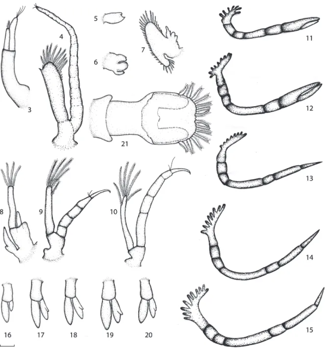
Antennule (Fig. 2.3): peduncle long, unsegmented. Endopod with 1 short apical plumose setae, exopod longer than endopod, with 4 apical naked setae.
Antenna (Fig. 2.4): biramous. Peduncle unsegmented. Exopod (or scale) unsegmented, with external disto-lateral spine, 6 naked setae, and 10 plumose setae on distal margin. Endopod (or flagellum) has almost double size of exopod, with numerous joints and 3 short naked setae.
Mandible (Fig. 2.5): rudimentary, without palp. Incisor and molar processes not clearly defined.
Maxillula (Fig. 2.6): Rudimentary, unsegmented and smooth endopod. Coxal and basal endits with 3 and 4 distal protuberances.
Maxilla (Fig. 2.7): biramous. Protopod rudimentary. Endopod with 1 short curved naked setae. Exopod (escaphognathite) large, fringed by 36 plumose setae.
Maxilliped 1 (Fig. 2.8): biramous. Protopod unsegmented, with a bilobed, smooth epipodite. Endopod short, unsegmented, smooth. Exopod long, unsegmented, with 4 terminal plumose setae and 1 subterminal.
Maxilliped 2 (Fig. 2.9): biramous. Protopod unsegmented, smooth. Endopod 5-segmented, with 2 naked setae on propodus, dactylus hooked. Exopod unsegmented, with 4 terminal plumose setae and 1 subterminal.
Maxilliped 3 (Fig. 2.10): biramous. Protopod unsegmented. Endopod 5-segmented, with 2 naked setae on distal margin of propodus. Dactilus hooked. Exopod unsegmented, smaller than the endopod, with 6 plumose setae.
Pereiopods 1-2 (Fig. 2.11-12): well
developed, but not functional buds. Uniramous. Endopod glabrous, chelate, with distinct segmentation. Pereiopod 2 larger than pereiopod 1.
Pereiopods 3-5 (Fig. 2.13-15): all as well
developed buds. Uniramous, glabrous, with clear segmentation between joints.
Pleopods 1-5 (Fig. 2.16-20): All buds
birramous and glabrous. Pleopod 1 the
smallest; pleopods 2-5 with a rudimentary inner appendix in exopod.
Uropod (Fig. 2. 21): Not yet freed. Bud
visible through the telson cuticle.
Telson (Fig. 2.21): fan-like. Posterior
margin broadly convex, with 7 + 7 plumose setae and pair of minute naked setae placed in a very faint median notch.
Discussion
According to Jalihal et al. (1993),
the larval development of Macrobrachium
brasiliense corresponds to Type II: partially
abbreviated development, with all pereiopods uniramous, well developed pleopods, and
a fan-shaped telson in first stage. Other
characteristics noted by Jalihal et al. (1993)
for species with this kind of development
(e.g., medium body size, distribution in upper
reaches ofrivers, and absence of marked sexual dimorphism) fit M.brasiliense well.
The larval characters of M. brasiliense
described here suggests a closer relationship with M. nattereri (Heller, 1862) and M. iheringi (Ortman, 1897), both Neotropical
species. These species have a morphologically similar first stage with uniramous pereiopods, biramous pleopods, and rounded telson (see Tab. 1). In addition, characteristics mentioned by Bueno and Rodrigues (1995) for M. iheringi from São Paulo State, as an
unsegmented antennal scale, the numerous of marginal plumose setae in scaphognathite and six distinct abdominal somites are present in
M. brasiliense.
Magalhães (1989), comparing the description done by Vega-Pérez (1984), had already mentioned that M. nattereri and M. brasiliense are closely related. They both occur in the Amazon basin and, in many cases even adult specimens are not easily identified. Although their larval development is similar in many respects, some differences can be mentioned in the first larval stage. In M. nattereri, the endopod of antenna, maxillipeds 2 and 3, and the pereiopods are unsegmented, while in M. brasiliense, these structures are clearly segmented. Yet, in the description performed by Vega-Pérez (1984), M. brasiliense
has the 6th abdominal somite clearly separated from the telson, while in M. nattereri this separation is indistinct. Both species have 7 + 7 plumose setae on the posterior margin of telson, but in M. brasiliense this margin is clearly bilobed due to a distinct bulging of the disto-lateral corners (Magalhães, 1989).
However, the first zoea of M. brasiliense
studied here, from northwest of São Paulo State, some characteristics resemble those of M. nattereri: the segmentation of the 6th abdominal somite/telson is indistinct, the posterior margin of telson is not bilobed as was
described by Vega-Pérez (1984), and there is a pair of minute naked setae placed in a very faint median notch of telson, which is very similar to M. nattereri.
Pileggi and Mantelatto (2010), studying the molecular phylogeny of the genus
Macrobrachium based on direct optimization
analysis of 16S rDNA, used specimens of M. brasiliense from the São Paulo State (cities of
Bauru and Serra-Azul) and M. nattereri from
the Amazonas State (Manaus), and observed that the two species belong to sister groups, corroborating the conclusions drawn from larval morphology by Magalhães (1989) and now by the present study.
The wide distribution of M. brasiliense
throughout tropical South America suggests that the populations occurring so far apart would exhibit some degree of morphological variation. Since the specimens of M. brasiliense
studied by Vega-Pérez (1984) came from Paraná state, and in the present study, from São Paulo State, both Southeastern Brazil (Rio Paraná Basin), it would be interesting to verify how similar the larval morphology of the Central Amazonian population of M. brasiliense is to that one’s already studied, as
suggested by Magalhães (1989).
Acknowledgments
This research was supported by UNIRP (Centro Universitário de Rio Preto). The authors are indebted to Dr. Adilson Fransozo and Dr. Maria Lucia Negreiros-Fransozo for laboratorial assistance for drawings. The samplings were performed according to Brazilian State and Federal laws concerning wild animals.
References
Bueno, S.L.S. and Rodrigues, S.A. 1995. Abbreviated larval development of
the freshwater prawn, Macrobrachium
iheringi (Ortmann, 1897) (Decapoda,
Palaemonidae), reared in the laboratory.
Crustaceana, 68(6): 665-686.
Clark, P.F.; Calazans, D.K. and Pohle, G.W. 1998. Accuracy and standardization of Brachyuran larval descriptions. Invertabrate Reproduction and Development, 33: 127-144.
Coelho, P.A. and Ramos-Porto, M. 1985. Camarões de água doce do Brasil: Distribuição geográfica. Revista Brasileira de Zoologia, 2: 405-410.
García-Dávila, C.R. and Magalhães, C. 2003. Revisão taxonômica dos camarões de água doce (Crustacea, Decapoda, Palaemonidae, Sergestidae) da Amazônia Peruana. Acta Amazonica, 33(4): 663-686.
Holthuis, L.B. 1952. A general revision of the Palaemonidae (Crustacea, Decapoda, Natantia) of the Americas. II. The subfamily Palaemonidae. Occasional Papers of the Allan Hancock Foundation,12: 1-396.
Jalihal, D.R.; Sankolli, K.N. and Shenoy, S. 1993. Evolution of larval developmental patterns and the process of freshwaterization
in the prawn genus Macrobrachium
Bate, 1868 (Decapoda, Palaemonidae).
Crustaceana, 65: 365-376.
Kensley, B. and Walker, I. 1982. Palaemonid shrimps from the Amazon Basin, Brazil (Crustacea: Decapoda: Natantia).
Smithsonian Contributions to Zoology, 362: 1-18.
Lopez, B. and Pereira, G. 1996. Inventario de los crustáceos decapodos de las zonas alta y media del delta del Rio Orinoco, Venezuela.
Acta Biologica Venezuelica, 16(3):45-64. Maciel, C.R. and Valenti, W.C. 2009. Biology,
Fisheries, and Aquaculture of the Amazon
River Prawn Macrobrachium amazonicum:
A Review. Nauplius, 17(2): 61-79.
Magalhães, C. 1988. The larval development of palaemonid shrimps from the Amazon region reared in the laboratory. II. Extremely abbreviated development in Euryrhyncus
Miers, 1877 (Decapoda, Euryrhynchinae).
Crustaceana, 55(1): 39-52.
Magalhães, C. 1989. The larval development of palaemonid shrimps from the Amazon region reared in the laboratory. Abbreviated
development of Macrobrachium nattereri
Amazoniana, 10: 379-392.
Magalhães, C. 2000. Abbreviated larval
development of Macrobrachium jelskii
(Miers, 1877) (Crustacea: Decapoda: Palaemonidae) from the Rio Solimões floodplain, Brazil, reared in the laboratory.
Nauplius, 8(1): 1-14.
Magalhães, C. 2002. A rapid assessment of the decapod fauna in the Rio Tahuamanu and Rio Manuripi Basins, with new records of shrimps and crabs for Bolivia (Crustacea, Decapoda, Palaemonidae, Sergestidae, Trichodactylidae). Revista Brasileira de Zoologia, 19(4): 1091-1103.
Magalhães, C.; Bueno, S.L.S.; Bond-Buckup, G.; Valenti, W.C.; Silva, H.L.M.; Kiyohara, F.; Mossolin, E.C. and Rocha, S.S. 2005. Exotic species of freshwater decapod crustaceans in the state of Sao Paulo, Brazil: records and possible causes of their introduction. Biodiversity andConservation,
14: 1929-1945.
Mantelatto, F.L. and Barbosa, L.R. 2005. Population structure and relative growth of freshwater prawn Macrobrachium brasiliense
(Decapoda, Palaemonidae) fromSão Paulo State, Brazil. Acta Limnologica Brasiliensia, 17(3): 245-255.
Mejía-Ortíz, L.M.; Hartnoll, R.G. and López-Mejía, M. 2010. The abbreviated
larval development of Macrobrachium
totonacum Mejía, Alvarez & Hartnoll, 2003 (Decapoda, Palaemonidae), reared in the laboratory. Crustaceana, 83(1): 1-16. Melo, G.A.S. 2003. Manual de identificação
dos Crustacea Decapoda de água doce do Brasil. Loyola, São Paulo, 420 p.
Murphy, N.P. and Austin, C.M. 2005. Phylogenetic relationships of the globally distributed freshwater prawn genus
Macrobrachium (Crustacea: Decapoda: Palaemonidae): biogeography, taxonomy and the convergent evolution of abbreviated larval development. Zooloogica Scripta, 34(2): 187-197.
Pereira, M.G.C. and Chacur, M.M. 2009. Estrutura populacional de Macrobrachium brasiliense (Crustacea, Palaemonidae) do Córrego Escondido, Batayporã, Mato
Grosso do Sul, Brasil. Revista de Biologia Neotropical, 6(1): 75-82.
Pileggi, L.A. and Mantelatto, F.L. 2010. Molecular phylogeny of the freshwater
prawn genus Macrobrachium (Decapoda,
Palaemonidae), with emphasis on the relationships among selected American species. Invertebrate Systematics, 24(1): 194-208.
Pohle, G.W. and Telford, M. 1981. Morphology and classification of decapod crustacean larval cerdae: a scanning electron microscope study of Dissodactylus crinitichelis Moreira, 1901 (Brachyura: Pinnotheridae). Bulletin of Marine Science,
31: 736-752.
Rodriguez, G. 1982. Freshwater shrimps (Crustacea, Decapoda, Natantia) of the Orinoco basin and the Venezuelan Guayana. Journal of Crustacean Biology, 2(3): 378-391.
Taddei, F.G. 2006. Biologia populacional, reprodutiva e crescimento dos camarões Palemonídeos Macrobrachium jelskii (Miers, 1877) e Macrobrachium brasiliense (Heller, 1868) (Crustacea: Caridea) na região nordeste do Estado de São Paulo. PhD Thesis. Instituto de Biociências, UNESP, Botucatu, São Paulo. 217 p.
Valencia, D.M. and Campos, M.R. 2007. Freshwater prawns of the genus
Macrobrachium Bate, 1868 (Crustacea: Decapoda: Palaemonidae) of Colombia.
Zootaxa, 1456:1-44.
Vega-Pérez, L.A. 1984. Desenvolvimento larval de Macrobrachium heterochirus (Wiegmenn,
1836), Macrobrachium amazonicum
(Heller, 1862) e Macrobrachium brasiliense
(Heller, 1862) (Crustacea, Decapoda, Palaemonidae), em laboratório. PhD Thesis, Universidade de São Paulo, São Paulo. 277 p. (Unpublished)