1. Head of the Brachial Plexus Surgery and Microsurgery Unit; Senior Consultant at the Plastic and Reconstructive Surgery Department, São José Hospital, Lisbon, Portugal
2. Consultant at the Plastic and Reconstructive Surgery Department, São José Hospital, Lisbon, Portugal
3. Instructor at the Anatomy Department, Medical Sciences Faculty, New University of Lisbon
4. Senior Consultant at the Neurosurgery Department at São José Hospital, Lisbon, Portugal
5. Senior Consultant at the Orthopedic Department and Vertebro-medullary Unit at São José Hospital, Lisbon, Portugal
Neurogenic thoracic outlet syndrome
associated with cervical rib
ACTA REUMATOL PORT. 2013;38:98-103
AbstRAct
A true neurogenic thoracic outlet syndrome (TOS) as-sociated with a cervical rib is considered extremely rare.
The authors present their experience with 5 cases of true neurogenic TOS associated with a cervical rib. All patients were female and had a cervical rib confirmed radiographically pre-operatively. Average age was 34,8 years. Although all patients had been treated with se-veral combinations of diverse drugs and a rehabilitation program before referral to surgery, all described their pain as intense and debilitating before surgical treat-ment. All patients had pre-operative electromyographic abnormalities.
Patients were operated on via a supraclavicular ap-proach and the cervical rib was resected. No intra-ope-rative or postopeintra-ope-rative complications were noted.
Two years postoperatively, all patients mentioned im-provement. However, only 2 were symptomless, and on no medication. In one patient there was significant improvement, and in the remaining 2 patients some re-sidual pain persisted that had to be dealt with phar-macologically. All patients were able to resume their daily life activities.
Recovery was poorer in the 2 patients that had been referred to surgery after a longer period of time since the beginning of symptoms.
Keywords:Thoracic outlet syndrome; Brachial plexus; Cervical rib; Nerve compression syndrome; Surgery.
Gerardo Millan1, Diogo Casal2,3, Amets Sagaribay4, Valdemar Marques4, J. Estrela Martins5
Resumo
Os síndromes de desfiladeiro torácico neurogénicos pu-ros associados a uma costela cervical são considerados muito raros.
Os autores apresentam a sua experiência no trata-mento de 5 doentes nestas circunstâncias. Os doentes eram todos do sexo feminino e tinham uma costela cer-vical confirmada radiologicamente pré-operatoria-mente, bem como alterações electromiográficas. A ida-de média era ida-de 34,8 anos. Embora todas as pacientes tivessem sido tratadas com uma combinação de fárma-cos e com um programa de reabilitação antes de serem referenciadas para cirurgia, todas descreviam uma dor intensa, incapacitante e persistente. As doentes foram operadas por abordagem supraclavicular, tendo sido ressecada a costela cervical.
Não se registaram complicações intra ou pós opera-tórias. Dois anos após a cirurgia, todas as doentes apre-sentavam melhoria clínica. Contudo, apenas 2 estavam completamente assintomáticas, sem qualquer medica-ção. Numa doente registou-se melhoria significativa e nas duas restantes havia persistência de dor residual que tinha de ser controlada farmacologicamente. A cuperação foi pior nas duas doentes que tinham sido re-ferenciadas para cirurgia ao fim de mais tempo desde o início da sintomatologia. No entanto, todas as pacien-tes retomaram as suas actividades quotidianas.
Palavras-chave: Síndrome do desfiladeiro torácico; Ple-xo braquial; Costela cervical; Síndrome compressivo nervoso; Cirurgia.
IntRoductIon
Thoracic outlet syndrome (TOS) is defined as a group of symptoms that result from the entrapment of the bra-chial plexus and/or subclavian vessels in the thoracic ou-tlet region, that is to say, between the neck and the
ÓRgÃO OFICIAL DA SOCIEDADE PORTUgUESA DE REUMATOLOgIA
99
Gerardo Millan e col.
Peet5-7. However, it has also been referred to as scalenus
anticus syndrome, cervical rib syndrome,
costo-clavi-cular syndrome, and hyperabduction syndrome7,8.
Most authors agree that the TOS is the most over-looked and misdiagnosed nerve entrapment syndrome in the upper limb, as well as one of the most debilita-ting, and certainly one of the most difficult to
mana-ge8-11. The complexity in the diagnosis is due to the
lack of any specific clinical or ancillary confirmatory tests1,7,11,12. The reported incidence of TOS varies be
-tween 3 to 80 cases /1.000 inhabitants, making it a signi ficant pathology for anyone who deals with upper limb pathology1,13.
TOS has been the subject of various classifications1,
being the Wilbourn’s classification the most widely used in clinical practice7. According to this
classifica-tion, TOS can be of two main types: vascular and neu-rogenic4. The vascular type is subdivided into arterial
and venous. The neurogenic type can be “true” neuro-genic or “disputed” neuroneuro-genic, if nerve conduction
studies show changes or not, respectively7. Vascular
TOS corresponds to about 1% of cases, whereas the “disputed” TOS accounts for up to 97% of patients
diagnosed with this syndrome7,12. True neurogenic
TOS is rare, having a prevalence of around 1 in 1 mil-lion patients7,11. In these patients, there is usually an
anatomical anomaly causing brachial plexus com-pression, namely a cervical rib, a prolonged transver-se process of the transver-seventh cervical vertebra, an anoma-lous first rib, first rib or clavicle fracture’s, a scalenus minimus, subclavius tendon anomalies, anomalies of scalene muscle development or insertion, and thick fi-brous bands in the thoracic outlet region7,11,13,14. How
-ever, a true neurogenic TOS associated with a cervical rib is considered extremely rare7,11,13,14. For example,
among the more than 1000 operations performed for neurogenic TOS during a period of over 28 years in a tertiary referral centre, Sanders et al. found only 37 pa-tients with cervical ribs13.
In the present work, the authors present their ex-perience with 5 cases of true neurogenic TOS associa-ted with a cervical rib.
clInIcAl cAses
The authors retrospectively reviewed the charts, and clinical images of 5 patients referred to either the Bra-chial Plexus, Vertebro-medullary or Neurosurgery Outpatient Clinic at São José Hospital (Lisbon,
Portu-gal). The basic demographic features, clinical picture and outcome one year after surgery of those 5 patients are described in Table I.
All patients were female and had a cervical rib con-firmed radiographically pre-operatively (Fig. 1). Ave-rage age was 34.8 ±19.6 years, ranging from 18 to 65 years. All patients had been treated conservatively with several combinations of drugs (analgesics, anti-inflam-matory drugs, muscle relaxants, and antidepressants), and a rehabilitation program before referral to the Cli-nic. The average time from the onset of symptoms to referral to our Clinic was 2.9 ± 2.4 years, varying from 1 to 7 years.
Three patients experienced neck and upper limb pain and paresthesias, whereas 2 patients had com-plaints only at the upper limb. All patients described the pain as intense, debilitating and persisting. Two pa-tients were unable to work due to their symptoms, and one of them (patient 4) had been forced to give up gym-nastics which she had been performing competitively for the 10 years before the onset of symptoms. Other patient (patient 3), although retired, complained that she could no longer do most of her daily life tasks. All patients had positive provocative tests (Adson’s, hyper-abduction, costo-clavicle, Halsted’s, Roos’s, Wright and upper limb tension tests). Diminished strength in the intrinsic hand muscles was noted in 2 patients: in the territory of the ulnar nerve in one patient, and in the territory of both the ulnar and median nerve in ano ther patient. Hand muscle wasting was observed in 2 of the-se patients. All patients had electromyographic abnor-malities, as depicted in Table I.
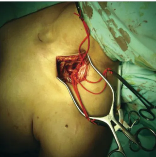
Patients were operated on via a supraclavicular ap-proach, isolating the constituents of the brachial ple-xus, the subclavian artery and vein and the cervical rib (Figs. 2 and 3). This rib was resected, freeing the bra-chial plexus (Fig. 4). No intra-operative or postopera-tive complications were noted, being the patients dis-charged home a few days after the surgery (3 to 5 days).
Patients were followed up for a minimum of two years after surgery.
defi-ÓR
gÃO
OFI
CI
AL
D
A
S
OCI
ED
AD
E
P
ORT
U
gU
ES
A
D
E
REU
MA
T
OL
OgI
A
1
0
0
n
e
u
roG
e
n
ic
t
horaci
c
ou
t
le
t
s
y
n
droMe
Time from the development
Age Affected Complaints Electrodiagnostic surgery to Outcome two
Patient (years) Sex Occupation Side of symptoms Physical exam tests surgery (years) years after
1 29 F Cruise ship Left Neck and upper Positive provocative tests; Chronic denervation 2 Occasional hand pain stewardess limb pain and Atrophy and diminished in the territory of managed with pain
paresthesias; strength of intrinsic the inferior trunk killers;
Unable to work hand muscles of the brachial plexus Partial recovery of strength and intrinsic hand muscles bulk; Resumed working 2 43 F Office clerk Left Debilitating neck Positive provocative tests Chronic denervation 3 Residual hand pain
and upper limb in the territory of the managed with pain
pain and inferior trunk of the killers;
paresthesias; brachial plexus Resumed working
Unable to work
3 65 F Retired Right Debilitating neck Positive provocative tests; Chronic denervation 7 Residual hand pain and upper limb diminished strength in in the territory of managed with pain pain and the territory of the ulnar the middle and killers;
paresthesias; and median nerves; inferior trunks of Partial recovery of Allodynia in the atrophy of intrinsic the brachial plexus strength and
ulnar nerve hand muscles intrinsic hand
territory; muscles bulk;
Unable to Resumed most
perform most daily life activities
daily life activities
4 19 F Student Right Intense upper Positive provocative Denervation in the 1.5 Symptomless limb pain and tests; diminished strength territory of the
ÓRgÃO OFICIAL DA SOCIEDADE PORTUgUESA DE REUMATOLOgIA
101
Gerardo Millan e col.
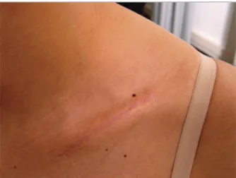
cits. All surgical wounds healed uneventfully (Fig. 5).
dIscussIon
Cervical ribs are usually asymptomatic and thus found incidentally on routine image exams, being present in 0,01 to 0,5 % of the general population1,13,15. These
su-pernumerary ribs are more common in women, being bilateral in more than half of cases7,13. However, the
cervical rib is considered a risk factor for the
develop-ment of TOS following cervical trauma, particularly after whiplash injuries12,13. In fact, it has been
propo-sed that TOS is due to a combination of a constitutio-nal tendency coupled to muscle dysfunction and re-petitive trauma4,5.
The existence of a higher placed bone piece, usual-ly connected to the first thoracic rib directusual-ly or through a thick fibrous band, narrows the thoracic outlet. Hen-ce, people with these anatomical variants are more pro-ne to TOS, especially if they do sports or jobs that de-mand prolonged arm hyper-abduction, like
swim-ming, gymnastics or weight throwing1,7,16. Supporting
this way of thinking is the report by Boles et al. of 15 TOS patients in a single family, with a cervical rib or
an apophysomegaly of the seventh cervical vertebra17.
Interestingly, in our series one patient was an active
fIguRe 1.Radiograph showing a right cervical rib (asterisk). fIguRe 2.Introperative view of the supraclavicular contents of the thoracic outlet in a patient with a cervical rib.
fIguRe 3.Photograph of the thoracic outlet in a close-up view showing the intimate relation between the cervical rib and the subclavian artery and the lower portion of the brachial plexus. 1- cervical rib; 2- subclavian artery; 3- subclavian vein; 4- inferior trunk of the brachial plexus; 5- middle trunk of the brachial plexus, 6- superior trunk of the brachial plexus; 7- trapezius muscle, 8- subclavius muscle; 9- anterior scalene muscle, 10- clavicle; 11- sternocleidomastoid muscle.
gymnast before the development of symptoms. In the cases we reviewed, only female patients were found. This is in accordance with the literature that usually states that TOS is almost twice as common in women than in men7,8,18. This sex predilection is
attri-buted to the fact that usually women present compa-ratively weaker muscles which makes their scapulas to be placed lower, predisposing to the compression of the structures involved in TOS1,7. Authors usually agree
that most TOS patients present between 20 to 50 years7,8,18. In our series this was also observed, as all
patients were in this age interval except for one. The differential diagnosis of TOS is extensive, in-cluding cervical disc disease; osteophytes; neoplasms (e.g. Pancoast tumor, nerve sheath tumors, spinal cord tumors) peripheral nerve entrapments (ulnar and/or median nerve entrapment); brachial plexitis; shoulder pathology (e.g. rotator cuff injuries), muscular spasms; fibromyalgia; multiple sclerosis; coronary artery di-sease; vasculitis (e.g. Takayusu’s arteritis); Raynaud’s phenomenon; complex regional pain syndrome; ve-nous thrombosis, micro-embolism; hand–arm
vibra-tion syndrome; and myofascial syndrome1,8,19.
The diagnosis of TOS is made by the patient
histo-ry associated with physical examination1,8. The most
common complaints are chronic pain of insidious on-set involving the shoulder girdle, neck and upper back, combined with paresthesias and hypoesthesia in the medial arm and forearm, and in the territory of the
ul-nar nerve and/or the median nerve1,8,19. Provocative
tests (e.g. Adson’s, hyper-abduction, costo-clavicle, Halsted’s, Roos’, Wright and upper limb tension tests)
are frequently positive in the normal population, and are neither sensitive nor specific for TOS1,4,8,19. Their
value is therefore limited1,4,8,19.
Regarding ancillary diagnostic tests, a chest graphy in an apical lordotic view or a cervical radio-graphy may allow identification of bone abnormalities that may predispose to TOS, narrowing down the
dif-ferential diagnosis20. Ultrasonography, computed
to-mography scans and magnetic resonance imaging, so-metimes performed in association with postural ma-neuvers, are helpful in analyzing the dynamically in-duced compression, as well as places of neurological
compression7,19,20. Electromyography and nerve
con-duction velocity tests are normal in the large majority of patients with clinical signs of TOS, which led some authors to argue that TOS is frequently underdiagno-sed in the primary care setting and over-diagnounderdiagno-sed in patients demanding compensation from insurance companies7,13.
It is generally accepted that the first line of treat-ment of TOS should be conservative, including com-binations of several drugs (analgesics, anti-inflamma-tories, muscle relaxants, antidepressants, and particu-larly the anticonvulsants gabapentin and pregabalin), avoiding activities and positions that aggravate symp-toms, and rehabilitation with strengthening of
pecto-ral musculature7. The improvement after conservative
treatment varies from 50 to 90% and depends on its etio-logy, being less efficient in “true” neurogenic TOS1-3.
Sur-gery should be considered after failure of appropriate conservative treatment of 6 months’ duration1-3,7.
Sur-gical treatment involves surSur-gical decompression by cervical rib excision and/or first rib excision; resection of cervical muscles, brachial plexus neurolysis; and, when necessary, vascular reconstruction. The presen-ce of a presen-cervical rib is not in itself an indication for sur-gery, unless there is failure in conservative treatment or debilitating symptoms1,7,11,19.
Surgical treatment is considered successful in ap-proximately 80% of selected TOS cases, being deemed unsatisfactory in around 20% of patients in the best series. In contrast, Sanders et al., reviewing the largest series of cervical ribs associated with TOS (n=37), con-sidered their long term results as good to excellent in only 59%, and fair and poor in 13% and 28%, respec-tively13. These results are not significantly different
from the ones we observed in the present study, in which, two years postoperatively, all patients had re-gistered some improvement. However, in only 2 of the patients (40%) was the recovery complete. In 1 patient
ÓRgÃO OFICIAL DA SOCIEDADE PORTUgUESA DE REUMATOLOgIA
103
Gerardo Millan e col.
(20%) there was significant improvement, and in the remaining 2 patients (40%) some residual pain per-sisted that had to be dealt with pharmacologically. Ho-wever, surgery allowed all patients to resume their pre-vious daily activities and occupations. Recovery was poorer in the 2 patients that had been referred to sur-gery after a longer period of time since the beginning of symptoms (3 and 7 years), which is also according to the literature7,11.
Therefore, to maximize the recovery of TOS patients associated with cervical ribs, it is reasonable to suggest that these patients should be managed by a multidis-ciplinary team involving a rheumatologist, a pain spe-cialist, a surgeon, and a physiotherapist, with possible advice from a psychologist or a psychiatrist, in order to maximize recovery and facilitate a rapid return to work5,2.
coRRespondIng AuthoR Diogo Casal
Serviço de Cirurgia Plástica e Reconstrutiva, Rua José António Serrano, 1150-199, Lisbon, Portugal E-mail: diogo_bogalhao@yahoo.co.uk
RefeRences
1. dos Reis Neto ET, Pucinelli ML, Silva de Souza AW, Sato EI. Thoracic outlet syndrome (TOS) mimicking Takayasu’s arteri-tis—case report. Acta Reumatol Port 2009;34:96-101. 2. Watson LA, Pizzari T, Balster S. Thoracic outlet syndrome part
1: clinical manifestations, differentiation and treatment path-ways. Man Ther 2009;14:586-595.
3. Watson LA, Pizzari T, Balster S. Thoracic outlet syndrome part 2: conservative management of thoracic outlet. Man Ther 2010;15:305-314.
4. McGillicuddy JE. Thoracic outlet syndrome. In: Chung KC, Yang LJ, McGillicuddy JE, editors. Practical Management of Pe-diatric and Adult Brachial Plexus Palsies. First ed. United Sta-tes of America: Elsevier, 2012:318-336.
5. Vanti C, Natalini L, Romeo A, Tosarelli D, Pillastrini P. Conser-vative treatment of thoracic outlet syndrome. A review of the literature. Eura Medicophys 2007;43:55-70.
6. Peet RM, Henriksen JD, Anderson TP, Martin GM. Thoracic-ou-tlet syndrome: evaluation of a therapeutic exercise program. Proc Staff Meet Mayo Clin 1956;31:281-287.
7. Meyer R, Jones KJ. Thoracic outlet compression syndrome. In: Wolfe SW, editor. Green\’s operative hand surgery. 6th ed. Phi-ladelphia: Elsevier Churchill Livingstone, 2011:1015-1034. 8. Cooke RA. Thoracic outlet syndrome—aspects of diagnosis in
the differential diagnosis of hand-arm vibration syndrome. Oc-cup Med (Lond) 2003;53:331-336.
9. Muizelaar JP, Zwienenberg-Lee M. When it is not cervical radi-culopathy: thoracic outlet syndrome—a prospective study on diagnosis and treatment. Clin Neurosurg 2005;52:243-249. 10. Sheth RN, Belzberg AJ. Diagnosis and treatment of thoracic
ou-tlet syndrome. Neurosurg Clin N Am 2001;12:295-309. 11. Janjua RM, Tender GC, Tiel RL, Kline DG. Thoracic outlet
syn-drome. In: Slutsky DJ, Hentz VR, editors. Peripheral nerve sur-gery: practical applications in the upper extremity. First ed. Phi-ladelphia: Elesevier Churchill Livingstone, 2006:285-297. 12. Sanders RJ, Hammond SL, Rao NM. Diagnosis of thoracic
ou-tlet syndrome. J Vasc Surg 2007;46:601-604.
13. Sanders RJ, Hammond SL. Management of cervical ribs and anomalous first ribs causing neurogenic thoracic outlet syn-drome. J Vasc Surg 2002;36:51-56.
14. Huang JH, Zager EL. Thoracic outlet syndrome. Neurosurgery 2004;55:897-902; discussion 02-3.
15. Kurihara Y, Yakushiji YK, Matsumoto J, Ishikawa T, Hirata K. The ribs: anatomic and radiologic considerations. Radiogra -phics 1999;19:105-19; quiz 51-52.
16. Nichols AW. The thoracic outlet syndrome in athletes. J Am Board Fam Pract 1996;9:346-355.
17. Boles JM, Missoum A, Mocquard Y, et al. [A familial case of tho-racic outlet syndrome. Clinical, radiological study with treat-ment (author’s transl)]. Sem Hop 1981;57:1172-1176. 18. Sallstrom J, Thulesius O. Non-invasive investigation of
vascu-lar compression in patients with thoracic outlet syndrome. Clin Physiol 1982;2:117-125.
19. Atasoy E. A hand surgeon’s further experience with thoracic outlet compression syndrome. J Hand Surg Am 2010;35(9): 1528-1538.
20. Demondion X, Herbinet P, Van Sint Jan S, Boutry N, Chantelot C, Cotten A. Imaging assessment of thoracic outlet syndrome. Radiographics 2006;26:1735-1750.