Article 4
The Case of the Missing Muscles
Mira Silbert Aumiller, OD, Wilmington VA Medical Center, Wilmington, Delaware Michael Gallaway, OD, Pennsylvania College of Optometry at Salus University, Philadelphia, Pennsylvania
Jo Ann T. Bailey, OD, St. Christopher’s Hospital for Children, Philadelphia, Pennsylvania
ABSTRACT
Background: Congenital absence of the superior oblique (SO) is a rare cause of strabismus.
It should be suspected in a patient with a presumed diagnosis of SO palsy presenting with amblyopia, an unusually large vertical deviation, and a signiicant horizontal deviation.
Case Report: We report a case of an infant presenting at ive months of age with a signiicant
right head tilt since birth and signiicant vertical and horizontal strabismus. The patient was not meeting developmental milestones. The infant was referred for surgery, where it was discovered that she had a congenital lack of SO muscles. After surgery, the patient was treated with amblyopia therapy and vision therapy and followed for six years. Surgical and non-surgical intervention led to a good visual outcome, enhanced achievement of developmental milestones, and improved cosmesis.
Conclusion: Although absence of SO muscles is a rare etiology for torticollis and strabismus,
it should be considered in cases of early and signiicant ocular misalignment. The presence of horizontal and vertical strabismus in a patient with a presumed congenital SO palsy should raise suspicion of an absent SO. Early intervention can make a signiicant diference for the patient in terms of both visual outcome and cosmesis. This patient is the youngest documented case of SO aplasia and the irst published case using surgery and vision therapy in combination.
Keywords: amblyopia, strabismus, superior oblique aplasia, superior oblique palsy, torticollis
Introduction
Historically, congenital absence of the superior oblique (SO) has been considered a rare cause of strabismus. However, it should be suspected in cases of a signiicant horizontal deviation in a patient with a pre-operative diagnosis of congenital SO palsy. This condition may be associated with craniofacial dystoses, such as Crouzon and Apert syndrome, which involve a premature growth arrest at one or more of the cranial sutures. Down syndrome and anencephaly have also been associated with absent SO muscles.1-3
Descriptions of an absent SO muscle exist as case reports in the literature. The largest series has been six patients (ages 1 to 8 years old) seen over a seven-year time period by Helveston4 who were all initially diagnosed
for SO palsy, which typically presents without a large horizontal deviation or signiicant sensory anomalies since the head tilt typically compensates for the vertical and torsional deviation. Helveston concluded at that time that the true frequency of SO aplasia would only come from a prospective study involving surgical exploration of all patients diagnosed with a congenital SO palsy.4
Case Report
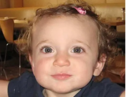
A ive-month-old female presented with a 15-20 degree right head tilt since birth (Figure 1). The parents also reported facial asymmetry, with smaller features on her left side. Physical therapy for the infant’s torticollis was initiated, and marked worsening of ocular alignment following some straightening of the head was reported. The infant’s visual acuity was 20/300 OD and 20/150 OS. The OS was tested irst due to parental suspicions. The infant began to fatigue upon testing of the OD, thus amblyopia was not ruled out. She also presented with 35∆
left exotropia with a V pattern and 5-10∆ left
hypertropia (Figure 2). The left hyperdeviation increased on left head tilt, consistent with a left SO palsy. Her systemic history was positive for reactive airway disease. The parents reported that the infant was unable to roll over, which is considered a 5-month milestone. She was referred for a surgical consult for a congenital SO palsy OS.
Surgery was performed at Wilmer Eye Institute at Johns Hopkins at 9 months of age. Under general anesthesia, fundus examination showed 3+ excyclotorsion of the fundus OD and 4+ OS. Forced ductions were performed, and there was no palpable SO tendon/muscle on the right and only a rudimentary one OS. Both inferior oblique (IO) muscles were Figure 1: 5 months old, note right head tilt
Figure 2: 7 months old, note 35∆ left exotropia and 5-10∆ left hypertropia in
primary gaze
exceedingly tight. A complete IO denervation and excision was performed OU, and the lateral recti were recessed by 6 mm OU (Figure 3). Adjustable sutures were used, and the patient was left with an ortho posture at near and a very small left esodeviation in the distance. The surgeon recommended initiating one drop of atropine 1% OD once a week for the possible mild amblyopia in the left eye. This was reduced a month later to every other week dosing in the OD as the parents noted an esotropia developing in the OS.
Three days following surgery, the parents reported that the patient began repeatedly to roll over and began to crawl shortly after. No associated craniofacial anomalies were found per a consult with a genetics doctor at Children’s Hospital of Pennsylvania. The parents reported improving symmetry of facial features within a year following the patient’s surgery (Figure 4).
At a two month post-operative visit with the surgeon, the patient had a completely resolved head tilt. No signiicant A- or V-pattern was noted, nor was there any gross misalignment in any direction of gaze. The surgeon opted to discontinue atropine penalization since there was equal objection by the patient to either eye being occluded, suggesting that vision was now relatively equal. However, when the
parents noted the left eye starting to turn in a month later, atropine penalization with one drop of atropine 1% OD every other week was reinstituted. This intermittent left esotropia was no longer present by the six month follow-up visit. Atropine penalization continued and was eventually tapered to 1 drop per month OD by 2.5 years of age. At this time, the patient was able to perform sensory testing and was able to achieve the stereo ly and 400” on the animals on the Randot stereo test. Her visual acuity was 20/30 by Snellen acuity in each eye, and she refracted to +0.50 bilaterally. No A- or V-pattern was noted.
By age four, she was still using one drop of atropine OD once per month as maintenance. Visual acuity was now slightly worse in the OD (20/30- Snellen) than OS (20/20-). Versions revealed a 2+ underacting LSO, 1+ underacting RSO, and 2+ underacting inferior oblique OU. Although the IO had been rendered totally non-functional due to the surgery, it appeared that the superior recti had compensated and were able to provide some elevation in the ield of the IOs. Cover test was 5-6∆
intermittent right hypotropia at distance and near with a peripheral fusion response. There was a V-pattern left ET without diplopia. She appreciated the stereo ly but no Randot shapes or circles. She suppressed the OD on Worth 4-dot beyond 4 ft. At this point, she had reversed ixation and was slightly amblyopic OD. Atropine was discontinued, and one hour patching OS was begun along with monocular ixation in a binocular ield (MFBF) activities designed to reduce suppression OD and to promote fusion.
not appreciate Randot shapes, she was able to demonstrate BI and BO fusion ranges on the VTS 3 Computer Orthopter. Cover test was a 4-5∆ intermittent right hypotropia at distance
and near. In-oice vision therapy (VT) was not feasible for the family at this time, so several options were presented to the parents: vertical prism, Home Therapy System (HTS) software program for home use, or no treatment and monitor. Due to the non-comitant nature of her strabismus and the risk of creating diplopia, the parents decided to discontinue treatment. The next follow-up was at age 5 years 9 months. No patching or other treatment had been done. Visual acuity had improved to 20/20- OD, 20/20 OS. Versions still showed a 2+ underacting LSO, 1+ underacting RSO, and 2+ underactions in the ields of the IOs. She also had a signiicant left ET in downgaze with suppression. Cover test was unchanged at near, but she was able to alternate at distance. The patient reported vertical diplopia in left gaze, and Maddox rod showed an increase to 7-8∆ right hypo in left gaze. Stereopsis was
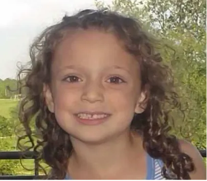
200” with circles, but there was no Randot stereopsis. She showed a vergence response objectively with up to 10 BO and 8 BI, but she suppressed beyond that. All treatment was formally discontinued (Figure 5).
Her last follow up was at age 6 years 3 months. All indings were unchanged, but she now reported diplopia in extreme downward gaze. Maddox rod in down gaze showed an increase to 13∆ esotropia and 8∆ right
hypotropia. Her parents reported that she was an excellent soccer player and was very accurate in kicking the ball. They were advised to encourage reading closer to primary gaze as well as to try a reading stand as her reading skills improved. Yoked prism was also discussed as an option if needed.
Discussion
Congenital SO paresis is the most common cause of a vertical deviation.5 Up to 90% of all
vertical misalignment is the result of SO palsy, and 30-66% of SO pareses are congenital in etiology.6 Most infants or young children with
SO palsy present for eye care with concerns about poor cosmesis and abnormal head position. SO palsy rarely causes amblyopia and suppression because the head tilt and torticollis counteract the torsional diplopia, helping to maintain binocular viewing and stereopsis.4
Parental concerns and symptoms in congenital SO absence are often indistinguishable from those with SO palsy. Examination of the patient with SO absence often reveals a large horizontal strabismus, a large hypertropia in primary position, amblyopia, pseudo-overaction of the contralateral superior oblique muscle, and spread of comitance secondary to restriction of the ipsilateral superior rectus.1 The presence
of a horizontal strabismus with amblyopia in Figure 5: 5 1/2 years old
a patient diagnosed with a SO palsy should raise suspicion of a congenital absence of this muscle.3,4
An abnormal head posture is a relatively common manifestation in children. The estimated incidence is 1.3%.7 In a series of 63
children evaluated for abnormal head posture by Nucci et al., the majority had congenital muscular torticollis. However, this presence of a tight neck does not rule out the possibility of neurologic or ophthalmic causes in addition to the congenital muscular torticollis. Forty percent of this series had an ocular etiology for their abnormal head posture. Although superior oblique muscle palsy was found to be the most common ocular etiology, there are numerous other causes such as lateral rectus palsy, nystagmus, Brown’s syndrome, Duane’s syndrome, and vertically non-comitant horizontal strabismus (A- or V-patterns). Therefore, it is important that when a child is being evaluated for an abnormal head position, the possibility of an ocular cause must be considered in addition to congenital muscular torticollis.7
Additionally, multiple reports exist in the literature correlating torticollis with facial asymmetry. The torticollis becomes obvious as soon as the infant can control the head.8 The
presence of muscular torticollis often leads to mid-facial hypoplasia (shortening and lattening) on the side of face opposite the paretic muscle.9 Thus, the patient will appear
to have a fuller mid-face on the side away from the head tilt.8 While the mechanism of the
development of the facial asymmetry is not understood, early treatment is advocated.10 In
the series of patients studied by Helveston, facial asymmetry was eliminated when treat-ment for the cause of the head tilt was successful.8 Although our patient had bilateral
aplasia of the superior obliques, she had facial asymmetry, with the left features somewhat smaller than the right. As demonstrated in this case, early surgical intervention allowed
our patient’s asymmetric facial features to normalize over time.
With the use of imaging, it has become easier to make the diagnosis of absence of the SO muscle in patients who were previously diagnosed with SO palsy. In 1999, qualitative magnetic resonance imaging (MRI) was performed on a series of 26 patients who all had the clinical diagnosis of SO palsy.11 MRI
identiied 6 of these 26 patients (23%) with an absent SO muscle. All six of these patients had a history of strabismus since childhood. Five of them had unilateral absent SO muscles, and one was bilateral. In this series, all of the patients with unilateral SO muscle absence maintained visual acuity of at least 20/25 in each eye. The patient with bilateral absence was 10 years old with worse visual acuity of 20/60 OD and 20/50 OS.11
In 2009, an MRI technique with much higher resolution was developed that could, for the irst time, clearly visualize the trochlear nerve.12 Since then, such high-resolution,
thin-section MRI studies have identiied trochlear nerve aplasia in patients with superior oblique muscle hypoplasia.13 It has been proposed
by multiple authors that patients with SO aplasia are included in what is described as congenital cranial dysinnervation disorders.12,13
These are deined as developmental abnormalities involving the hypoplasia or aplasia of the 4th cranial nerve with muscle denervation.12 A retrospective study in 2011
by Yang et al. evaluated 97 patients who were diagnosed with a congenital superior oblique palsy. Seventy-three percent of these patients were identiied as having an ipsilateral absent trochlear nerve and a variable degree of SO muscle hypoplasia.13
recession or resection of the horizontal recti for the coexisting horizontal deviations.4 There is
no data on success rates for these procedures. Careful follow-up is needed to monitor for the presence of amblyopia and to promote as much binocularity as possible. Atropine penalization can be thought of as a form of MFBF and can be done with children too young to participate in active VT.14 Atropine
has been studied extensively in the Amblyopia Treatment Studies and has been found to be well tolerated and very safe.15
As the child gets older, active VT can be helpful in developing fusion. While the spread of comitance demonstrates the plasticity of the ocular motor system, it is likely that some non-comitancy will remain. Allowing some suppression to remain may be helpful in avoiding signiicant diplopia in other positions of gaze.16 Establishment of at least peripheral
binocularity will help to maintain alignment over time as well as providing the sensory and motor beneits of stereopsis.17
We believe that our patient is the irst published case of absent SO muscles demon-strating the use of surgery and VT together to achieve binocular vision. Given her signiicant remaining non-comitancies (especially the V pattern esotropia), we decided not to pursue more aggressive treatment. The patient has monoixation syndrome with the classic triad of central suppression (Worth 4 dot), peripheral stereopsis (contour stereo on Randot), and peripheral motor fusion (vergence response on VTS and with hand held prisms).18 We hope
that this will allow a stable long-term prognosis, but she will need consistent follow-up.19
Conclusion
Although absence of SO muscles is a rare etiology for torticollis and strabismus, it should be considered in cases of early and signiicant ocular misalignment. Congenital absence of the superior oblique can cause a large vertical and horizontal deviation. Absence of
the superior oblique is more common than previously reported. Historically, few cases existed in the literature as it was diagnosed only through surgical exploration during what was thought to be a SO palsy procedure. Recent advances in MRI studies have demonstrated an increase in the diagnosis of superior oblique hypoplasia. Early intervention can make a signiicant diference for the patient in terms of both visual outcome and cosmesis. After a review of cases in the literature, we believe that our patient is the youngest to be diagnosed with absent SO muscles and the irst published case documenting the use of surgery and VT to achieve binocularity. Early intervention facilitated excellent visual acuity, some binocular vision, and good cosmesis. Considering our patient’s ability to roll over just 72 hours after her surgery, this case also highlights the integration of visual function and the achievement of developmental milestones.
References
1. Pinchof BS, Sandall G. Congenital absence of the superior oblique tendon in craniofacial dysostosis. Ophthalmic Surg 1985:16:375-7. http://1.usa.gov/1DU7l6t
2. Kerr NC. Management of vertical deviations secondary to other anatomical abnormalities. AmOrthoptic J 2011:61:39-48. http://bit.ly/1zp823r
3. Wallace DK, von Noorden GK. Clinical characteristics and surgical management of congenital absence of the superior oblique tendon. Am J Ophthalmol 1994:118:63-9. http://bit. ly/1DU7Nlf
4. Helveston EM, Giangiacomo JG, Ellis FD. Congenital absence of the superior oblique tendon. Tr Am Ophth Soc 1981:79:123-35. http://1.usa.gov/1Dcta2M
5. Holmes JM, Mutyala S, Maus TL, Grill R, et al. Pediatric third, fourth, and sixth nerve palsies: A population-based study. Amer J Ophthalmol 1999:127:388-92. http://bit.ly/1AaORQ6
6. Bixenman WW. Diagnosis of superior oblique palsy. J Clin Neuro-ophthalmol 1981:1:199-208. http://bit.ly/1KG3w73
7. Nucci P, Kushner BJ, Seraino M, Orzalesi N. A multi-disciplinary study of the ocular, orthopedic, and neurologic causes of abnormal head posture in children. Amer J Ophthalmol 2005:140:65e1-65e4. http://bit.ly/1uvPvXw
9. Goodman CR, Chabner E, Guyton DL. Should early strabismus surgery be performed for ocular torticollis to prevent facial asymmetry? J Pediatr Ophtalmol Strabismus 1995:32:162-6. http://1.usa.gov/1AaPGZb
10. Velez FG, Clark RA, Demer JL. Facial asymmetry in superior oblique muscle palsy and pulley heterotopy. JAAPOS 2000:4:233-9. http://bit.ly/1z0XjNC
11. Chan TK, Demer JL. Clinical features of congenital absence of the superior oblique muscle as demonstrated by orbital imaging. JAAPOS 1999:3:143-50. http://bit.ly/1CLNMAr
12. Kim JH, Hwang J. Absence of the trochlear nerve in patients with superior oblique hypoplasia. Ophthalmol 2010:117:2208-13. http://bit.ly/1Ca5uIx
13. Yang HK, Kim JH, Hwang J. Congential superior oblique palsy and trochlear nerve absence. Ophthalmol 2012:119:170-7. http://bit.ly/16KGzCK
14. The Pediatric Eye Disease Invetigator Group. The course of moderate amblyopia treated with atropine in children: experience of the amblyopia treatment study. Amer J of Ophthalmol 2003:136:630-9. http://bit.ly/1Fq5nMs
15. Repka MX, Cotter SA, Beck RW, Kraker RT, et al. A randomized trial of atropine regimens for treatment of moderate amblyopia in children. Ophthalmol 2004:111:2076-85. http:// bit.ly/1KG4osw
16. Birnbaum MH. Noncomitant strabismus: evaluation and management. J Am Optom Assoc 1984:55:758-64. http://1. usa.gov/1Dx4AtU
17. Press LJ. The story behind ‘Stereo Sue’ and a world-famous neurologist’s discovery of vision therapy. Optom Vis Devel 2006:37:55-7. http://bit.ly/1ESDeA5
18. Tyschen L. Can ophthalmologists repair the brain in infantile esotropia? Early surgery, stereopsis, monoixation syndrome, and the legacy of Marshall Parks. JAAPOS 2005:9:510-21. http://bit.ly/16KHfrM
19. Hunt MG, Keech RV. Characteristics and course of patients with deteriorated monoixation syndrome. JAAPOS 2005:9:533-6. http://bit.ly/1A5QH6l
Correspondence regarding this article should be emailed to Mira Silbert Aumiller, OD, at [email protected]. All statements are the authors’ personal opinions and may not relect the opinions of the representative organizations, ACBO or OEPF, Optometry & Visual Performance, or any institution or organization with which the authors may be ailiated. Permission to use reprints of this article must be obtained from the editor. Copyright 2015 Optometric Extension Program Foundation. Online access is available at
www.acbo.org.au, www.oepf.org, and www.ovpjournal.org.