Primary cardiac angiosarcoma:
case report of a rare neoplasia
Angiossarcoma cardíaco primário: relato de caso de uma rara neoplasia
Gabriela C. Abreu1; Maria Clara R. Gontijo1; Maria Clara L. Chaves1; Júlia W. Drumond1; Bárbara C. A. Marino2; Rodrigo C. Bernardes2; Alexandre Cobucci2; Cynthia K. Berenstein1, 3
1. Centro Universitário de Belo Horizonte (UNIBH), Minas Gerais, Brazil. 2. Hospital Madre Teresa, Minas Gerais, Brazil. 3. Instituto Roberto Alvarenga, Minas Gerais, Brazil.
First submission on 24/02/17; last submission on 14/03/17; accepted for publication on 31/07/17; published on 20/10/17
ABSTRACT
This article reports a case of primary cardiac angiosarcoma and a brief review is provided. A 44-year-old male patient was suspected of having myxoma in the right atrium. The tumor, on pathology examination, was shown to be a cardiac angiosarcoma. In the postoperative period, the patient developed a cardiac tamponade, requiring reoperation and evolving to death. Angiosarcomas are malignant tumors characterized by a devastating clinical course. They have a predilection for the right atrium, occurring between the third and ifth decades of life, with a male preponderance. Because of its rarity, the ideal treatment has not been identiied yet.
Key words: hemangiosarcoma; heart neoplasms; heart atria.
INTRODUCTION
Primary cardiac neoplasms are rare tumors of the human body, arising in tissues of inner lining, muscle layer or the pericardium(1). Metastases to the heart are more common: 40
times more prevalent than primary cardiac tumors(2).
In adults, 25% of primary cardiac tumors exhibit characteristics of malignancy, or behave as malignant(3, 4), with
95% being sarcomas, and 5% lymphomas(1). Angiosarcomas,
rhabdomyosarcomas, ibrosarcomas and undifferentiated sarcomas(5) have been reported; the irst are the most common.
CASE REPORT
A 44-year-old male patient experienced fatigue and dyspnea during cycling. After a period, he evolved to a picture of swelling in the face and upper chest (superior vena cava syndrome). A chest radiograph was taken, which showed no alteration, and the recorded electrocardiogram showed sinus rhythm. Exercise stress test displayed no ischemic changes. Transthoracic echocardiography (TTE) showed left and right ventricles with preserved dimensions, thickness and
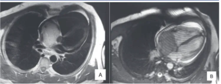
contractility. Left and right atria presented normal dimensions, but, inside the right atrium, close to the entry of the superior vena cava, a pedunculated mass with irregular borders was visualized, measuring 52 × 43 mm, causing obstruction in the atrium. A nuclear magnetic resonance (NMR) revealed a large sessile immobile heterogeneous mass, measuring about 77 × 54 mm, situated in the interior of the right atrium, apparently iniltrating inferior and superior venae cavae and invading tricuspid valve, interatrial septum and pericardium
(Figure 1). On T1-weighted sequences, the mass appeared to
be hypodense; on T2, presented hypersignal. On fat-suppressed sequences, there was no signal dropout, suggesting the mass was not a lipoma. After contrast infusion, heterogeneous late enhancement
FIGURE 1 − Cardiac NMR with the presence of sessile immobile heterogeneous mass, measuring about 77 × 54 mm, situated in the interior of the right atrium, apparently infiltrating inferior and superior venae cavae
NMR: nuclear magnetic resonance.
was perceived, with sparse areas of hyposignal inside. Due to those indings, the diagnostic possibilities were angiosarcoma, lymphoma, myxoma and myxosarcoma.
The patient underwent surgical excision of the tumor in the right atrium. It was completely removed, leaving the basal portion of the coronary sinus up to the tricuspid valve; the resection of the atrial roof was also necessary, due to the presence of a mass attached to it. The reconstruction of the interatrial septum was performed, employing a large patch of bovine pericardium for the roof. For reconstruction of the right atrium and venae cavae, 20 mm Dacron prostheses with anastomoses were used to the ends of superior and inferior venae cavae. The procedure occurred uneventfully. The patient presented junctional rhythm, and was supplied with a temporary pacemaker. Due to tumor removal, right atrium replacement with bovine patch, and increased thrombotic risk for the presence of tumor, unfractionated heparin was initiated 48 hours after surgery. The patient was discharged from the intensive care unit (ICU) after 48 hours, and was referred to the inpatient department.
In the ifth postoperative day, he had a cardiopulmonary arrest, and needed cardiopulmonary resuscitation. TTE performed after the event revealed cardiac tamponade, and the patient was urgently taken to the operating room, to undergo pericardiocentesis. A cardiopulmonary arrest occurred in the immediate postoperative phase, what resulted in the patient’s death the following day.
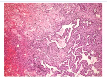
On pathological examination, the specimen was received fragmented, measuring 9.2 × 7 × 4 cm. There was a brownish nodule with hemorrhagic areas, measuring 4 cm in diameter, apparently iniltrating the cardiac wall (Figure 2). Microscopically,
it was identiied as a mesenchymal neoplasm, characterized by the proliferation of atypical endothelial cells with irregular nuclei, sometimes round sometimes spindle, without evident nucleolus, forming vascular spaces, papillae, and solid areas. Mitoses were frequent and necrosis was not observed. The lesion permeated cardiomyocytes, which presented clear cytoplasm and hypertrophic nucleus (Figures 3 to 7). There was iniltration of the visceral pericardium. The diagnosis was primary cardiac angiosarcoma.
FIGURE 3 − Well-differentiated area of angiosarcoma with vessels lined with atypical endothelium permeating myocardiocytes (HE, 40×)
HE: hematoxylin and eosin.
FIGURE 4 − Detail of the atypical vessels permeating myocardiocytes (HE, 400×)
HE: hematoxylin and eosin.
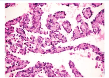
FIGURE 5 − Well-differentiated area with the formation of papillae (HE, 40×)
HE: hematoxylin and eosin. FIGURE 2 − Large reddish-brown mass, occupying and infiltrating the right atrium
B A
pain, conduction abnormalities, besides general symptoms, such as weight loss and asthenia(3, 8). The described patient presented
dyspnea, considered the most common symptom(9), evolving with
superior vena cava syndrome.
Malignant tumors are generally found in the right heart, particularly in the right atrium(8), near the atrioventricular groove
(80% of the cases)(7); in contrast, benign tumors are situated in
the left atrium(8). The differential diagnosis of the right atrial
mass includes benign entities, such as myxoma and thrombi, and malignant causes, such as primary cardiac angiosarcoma and other sarcomas(2).
Echocardiography is the main way to assess cardiac tumors. Transesophageal echocardiography (TEE) has 97% sensitivity to detect cardiac neoplasms(9) and delineate them(7). NMR can help
distinguish intracavitary thrombi from tumor masses(9). In the
current case, the patient was subjected to those examinations, but, despite the suggestion of a more aggressive lesion, cardiac myxoma was considered, probably because it is more common.
Grossly, angiosarcomas are large multilobulated dark brown or black tumors, measuring 2-10 cm. The tumor mass permeates the myocardium and can ill the atrial cavity. The pericardium is generally iniltrated, and in rare cases can be the primary site of the neoplasm(7, 9).
Histology can reveal different aspects, depending on the degree of differentiation. Well-differentiated areas are made by atypical pleomorphic endothelial cells, which form papillary structures or vascular channels. The poorly differentiated areas are formed by spindle anaplastic cells in solid pattern(7). The tumor presented
both well-differentiated and poorly differentiated areas.
Immunohistochemistry reveals positivity for CD31 (90%)
(Figure 8), CD34 (50%-74%) and friend leukemia integration 1
(FLI-1) (100%)(9). There can be positivity for cytokeratins in 35%
of the cases, although most of them are of the epithelioid variant, uncommon in the heart(7, 9). The most recent immunohistochemical
markers with high sensitivity and speciicity include nuclear factors ETS-related gene (ERG) and FLI-1(7). Values of Ki67 > 10%
have been correlated with poor prognosis(9).
Death can occur due to myocardium iniltration and its rupture, low obstruction or distant metastases. Around 66%-89% of the patients present metastases at diagnosis, more often in the lungs(7). Other main sites include thoracic lymph nodes,
mediastinum, and vertebral column(6).
The natural history of angiosarcomas is characterized by a devastating clinical course, even after surgery, chemotherapy and radiotherapy. Symptoms are normally late and more related
FIGURE 6 − Detail of the formation of papillae lined with atypical endothelium (HE, 400×)
HE: hematoxylin and eosin.
FIGURE 7 − Poorly differentiated solid area of the angiosarcoma (HE, 400×)
HE: hematoxylin and eosin.
DISCUSSION
Among the primary cardiac tumors, angiosarcoma is the most common malignant one, accounting for 30% of sarcomas(3).
Originated from mesenchymal tissue, it mostly occurs on the skin and soft parts, just 3% being primary of the heart and great
vessels(6). It affects a wide age range, but the peak incidence
is between the third and the ifth decades of life(7), with a male
preponderance in the ratio 2:1(8).
Case reports describe many of clinical manifestations related to site, size, and tumor extension, as well as to the presence or absence of metastases(7). The most common are arrhythmia,
to location than histological type, what makes early diagnosis dificult, impairing the eficacy of the treatment even more(10).
Unspeciic clinical presentation is also related to the degree of local involvement and the presence or absence of metastases(11).
All these characteristics lead to diagnosis at an advanced phase. Literature describes a uniformly adverse prognosis, with a median survival of just six months(8).
REFERENCES
1. Karigyo CJT, Silva FBF. Tumores cardíacos: uma breve revisão de literatura. Rev Med Res. 2014; 16(1): 27-34. Available at: ttp://www.crmpr. org.br/publicacoes/cientiicas/index.php/revista-do-medico-residente/ article/viewFile/528/513.
2. Bouma W, Lexis CP, Willems TP, et al. Successful surgical excision of primary right atrial angiosarcoma. J Cardiothorac Surg. 2011; 6: 47. PubMed PMID: 21477334.
3. Cataldi MS, Kruczan DD, Melo GR, et al. Angiossarcoma cardíaco primário. Rev SOCERJ. 2006; 19(1): 87-91. Available at: http://www. rbconline.org.br/artigo/angiossarcoma-cardiaco-primario/.
4. Bakaeen FG, Jaroszewski DE, Rice DC, et al. Outcomes after surgical resection of cardiac sarcoma in the multimodality treatment era. J Thoracic Cardiovasc Surg. 2009; 137(6): 1454-60. PubMed PMID: 19464464.
5. Higuchi ML, Aiello VD, Gutierrez PS. Coração. In: Brasileiro Filho GB, editor. Patologia. 7 ed. Rio de Janeiro: Guanabara Koogan; 2006. p. 408-56.
RESUMO
Este artigo ilustra um caso de angiossarcoma primário cardíaco acompanhado de uma revisão dessa rara neoplasia. Relatamos o caso de um paciente do sexo masculino, 44 anos, com suspeita de mixoma em átrio direito. O tumor, ao exame anatomopatológico, mostrou tratar-se de um angiossarcoma cardíaco. No pós-operatório, o paciente cursou com tamponamento cardíaco, necessitando ser reoperado e evoluindo a óbito. Angiossarcomas são tumores malignos caracterizados por curso clínico devastador. Apresentam predileção pelo átrio direito, e ocorrem entre a terceira e a quinta década de vida, com preponderância no sexo masculino. Devido a sua raridade, o tratamento ideal ainda não está bem estabelecido.
Unitermos: hemangiossarcoma; neoplasias cardíacas; átrios do coração.
FIGURE 8 − Immunohistochemistry positive for CD31 (100×)
Surgical treatment presents unsatisfactory results, regardless the employed techniques(11), since complete surgical excision
is not successful most of the cases due to the absence of dissection plane and invasion of myocardial tissue(7). Not even
the therapeutic combinations involving surgery, radiotherapy and/or chemotherapy have proven effective in the treatment of cardiac sarcomas(11). Surgical resection is indicated when there
is no evidence of metastasis and when myocardial resection is reparative(10), making it impossible in almost half of tumors,
being biopsy the only tissue sample collected(7). Chemotherapy and
radiotherapy can be adjuvant or preferential therapeutic options, but have limited use due to the patient’s clinical state. Cardiac transplantation became an alternative for treatment, however, survival does not differ from that in which it is not carried out(11).
Its failure is believed to be due to the use of immunosuppressive drugs, which predispose to tumor relapse and metastases(10).
This tumor challenges current methods of diagnosis and treatment; the ideal therapy is not deined. Moreover, post-operative survival seems not to differ in patients treated with or without surgery, regardless the extension of the surgical resection(11). Cardiac tamponade and pericardial effusion are
common complications(9) and causes of fatal outcomes in many
CORRESPONDING AUTHOR
Gabriela Carvalho Abreu
Centro Universitário de Belo Horizonte (UNIBH); Avenida Mário Werneck, 1.685, bloco B8, 4º andar; Secretaria do Instituto de Ciências Biológicas e da Saúde do UNIBH (ICBS-UNIBH); CEP: 30455-610; Belo Horizonte-MG, Brasil; e-mail: [email protected].
6. Lima MH, Weitzel LH, Pereira MA, Nascimento C, Vieira AM. Diagnóstico ecocardiográico de tumor primário do coração – a propósito de um caso de angiossarcoma. Rev Bras Ecocard. 2006; 19(1): 53-58. Available at: http://departamentos.cardiol.br/dic/publicacoes/revistadic/revista/2006/ Revista01/09_marcos_heber.pdf.
7. Burke A, Tavora F, Maleszewski M, Frazier A. Tumors of the heart and great vessels. AFIP Atlas of Tumor Pathology. 4 ed. Fasc 22. Silver Spring (MD): American Registry of Pathology; 2015. p. 265-83.
8. Kodali D, Seetharaman K. Primary cardiac angiosarcoma: case report. Sarcoma. 2006; 2006: 39130. PubMed PMID: 17251657.
9. Patel SD, Peterson A, Bartczak A, et al. Primary cardiac angiosarcoma – a review. Med Sci Monit. 2014; 20: 103-9. PubMed PMID: 24452054.
10. Corso RB, Kraychete N, Nardeli S, et al. Spontaneous rupture of a right atrial angiosarcoma and cardiac tamponade. Arq Bras Cardiol. 2003; 81(6): 611-3. PubMed PMID: 14963611.