Autores
Luciano da Silva Selistre1,2 Vandréa Carla de Souza1,3 Laurence Dubourg4 Mário Bernardes Wagner2,3 João Rubião Hoefel Filho2
David Saitovitch2
1 University of Caxias do Sul. 2 Pontifical Catholic University of Rio Grande do Sul.
3 Federal University of Rio Grande do Sul.
4 Civilian Hospital of Lyon.
Data de submissão: 08/04/2014. Data de aprovação: 04/12/2014.
Correspondência para: Luciano da Silva Selistre. Pontifical Catholic University of Rio Grande do Sul.
Rua Adelino Roldo, nº 310, Caxias do Sul, RS, Brazil. CEP: 95052-020. E-mail: [email protected] Tel: 55 54 9122-4798. Fax: 55 54 32022540.
INTRODUCTION
Contrast-induced nephropathy (CIN) is an important cause of acute kidney injury (AKI) in hospitalized patients. There are several risk factors associated with CIN after arterial in-fusion: high doses of iodine; diabetes mellitus (DM); old-age, chronic renal failure (CKD); female gender, heart failure (HF), association with nephrotoxic drugs, etc.1-6
CIN pathogenesis is related to direct toxic effect of contrast medium on the tubular epithelial cells and results from direct hemodynamic disturbances in renal blood flow. Renal tubules are less prone to injury
Contrast-induced nephropathy after computed tomography
Nefropatia induzida por contraste após tomografia computadorizada
Introdução: Nefropatia induzida por con-traste é a terceira causa de lesão renal aguda em pacientes hospitalizados. Ela é definida como: um aumento absoluto da creatinina sérica ≥ 0,5 mg/dL e relativo em
≥ 25%. Objetivo: Nós estudamos os fatores de risco associados à nefropatia do con-traste após tomografia computadorizada.
Métodos: Analisamos prospectivamente 400 pacientes submetidos ao contraste en-dovenoso na tomografia computadorizada.
Resultados: A incidência de nefropatia por contraste variou de 4 a 13,9%, conforme o critério de aumento da creatinina sérica. Diabetes e insuficiência cardíaca foram as-sociados significativamente no aumento ab-soluto da creatinina sérica (O.R.: 3,5 [95% CI: 1,92-6,36], p < 0,01, 2,61 [95% CI: 1,14-6,03%], p < 0,05, respectivamente).
Conclusão: Encontramos uma relação di-reta da infusão de contraste endovenoso na tomografia computadorizada e injúria renal, notadamente com diabetes e insuficiência cardíaca.
RESUMO
Palavras-chave: fatores de risco; nefropatia induzida por contraste; tomografia.
Introduction: Contrast induced ne-phropathy is the third most prevalent preventable cause of acute kidney in-jury in hospitalized patients. It defined as an absolute increase in serum creat-inine ≥ 0.5 mg/dL and relative ≥ 25% increase. Objective: We studied the risk factors to intravenous injection contrast nephropathy after computed
tomogra-phy. Methods: We studied 400 patients
prospectively. Results: The incidence
of contrast induced nephropathy, with an absolute or a relative increase were 4.0% and 13.9%, respectively. Diabetes and cardiac failure were independent risk factors for CIN a relative increase de serum creatinine (O.R.: 3.5 [95% CI: 1.92-6.36], p < 0.01, 2.61 [95% CI: 1.14-6.03%], p < 0.05, respectively).
Conclusions: We showed association between uses of intravenous injection contrast after computed tomography with acute injury renal, notably with diabetes and heart failure.
A
BSTRACTKeywords: contrast media; risk factors; tomography.
DOI: 10.5935/0101-2800.20150005
when isosmotic contrast medium is used as compared to low-osmolality contrast media. Intravascular contrast administration effects on renal blood flow were biphasic. The initial vasodilatation turns into the longest lasting phase of reduced renal blood flow, consequent to vasoconstriction and hypoxia. Moreover, there is a release of endogenous factors such as endothelin, adenosine, free radicals, Ca2+ ions, and the glomerular
filtration rate issue.6,7
We did not divide our sample into CKD groups according to the KIDGO criteria because of the small number of patients with eGFR < 60 mL/min/1.73 m2.
Potential risk factors for CIN were considered based on the concepts and terminology from the American College of Radiology (ACR):5 DM, neoplasia, HF, CKD,
female gender, low mean arterial blood pressure upon examination (MBP < 80 mmHg), CKD (eGFR < 60 mL/ min/1.73 m2), old-age (≥ 65 years), obesity (BMI ≥ 30
kg/m2), anemia (Hematocrit < 36%). We defined CIN
prophylaxis as the use of parenteral hydration with saline solution at a dose of 1 mL/kg/h 6 hours prior to the procedure, and continued up to 12 hours after it.
OUTCOMES
The primary outcomes was CIN incidence and asso-ciation with risk factors. Secondary outcome was SCr variation vis-à-vis contrast volume per 1.73 m2 of BSA.
STATISTICALANALYSIS
Our data was submitted to double entry, checking for inconsistencies.
We used backward stepwise linear and multivariable logistic regression, comparing the new variable to those previously reported. A p value< 0.05 was considered statistically significant. The analyses were performed using R for Windows, version 3.1.1 (R-Cran project) with the MASS package for Windows.
RESULTS
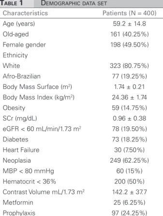
BASELINE CLINICAL CHARACTERISTICS
The baseline clinical characteristics of those 400 pa-tients are show on the Table 1. Upon inclusion in the cohort, the participants’ mean ages were 59.2 ± 14.8 years. Elderly patients and male gender accounted for 40.2% and 50.4% respectively, with Caucasian pre-dominance at 80.5%. Mean BMI was 24.36 ± 1.74 kg/m2, with underweight and obesity prevalences of
14.8% and 13.6%, respectively.
Most intravenous contrast-enhanced CT examinations were associated with malignancies (n = 249, 62.25%) in the chest, and chest-abdomen (n = 289, 72.25%). See details on Table 2. Mean contrast volume was of 142.2 ± 37.7 mL/1.73 m2 of BSA.
Of the entire sample, 25 patients (6.25%) took metformin on the contrast injection day. Only 97 (24.25%) patients received intravenous hydration (Table 1).
imaging has increased substantially in recent years. Studies have described a CIN incidence between 5 and 13% in outpatients after venous contrast injection to enhance CT scan images. These studies are limited by their retrospective design and patient selection bias.3,5,8,9
This study evaluated CIN incidence in hospitalized patients after CT scan with intravenous contrast injection, its relation with classic risk factors (DM, HF, old age, etc.) and contrast volume with variations in serum creatinine (SCr) levels.
METHODS
STUDY POPULATION
Our cohort study allocated 400 hospitalized patients from a single center (Hospital São Lucas PUCRS) between January 01, 2007 and March 31, 2008. All patients underwent CT scan with hyperosmolar in-travenous contrast (59.285 g, meglumine 15.1 g/100 mL, iodine content of 300 mg/mL, osmolality of 1650 mOsm/kg H2O, Telebrix 30 Laboratory Guebert).
Inclusion criteria for this study were: age over 18 years and hospitalization.
Exclusions criteria were: drugs that can interfere with the SCr assay (e.g.: cephalosporins, barbiturates, chemotherapeutic agents) and its secretion (e.g.: trimethoprim, cimetidine).
All patients signed consent forms. The local ethics committee approved this study.
STUDY EXECUTION
SCr values were obtained from a kinetic colorime-tric compensated Jaffe technique (Roche Modular, Meylan; compensation according to manufacturer’s recommendations). We evaluated the assay method’s inaccuracy (intra-assay coefficient was 0.7%; inter--assay coefficients were 4.0% at low SCr (0.51 - 0.71 mg/dL) and 1.5% at high SCr concentrations (6.5 mg/ dL), respectively. SCr was tested before and 48 hours after intravenous contrast injection.
To estimate GFR (eGFR), we used the CKD-EPI (Chronic Kidney Disease Epidemiology Collaboration) formula:6
141x min (SCr/k, 1)α max (SCr/k, 1)-1.209 x 0.993Age
Characteristics Patients (N = 400)
Age (years) 59.2 ± 14.8 Old-aged 161 (40.25%) Female gender 198 (49.50%) Ethnicity
White 323 (80.75%) Afro-Brazilian 77 (19.25%) Body Mass Surface (m2) 1.74 ± 0.21
Body Mass Index (kg/m2) 24.36 ± 1.74
Obesity 59 (14.75%) SCr (mg/dL) 0.96 ± 0.38 eGFR < 60 mL/min/1.73 m2 78 (19.50%)
Diabetes 73 (18.25%) Heart Failure 30 (7.50%) Neoplasia 249 (62.25%) MBP < 80 mmHg 60 (15%) Hematocrit < 36% 200 (50%) Contrast Volume mL/1.73 m2 142.2 ± 37.7
Metformin 25 (6.25%) Prophylaxis 97 (24.25%)
TABLE 1 DEMOGRAPHICDATASET
eGFR: Estimated glomerular filtration rate; SCr: Serum creatinine; MBP: Mean arterial blood pressure.
Localization N
Median volume contrast per 1.73
m2 [IQR]*
Cranial 31(7.75%) 64.0 [54.5; 120.0] Abdomen 64 (16%) 152.0 [105.5; 198.5] Thorax 162 (40.5%) 156.0 [49.5; 229.5] Thoracoabdominal 127 (31.75%) 198.5 [134.5; 249.0] Others 16 (4%) 132.0 [30.0; 254.0]
TABLE 2 TYPEOFCOMPUTERIZEDTOMOGRAPHYAND
VOLUMECONTRAST
We found an increase in baseline SCr of 25% in 61 (15.25%) patients and an absolute increase of 0.5 mg/dL in only 15 (3.75%) patients in our sample (Table 3).
PROCEDURESANDVARIATIONINRENALFUNCTION
MULTIVARIABLELOGISTICREGRESSION
After using intravenous contrast for CT, we found an association between absolute increase in SCr ≥ 0.5 mg/dL (Table 4) and ≥ 25% (Table 5) and the follo-wing factors: old age, DM, female gender, obesity, HF, CKD, neoplasia and anemia.
Multivariate analysis revealed a relationship between an absolute increase in SCr ≥ 0.5 mg/dL and
Outcome
Creatinine (mg/dL)
Baseline 0.9 ± 0.38 48 hours 1.0 ± 0.47 eGFR (mL/min/1.73 m2)
Baseline 93.43 ± 14.8 48 hours 91.95 ± 14.7 Occurrence of CIN
SCr increases ≥ 25% 61 (15.75%) SCr increases ≥ 0.5 mg/dL 15 (3.75%)
TABLE 3 INCIDENCEOFCONTRASTNEPHROPATHYAND
MARKERSOFRENALINJURY
eGFR: Estimated glomerular filtration rate; SCr: Serum creatinine; MBP: Mean arterial blood pressure.
TABLE 4 RISKFACTORSTOCONTRASTNEPHROPATHY
(SCRINCREASES ≥ 0.5 MG/DL)
Risk factors OR 95% CI p
Old-age 6.3 1.8 to 22.5 < 0.01 Diabetes 10.2 3.4 to 31.0 < 0.01 Female gender 0.9 0.3 to 2.5 0.8 Obesity 1.4 0.4 to 5.5 0.5 Heart failure 13.8 4.5 to 42.0 < 0.01 eGFR < 60 mL/
min/1.73 m2 3.9 1.3 to 11.0 < 0.05
Neoplasia 0.4 0.1 to 1.1 0.6 MBP < 80 mmHg 0.4 0.0 to 2.0 0.4 Hematocrit < 36% 0.6 0.2 to 1.9 0.4
SCr: Serum creatinine; eGFR: Estimated glomerular filtration rate; MBP: Mean arterial blood pressure; CI: Confidence interval; OR: Odds ratio.
DM (O.R.: 10.22 [95% CI: 3.37-30.92], p < 0.01); old-age (OR 6.27 [95% CI: 1.74-22.57], p < 0.05) and HF (3.9 [95% CI: 1.36-11.00], p < 0.01) (Table 4).
The relative variation (Table 5) of SCr was associated with diabetes (O.R.: 3.5 [95% CI: 1.92-6.36], p < 0.01) and HF (OR 2.61 [95% CI: 1.14-6.03%], p < 0.05). However, it was not significant vis-à-vis old age and CKD (Table 5).
Regardless of reports in the medical literature, we did not find associations between female gender, obesity, neoplasia, MBP < 80 mmHg, anemia and CIN (Tables 4 and 5).
MULTIVARIATE ANALYSIS REGRESSION MODEL AND THE
IMPACTONRENALFUNCTIONFLUCTUATION
DM and HF had a significant increase of 22% and 23% (p < 0.01) per 112 and 114 mL of intravenously injected contrast agent per 1.73 m2 of BSA,
respec-tively (model Ϋ2 and Ϋ3). Patients with eGFR < 60
mL/min/1.73 m2 and the elderly in the sample did
not show significant variation vis-à-vis contrast agent dose (model Ϋ4 and Ϋ5).
DISCUSSION
CIN-related papers have been published since the 50’s, notably after arterial contrast injection star-ted. However, only a handful of studies have inves-tigated CIN with intravenous contrast injection for CT.1-5,10 These studies described similar risk factors
for patients undergoing CT and angiographic exams. Nyman et al.10 reported a CIN incidence of 6.4%
af-ter CT and higher CIN incidences in patients with im-paired GFR.
Our results show CIN incidence after CT of 3.75 and 15.75%, with CIN defined as the absolute or relative increase of SCr, respectively. Thomsen et al.11
described that these two definitions of CIN are not interchangeable, because SCr is not an adequate marker for CIN. Thus, > 50% of renal function must be lost
before an elevation in SCr is detected. In addition, SCr does not accurately depict GFR until a steady state has been reached, which may require several days12 - this
could explain the different CIN incidence found in our study. The Acute Kidney Injury Network (AKIN) suggested two separate CIN endpoints using both absolute and relative SCr alterations.6 Their proposed
diagnostic criteria for AKI include an absolute increase in the SCr level of ≥ 0.3 mg/dL. However, calculations by Waikar & Bonventre13 showed that increases in
SCr of 0.3 mg/dL are only significant when they occur within 24 h; and 0.5 mg/dL at 48 h after CT may be a more appropriate cut-off point. Moreover, the medical literature is based on the concepts and terminology from the American College of Radiology (ACR) in reference to CIN studies, this report will do the same.5
However, we recognize that the clinical effects of slightly different definitions of CIN and AKI have yet to be clarified.6
Our results confirmed significantly classical risk factors to CIN after CT as being: CKD, DM and HF. Mehran et al.14 showed an incidence of 8.8% and
5.2%, after arterial injection in patients with CKD and DM, respectively. In patients with CKD, HF and the elderly, SCr rises more steeply when hemodynamic changes occur or contrast is administered.4,7
Our study demonstrated a statistically significant association, although low, between contrast medium volume and CIN, notably in DM and HF. It is opposite to the findings reported by other publications.8,10,15-17
Nyman et al.10 suggested a dose in grams of iodine numerically equal to the eGFR value in mL/min during percutaneous coronary intervention. These authors
described a CIN frequency of 12% at an iodine dose (in grams)/GFR ratio of 1.1. Our study demonstrated the risk of GFR reduction by checking SCr, especially among patients with diabetes, CKD and HF.
Other relevant information from our data was: lowest prescription of preventive hydration before TC (27.75%) and higher intake of biguanide (metformin) on the contrast injection day (6.25%). The European Guidelines to CIN described that 75% of CIN studies reported some form of hydration as a prevention
approach.18 They recommended expansion volume
before contrast with saline or bicarbonate solution. Biguanide (metformin) has the possibility of worsening CIN, with an associated increased risk of lactic acidosis. However, there are no direct studies on the subject.18 Prevention guidelines are based on the expert
TABLE 5 RISKFACTORSTOCONTRASTNEPHROPATHY
(SCRINCREASES ≥ 25%)
TABLE 6 IMPACTON SCRVARIATION (PERCENTAGEPER
100 ML/1.73 M2OFCONTRAST)
Risk factors OR 95% CI p
Old-age 1.0 0.6 to 1.8 0.9 Diabetes 3.5 1.9 to 6.4 < 0.01 Female gender 1.5 0.8 to 2.6 0.1 Obesity 1.7 0.8 to 3.3 0.1 Heart failure 2.6 1.1 to 5.9 < 0.05 eGFR < 60 mL/min/1.73 m2 0.5 0.2 to 1.0 0.1
Neoplasia 0.8 0.4 to 1.3 0.3 MBP < 80 mmHg 0.3 0.0 to 1.2 0.2 Hematocrit < 36% 0.8 0.5 to 1.4 0.5
SCr: Serum creatinine; eGFR: Estimated glomerular filtration rate; MBP: Mean arterial blood pressure; CI: Confidence interval; OR: Odds ratio.
Risk factors Model p
Entire population Ϋ1 = 116Χ + 0.07 0.3 Diabetes Ϋ2 = 112Χ + 0.22 < 0.01 Heart Failure Ϋ3 = 114Χ + 0.23 < 0.01 eGFR < 60 mL/min/1.73 m2 Ϋ4 = 118Χ + 0.01 0.8
Old-age Ϋ5 = 116Χ + 0.09 0.2
consensus about metformin pharmacokinetics and CIN pathophysiology.4,6,18 In this study, we did not find any
association between CIN and the use of metformin or lack of expansion volume (data not shown in the study).
The main strength of the meta-analysis is the large number of patients included (n = 400), resulting in an estimate of the CIN incidence after contrast-enhanced CT. Moreover, we have chosen a logistic model by default for all analyses to cope statistically with patient heterogeneity, resulting in a conservative incidence estimate compared to a fixed effects model.
The limitations of our study are mainly the facts that it was carried out in a single center and the impossibility of monitoring these patients to determine other possible outcomes such as death or dialysis.
CONCLUSION
Despite the difficulties due to the variability of this population, this study is one of the few prospective publications that have shown the use of intravenous contrast after CT as a variation factor associated with acute kidney injury. This condition is stronger in pa-tients with diabetes and heart failure.
CONFLICTINGINTERESTS
The authors declare that they have no conflicting interests.
REFERENCES
1. Barrett BJ, Katzberg RW, Thomsen HS, Chen N, Sahani D, Soulez G, et al. Contrast-induced nephropathy in patients with chronic kidney disease undergoing computed tomogra-phy: a double-blind comparison of iodixanol and iopami-dol. Invest Radiol 2006;41:815-21. PMID: 17035872 DOI: http://dx.doi.org/10.1097/01.rli.0000242807.01818.24 2. Elicker BM, Cypel YS, Weinreb JC. IV contrast
adminis-tration for CT: a survey of practices for the screening and prevention of contrast nephropathy. AJR Am J Roentgenol 2006;186:1651-8. PMID: 16714655 DOI: http://dx.doi. org/10.2214/AJR.05.0407
3. Haveman JW, Gansevoort RT, Bongaerts AH, Nijsten MW. Low incidence of nephropathy in surgical ICU patients recei-ving intravenous contrast: a retrospective analysis. Intensive Care Med 2006;32:1199-205. DOI: http://dx.doi.org/10.1007/ s00134-006-0198-2
4. Katzberg RW, Haller C. Contrast-induced nephrotoxicity: cli-nical landscape. Kidney Int Suppl 2006:S3-7. PMID: 16612398 DOI: http://dx.doi.org/10.1038/sj.ki.5000366
5. Rao QA, Newhouse JH. Risk of nephropathy after intravenous administration of contrast material: a critical literature analy-sis. Radiology 2006;239:392-7. PMID: 16543592 DOI: http:// dx.doi.org/10.1148/radiol.2392050413
6. Group KDIGOKCW. KDIGO 2012 clinical practice guideline for the evaluation and management of chronic kidney disease. Kidney Int Suppl 2013;3:1-150.
7. Geenen RW, Kingma HJ, van der Molen AJ. Contrast-indu-ced nephropathy: pharmacology, pathophysiology and pre-vention. Insights Imaging 2013;4:811-20. DOI: http://dx.doi. org/10.1007/s13244-013-0291-3
8. Kooiman J, Pasha SM, Zondag W, Sijpkens YW, van der Mo-len AJ, Huisman MV, et al. Meta-analysis: serum creatinine changes following contrast enhanced CT imaging. Eur J Ra-diol 2012;81:2554-61. PMID: 22177326 DOI: http://dx.doi. org/10.1016/j.ejrad.2011.11.020
9. Krol AL, Dzialowski I, Roy J, Puetz V, Subramaniam S, Coutts SB, et al. Incidence of radiocontrast nephropathy in patients undergoing acute stroke computed tomography angiogra-phy. Stroke 2007;38:2364-6. DOI: http://dx.doi.org/10.1161/ STROKEAHA.107.482778
10. Nyman U, Almén T, Aspelin P, Hellström M, Kristiansson M, Sterner G. Contrast-medium-Induced nephropathy correlated to the ratio between dose in gram iodine and estimated GFR in ml/min. Acta Radiol 2005;46:830-42. PMID: 16392608 DOI: http://dx.doi.org/10.1080/02841850500335051
11. Thomsen HS, Morcos SK, Erley CM, Grazioli L, Bonomo L, Ni Z, et al.; Investigators in the Abdominal Computed Tomogra-phy: IOMERON 400 Versus VISIPAQUE 320 Enhancement (ACTIVE) Study. The ACTIVE Trial: comparison of the effects on renal function of iomeprol-400 and iodixanol-320 in pa-tients with chronic kidney disease undergoing abdominal com-puted tomography. Invest Radiol 2008;43:170-8. DOI: http:// dx.doi.org/10.1097/RLI.0b013e31815f3172
12. Bruce RJ, Djamali A, Shinki K, Michel SJ, Fine JP, Pozniak MA. Background fluctuation of kidney function versus contrast-in-duced nephrotoxicity. AJR Am J Roentgenol 2009;192:711-8. PMID: 19234268 DOI: http://dx.doi.org/10.2214/AJR.08.1413 13. Waikar SS, Bonventre JV. Creatinine kinetics and the definition
of acute kidney injury. J Am Soc Nephrol 2009;20:672-9. DOI: http://dx.doi.org/10.1681/ASN.2008070669
14. Mehran R, Aymong ED, Nikolsky E, Lasic Z, Iakovou I, Fahy M, et al. A simple risk score for prediction of contrast-induced nephropathy after percutaneous coronary intervention: develo-pment and initial validation. J Am Coll Cardiol 2004;44:1393-9. PMID: 15464318
15. Katzberg RW, Newhouse JH. Intravenous contrast medium-indu-ced nephrotoxicity: is the medical risk really as great as we have come to believe? Radiology 2010;256:21-8. PMID: 20574082 16. Karlsberg RP, Dohad SY, Sheng R; Iodixanol Peripheral
Com-puted Tomographic Angiography Study Investigator Panel. Contrast medium-induced acute kidney injury: comparison of intravenous and intraarterial administration of iodinated con-trast medium. J Vasc Interv Radiol 2011;22:1159-65. DOI: http://dx.doi.org/10.1016/j.jvir.2011.03.020
17. Nyman U, Almén T, Jacobsson B, Aspelin P. Are intravenous injections of contrast media really less nephrotoxic than intra--arterial injections? Eur Radiol 2012;22:1366-71.