Prevalence of developmental defects of enamel
in children and adolescents with asthma*
,**
Prevalência de defeitos do desenvolvimento do esmalte dentário em crianças e adolescentes com asma
Rodrigho Pelisson Guergolette, Cássia Cilene Dezan,
Wanda Terezinha Garbelini Frossard, Flaviana Bombarda de Andrade Ferreira, Alcindo Cerci Neto, Karen Barros Parron Fernandes
Abstract
Objective: This study aimed to evaluate the prevalence of developmental defects of enamel (DDEs) in relation to asthma severity, symptom onset and pharmacological treatment in pediatric asthma patients. Methods: Children and adolescents (68 asthma patients and 68 controls), 5-15 years of age and residents of the city of Londrina, Brazil, were enrolled in the study. Medical and dental histories were collected through the use of a structured questionnaire. Each participant underwent a dental examination in which the examiner employed the DDE index. Results: Of the 68 asthma group subjects, 61 (89.7%) presented dental enamel defects, compared with only 26 (38.2%) of those in the control group. Using multivariate logistic regression analysis, we estimated the risk of DDEs in permanent dentition to be 11 times higher in pediatric subjects with asthma than in those without (OR = 11.88, p = 0.0001). The occurrence of dental enamel defects correlated with greater asthma severity (p = 0.0001) and earlier symptom onset (p = 0.0001). However, dental enamel defects did not correlate with the initiation of treatment (p = 0.08) or the frequency of medication use (p = 0.93). Conclusions: Pediatric patients with severe, early-onset asthma are at increased risk of dental enamel defects and therefore require priority dental care.
Keywords: Asthma/prevention & control; Bronchodilator agents; Adrenal cortex hormones/therapeutic use; Dental enamel; Amelogenesis.
Resumo
Objetivo: Avaliou-se a prevalência de developmental defects of enamel (DDEs, defeitos de desenvolvimento do esmalte dentário) em pacientes pediátricos com asma e sua relação com a severidade da asma, o início dos sintomas e o tratamento medicamentoso. Métodos: Os participantes do estudo eram residentes do município de Londrina (PR), com 5 a 15 anos, sendo 68 asmáticos e 68 controles. Foram levantados dados retrospectivos da história médica e de saúde bucal da população do estudo através de um questionário estruturado. Todos os participantes foram submetidos a um exame dental. Para a avaliação dos defeitos de desenvolvimento do esmalte dentário, utilizou-se o Índice DDE. Resultados: Neste estudo, foi observado que 61 (89,7%) dos 68 pacientes asmáticos apresentavam defeitos de desenvolvimento do esmalte dentário quando comparado à ocorrência em 26 (38,2%) dos no grupo controle. Através da análise multivariada por regressão logística, foi observado que um paciente pedi-átrico com asma apresenta risco aumentado em 11 vezes para o aparecimento de defeitos de desenvolvimento do esmalte em dentes permanentes (OR = 11,88, p = 0,0001). Além disso, foi observado uma associação entre defeitos do esmalte dentário e maior severidade da asma (p = 0,0001) e início dos sintomas mais precoce (p = 0,0001). Não se observou associação entre o início do tratamento (p = 0,08) ou frequência de uso da medicação (p = 0,93) com o aparecimento de defeitos de desenvolvimento do esmalte dentário. Conclusões: Pacientes pediátricos com asma apresentam risco aumentado para a ocorrência de defeitos de desenvolvimento do esmalte dentário relacionado à severidade da asma e início dos sintomas e, portanto, necessitam de atenção odontológica prioritária.
Descritores: Asma/prevenção & controle; Broncodilatadores; Corticosteroides/uso terapêutico; Esmalte dentário; Amelogênese.
* Study carried out at Londrina State University – UEL – and the University of Northern Paraná – UNOPAR – Londrina, Brazil. Correspondence to: Alcindo Cerci Neto. Rua Piauí, 61, apto. 102, Centro, CEP 86010-420, Londrina, PR, Brasil.
Tel 55 43 3323-9784. Fax 55 43 3323-9784. E-mail: [email protected]
Financial support: This study received financial support from the Universidade do Norte do Paraná (UNOPAR, University of Northern Paraná) and the Fundação Nacional de Desenvolvimento do Ensino Superior Particular (FUNADESP, National Foundation for the Development of Privately-held Institutions of Higher Education).
Submitted: 12 March 2008. Accepted, after review: 11 September 2008.
tion, celiac disease and respiratory disorders, are associated with dental enamel defects.(11-14)
However, due to a lack of medical and dental records providing data related to young children, it can be difficult to establish the relationship between systemic factors and developmental defects of enamel (DDEs).(15) Therefore,
epide-miological studies of enamel defects in children with systemic disease are quite important for public health, since they can identify possible etiological factors responsible for the occurrence of the enamel defects,(10) as well as
identi-fying populations that merit priority preventive interventions.(16) Therefore, this study aimed to
evaluate the prevalence of DDEs in children with asthma, drawing correlations with specific char-acteristics of the disease (symptom onset and asthma severity) and its treatment (timing of treatment initiation and frequency of medica-tion use).
Methods
This was a cross-sectional study involving children and adolescents (5-15 years of age) in two groups: asthma (n = 68) and control (n = 68).
The asthma group consisted of children and adolescents randomly selected from among patients treated via the Programa Respira Londrina (Breathe Londrina Program) or at the Londrina State University Hospital, located in the city of Londrina, Brazil. All asthma group subjects were either under continuous treatment with corticosteroids or were using bronchodila-tors for acute attacks. The control group subjects, who were selected from Londrina public schools, were using no chronic medication and had no systemic diseases. The control group subjects were matched for age and gender with the asthma group subjects. However, any child or adolescent selected from a public school and later found to have asthma was then included at the asthma group.
Introduction
Asthma is a major public health problem,(1) and
the prevalence of asthma has recently presented a marked increase in many countries.(2)
Some studies have reported that the preva-lence of dental caries is higher and that salivary flow levels are lower in children with asthma than in those without asthma.(3-5) There have
also been reports suggesting that respiratory disorders are associated with dental enamel defects.(6) However, such reports have presented
no epidemiological data regarding its preva-lence in children with asthma—nor have there been any studies investigating the potential role that pharmacological treatment plays in this association.
Dental enamel defects are a frequent finding in primary and permanent dentition. These defects are generally classified as enamel hypo-plasia or enamel hypomineralization. Enamel hypoplasia is a quantitative defect, whereas enamel hypomineralization is a qualitative defect characterized by abnormal enamel trans-lucency and is therefore also known as enamel opacity.(7)
Enamel formation occurs in three stages: matrix formation, during which proteins involved in amelogenesis are produced; calcification, during which mineral content is acquired and the proteins are removed; maturation, during which the enamel is calcified and the remaining proteins are removed. Disorders in the early stages of enamel development evoke enamel hypoplasia, clinically detectable as fissures or enamel loss.(8) In contrast, disorders occurring in
the calcification or maturation stage can cause hypomineralization.(9)
Dental enamel defects have been associated with a broad spectrum of etiologies, including systemic, genetic, local and environmental factors.(10) Some authors have stated that systemic
conditions, such as prenatal or perinatal illness, low birth weight, regular antibiotic
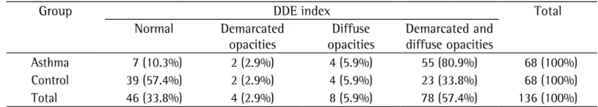
consump-Table 1 - Distribution of developmental defects of enamel index in the study sample.
Group DDE index Total
Normal Demarcated opacities
Diffuse opacities
Demarcated and diffuse opacities
Asthma 7 (10.3%) 2 (2.9%) 4 (5.9%) 55 (80.9%) 68 (100%)
Control 39 (57.4%) 2 (2.9%) 4 (5.9%) 23 (33.8%) 68 (100%)
Total 46 (33.8%) 4 (2.9%) 8 (5.9%) 78 (57.4%) 136 (100%)
examinations, the children were placed in a conventional dental chair. The teeth were dried for 30 s, after which they were examined with the aid of artificial light and a small mirror.
In order to perform a clinical assessment of possible mouth breathing, the examiner used the test described by Menezes et al. (2006) (18) in
which patients are asked to hold water in their mouth, lips closed and without swallowing, for 3 min. Patients who swallow the water or open their lips during this period are presumed to have reduced nasal breathing capacity.
Statistical analysis
Statistical analysis was performed using the program Statistical Package for the Social Sciences (SPSS Inc., Chicago, IL, USA). For all statistical tests, a confidence interval of 95% and significance level of 5% (p < 0.05) were adopted. In order to compare the means of the two groups, we used the Student’s t-test for independent samples and for variables with normal distribution.
The chi-square test, followed by Yates’ correction, was used in order to compare the prevalence of DDEs in the asthma group with that observed in the control group.
The exclusion criteria were as follows: presenting any other systemic disease; using a fixed orthodontic appliance; and presenting extensive carious lesions that might mask dental enamel defects.
This study was approved by the Ethics Committee of the University of Northern Paraná, as well as by the administrations of the Breathe Londrina Program and the Londrina State University Hospital das Clínicas. The parents or legal guardians of the participants received infor-mation regarding the purpose of the study, and all gave written informed consent prior to the clinical examinations. All participants received instructions related to oral health maintenance, and each was given a new toothbrush at the end of the clinical examination.
Two examiners were involved in the study, the first being responsible for data collection (patient medical and dental history), and the second being responsible for the evaluation of dental enamel defects.
A questionnaire related to personal medical and dental history, formulated by the authors of the study, was completed by all of the participating children and adolescents, with the assistance of their parents or legal guardians. After the questionnaire had been completed, participants were identified only by assigned numbers and as belonging to the asthma or control group. The examiner performing the clinical diagnosis of dental enamel defects was blinded to the group to which any given child belonged.
Clinical examinations were carried out at the Dental Clinic of the University of Northern Paraná. All examinations were conducted by the same examiner, who had been previously trained and certified as a reference examiner using the modified DDE index,(17) as recommended by
World Health Organization for the evaluation of enamel defects. In this index, all enamel defects, regardless of location or size, are recorded, being classified as follows: absent (normal condition); demarcated opacities (white or yellow—single or multiple); diffuse opacities (parallel lines or with a patchy distribution); and hypoplasia (pits, grooves, or larger areas of missing enamel— single or multiple).
The kappa value obtained to estimate the consistency of the fieldwork examiner was k = 0.84 for the DDE diagnosis. During clinical
Table 2 - Occurrence, adjusted for gender and age, of dental enamel defects in the permanent teeth of children and adolescents presenting asthma, allergic rhinitis or mouth breathing.
Variable OR 95% CI p
Asthma 11.88 4.38-32.19 0.0001
dentition was higher in the asthma group than in the control group (p = 0.0001).
Demarcated diffuse opacities, which were observed in 34 asthma group subjects (54%) and 16 control group subjects (20.8%), constituted the most prevalent enamel defect observed in this population (Table 1).
A representative photograph of the DDEs observed in one child with asthma is shown in Figure 1.
Logistic regression revealed that the risk of dental enamel defects is approximately 11 times higher for children with asthma (Table 2).
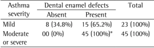
The occurrence of dental enamel defects correlated with asthma severity (rφ: 0.51, p = 0.0001), as well as with symptom onset (rφ: 0.67, p = 0.0001), as shown in Tables 3 and 4, respectively.
The occurrence of dental enamel defects was not found to correlate with the initiation of treat-ment (rφ: 0.27, p = 0.08) or with the frequency of medication use (rφ: 0.05, p = 0.93).
Discussion
In the present study, the prevalence of DDEs was found to be high among Brazilian pediatric patients with asthma, which was estimated to increase the risk of dental enamel defects in permanent dentition by 11 times.
Our findings are in agreement with those of another group of authors,(11) who observed a
positive correlation between respiratory disor-ders, such as asthma, and the presence of demarcated opacities in first permanent molars. Other authors have also suggested that asthma is associated with the occurrence of DDEs in permanent teeth.(19,20)
Despite the fact that some studies have iden-tified asthma as a potential etiological factor in the occurrence of dental enamel defects,(11) there
have been no studies investigating the possible interactions between such occurrence and the onset of asthma symptoms or the timing of treatment.
In our study, we found that the occurrence of dental enamel defects presented a statisti-cally significant correlation with asthma severity, as well as with symptom onset, the risk being greater in pediatric patients with moderate/ severe asthma, especially in those presenting symptoms before 3 years of age. One group of authors reported that the risk of dental enamel Logistic regression was used in order to assess
the risk of dental enamel defects in a multivar-iate analysis. The model included the variables asthma, allergic rhinitis and mouth breathing, all adjusted for age and gender.
The phi coefficient was used in order to determine whether the prevalence of dental enamel defects correlated with asthma severity, symptom onset, timing of the initiation of treat-ment and frequency of medication use. In order to perform this analysis, those variables were considered dichotomous: asthma severity (mild or moderate/severe); symptoms onset (before or after 3 years of age); initiation of treatment (before or after 3 years of age); and frequency of medication use (continuous and for acute attacks only).
Results
Of the 68 children and adolescents in the asthma group, 35 (51.5%) were girls and 33 (48.5%) were boys, compared with 42 (61.8%) and 26 (38.2%), respectively, of those in the control group. There were no gender-related differences between the asthma and control groups. The mean age in the asthma group was 9.61 ± 0.31 years, compared with 9.79 ±
0.18 years in the control group (p > 0.05). Permanent-tooth DDEs were observed in 61 (89.7%) of the asthma group subjects and 26 (38.2%) control group subjects. The teeth were free of dental enamel defects in only 7 (10.3%) of the asthma group subjects and 42 (61.8%) of the control group subjects. Therefore, the prev-alence of dental enamel defects in permanent
Table 3 - Prevalence of dental enamel defects according to asthma severity.
Asthma severity
Dental enamel defects Total Absent Present
Mild 8 (34.8%) 15 (65.2%) 23 (100%) Moderate
or severe
00 (0%) 45 (100%)* 45 (100%)
*p = 0.0001 vs. mild.
Table 4 - Prevalence of dental enamel defects according to symptom onset.
Symptom onset Dental enamel defects Absent Present Before 3 years of age 01 (1.8%) 55 (98.2%)* After 3 years of age 07 (58.3%) 05 (41.7%)
5. Ersin NK, Gülen F, Eronat N, Cogulu D, Demir E, Tanaç R, et al. Oral and dental manifestations of young asthmatics related to medication, severity and duration of condition. Pediatr Int. 2006;48(6):549-54.
6. Kellerhoff NM, Lussi A. Molar-incisor hypomineralization [Article in French, German]. Schweiz Monatsschr Zahnmed. 2004;114(3):243-53.
7. Jälevik B, Norén JG. Enamel hypomineralization of permanent first molars: a morphological study and survey of possible aetiological factors. Int J Paediatr Dent. 2000;10(4):278-89.
8. Frazão P, Peverari AC, Forni TI, Mota AG, Costa LR. Dental fluorosis: comparison of two prevalence studies [Article in Portuguese]. Cad Saude Publica. 2004;20(4):1050-8. 9. Hoffmann RH, De Sousa Mda L, Cypriano S. Prevalence
of enamel defects and the relationship to dental caries in deciduous and permanent dentition in Indaiatuba, São Paulo, Brazil [Article in Portuguese]. Cad Saude Publica. 2007;23(2):435-44.
10. Dini EL, Holt RD, Bedi R. Prevalence of caries and developmental defects of enamel in 9-10 year old children living in areas in Brazil with differing water fluoride histories. Br Dent J. 2000;188(3):146-9. 11. Jälevik B, Norén JG, Klingberg G, Barregård L. Etiologic
factors influencing the prevalence of demarcated opacities in permanent first molars in a group of Swedish children. Eur J Oral Sci. 2001;109(4):230-4.
12. Beentjes VE, Weerheijm KL, Groen HJ. Factors involved in the aetiology of molar-incisor hypomineralisation (MIH). Eur J Paediatr Dent. 2002;3(1):9-13.
13. Weerheijm KL, Jälevik B, Alaluusua S. Molar-incisor hypomineralisation. Caries Res. 2001;35(5):390-1. 14. Wierink CD, van Diermen DE, Aartman IH, Heymans HS.
Dental enamel defects in children with coeliac disease. Int J Paediatr Dent. 2007;17(3):163-8.
15. Suckling GW, Herbison GP, Brown RH. Etiological factors influencing the prevalence of developmental defects of dental enamel in nine-year-old New Zealand children participating in a health and development study. J Dent Res. 1987;66(9):1466-9.
16. Lunardelli SE, Peres MA. Prevalence and distribution of developmental enamel defects in the primary dentition of pre-school children. Braz Oral Res. 2005;19(2):144-9. 17. A review of the developmental defects of enamel index
(DDE Index). Commission on Oral Health, Research &
Epidemiology. Report of an FDI Working Group. Int Dent J. 1992;42(6):411-26.
18. De Menezes VA, Leal RB, Pessoa RS, Pontes RM. Prevalence and factors related to mouth breathing in school children at the Santo Amaro project-Recife, 2005. Braz J Otorhinolaryngol. 2006;72(3):394-9. 19. van Amerongen WE, Kreulen CM. Cheese molars:
a pilot study of the etiology of hypocalcifications in first permanent molars. ASDC J Dent Child. 1995;62(4):266-9.
20. Narang A, Maguire A, Nunn JH, Bush A. Oral health and related factors in cystic fibrosis and other chronic respiratory disorders. Arch Dis Child. 2003;88(8):702-7. 21. Mathu-Muju K, Wright JT. Diagnosis and treatment
of molar incisor hypomineralization. Compend Contin Educ Dent. 2006;27(11):604-10; quiz 611.
22. Cerci Neto A, Zamboni MM, Holanda MA. Open letter in favor of the creation of asthma programs in Brazil. J Bras Pneumol. 2007;33(2):ix-x.
defects is higher in children with poor health during the first three years of life (the critical period for crown formation of first permanent molars, permanent incisors and canines).(21) In
addition, it has been suggested that any health problem prior to 5 years of age can modu-late ameloblast activity and therefore impair amelogenesis.(6) One study showed that 67% of
Dutch children who present DDEs suffer from respiratory disease.(19) Since ameloblasts are
highly sensitive to oxygen supply,(6,7) we can
hypothesize that pediatric asthma patients with dental enamel defects have probably experi-enced previous episodes of oxygen deprivation. Therefore, these defects might be related to the disease itself rather than to its treatment.
Further studies of the prevalence of dental enamel defects in patients with asthma are needed. It has been recommended that admin-istrators of asthma programs be alerted to any such epidemiological data.(22) Longitudinal
studies could establish the mechanisms by which respiratory disorders affect enamel formation.
Dental enamel is an atypical tissue. Unlike other hard tissues, dental enamel, once formed, cannot remodel. Since changes during its forma-tion are permanent and are recorded in the tooth surface, dental enamel defects can consti-tute a biological marker of adverse events during development, and this marker could have clinical and epidemiological applications.(7)
The prevalence of dental caries is high among children with DDEs. Therefore, we suggest that priority dental programs be provided for this population in order to reduce aesthetic and dento-facial abnormalities, as well as to lower the prevalence of dental caries. In addition, we recommend that dentists be included in multidis-ciplinary teams affiliated with asthma programs.
References
1. Steinbacher DM, Glick M. The dental patient with asthma. An update and oral health considerations. J Am Dent Assoc. 2001;132(9):1229-39.
2. Lugogo NL, Kraft M. Epidemiology of asthma. Clin Chest Med. 2006;27(1):1-15, v.
3. Reddy DK, Hegde AM, Munshi AK. Dental caries status of children with bronchial asthma. J Clin Pediatr Dent. 2003;27(3):293-5.
About the authors
Rodrigho Pelisson Guergolette
Graduate Student. School of Dentistry, University of Northern Paraná – UNOPAR – Londrina, Brazil.
Cássia Cilene Dezan
Associate Professor. School of Dentistry, Londrina State University – UEL – Londrina, Brazil.
Wanda Terezinha Garbelini Frossard
Associate Professor. School of Dentistry, Londrina State University – UEL – Londrina, Brazil.
Flaviana Bombarda de Andrade Ferreira
Associate Professor. School of Dentistry, University of Northern Paraná – UNOPAR – Londrina, Brazil.
Alcindo Cerci Neto
Adjunct Professor. School of Medicine, Londrina State University – UEL – Londrina, Brazil.
Karen Barros Parron Fernandes