319
NEOPLASIA IN HORSESHOE KIDNEY WITH PYELIC FUSION Case Report
International Braz J Urol
Official Journal of the Brazilian Society of Urology
Vol. 30 (4): 319-320, July - August, 2004
NEOPLASIA IN HORSESHOE KIDNEY WITH PYELIC FUSION AND
CROSSED SINGLE URETER
RENATO P. COSTA, CARLOS H. SCHAAL, FÁBIO C. NAVARRO
Section of Urology, Amaral Carvalho Hospital, Jau, São Paulo, Brazil
ABSTRACT
Horseshoe kidney with pyelic fusion and crossed single ureter is a rare anomaly, with only 3 cases described in the literature. Such anomaly can be accompanied by other abnormalities, such as congenital scoliosis and situs inversus totalis. We present one case of this malformation associ-ated with malignant neoplasia, treassoci-ated with partial nephrectomy.
Key words: kidney; anomalies; kidney neoplasms Int Braz J Urol. 2004; 30: 319-20
INTRODUCTION
Horseshoe kidney with pyelic fusion and crossed single ureter is a rare anomaly, with only 3 cases found in a research of literature through Medline. We present one case of this malformation associated with malignant neoplasia, treated with partial nephrectomy.
CASE REPORT
Female, 26-year old patient, complaining of colic on right lumbar region, underwent investiga-tion by plain x-ray of abdomen that revealed no ab-normality. An ultrasound of urinary tract was per-formed, showing a solid expansive lesion measuring 8 x 4 cm in upper pole and middle portion of the right kidney, associated with horseshoe kidney. Abdomi-nal computerized tomography (CT) (Figure-1) con-firmed sonographic findings, demonstrating pyelic fusion associated with crossed single ureter as well. No distant lesions were observed during stag-ing. A cystoscopy was performed evidencing single ureteral meatus on the right side. Considering this condition, radical nephrectomy was indicated through
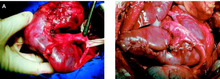
bilateral subcostal incision, with partial nephrectomy being feasible, with cold ischemia during 30 minutes (Figure-2). No macroscopically involved lymph node was observed intraoperatively.
The pathological examination of the surgical specimen revealed clear cell carcinoma, Furhman nuclear grade I, measuring 8.3 x 5.5 x 4.5 cm, weight-ing 200 g, infiltratweight-ing the renal capsule, but with mar-gins free of neoplasia. The pelvis and renal vein were not involved.
Patient has been followed for 8 months with-out any signs of recurrence.
COMMENTS
320
NEOPLASIA IN HORSESHOE KIDNEY WITH PYELIC FUSION
scoliosis (2,3), however they were not observed in this case.
Even though the development of neoplasia in this malformation is rare, we believe that the out-come is probably similar to other malignant renal tu-mors. Surgical planning is fundamental for a success-ful treatment.
REFERENCES
1. Mungan NA, Gundogdu S, Erdem Z, Akduman B, Erdem O, Yesilli C: Horseshoe kidney with pyelic fu-sion and crossed single ureter. J Urol. 2003; 170: 175-6.
2. Cass AS, Vitko RJ: Unusual variety of crossed renal
ectopy renal with only one ureter. J Urol. 1972; 107: 1056-8.
3. Aragona F, Seretta V, Fiorentini L, Marconi A, Spinelli C: Combined renal and pyelic fusion with crossed ectopia of single ureter. Urology. 1986; 28: 339-41.
Received: December 4, 2003 Accepted after revision: April 26, 2004
Correspondence address:
Dr. Renato Prado Costa Rua Luiz Paiva, 100 Jaú, SP, 17210-090, Brazil Fax: + 55 14 3624-5155 E-mail: [email protected]
Figure 1 – A) Abdominal computerized tomography following oral and venous administration of contrast agents, demonstrating hypodense and heterogeneous lesion with peripheral enhancement in upper pole and middle portion of the right kidney (8.3 x 5.5 x 4.5 cm). B) Abdominal computerized tomography with oral and venous administration of contrast agent, demonstrating pyelic fusion.
A
B
Figure 2 – A) Intraoperative aspect of horseshoe kidney with tumoral lesion in the upper pole of right kidney. B) Final surgical aspect, after right partial nephrectomy.