313
ADENOMATOID TUMOR OF SUPRA-RENAL GLAND Case Report
International Braz J Urol
Official Journal of the Brazilian Society of Urology
Vol. 30 (4): 313-315, July - August, 2004
ADENOMATOID TUMOR OF SUPRA-RENAL GLAND
NANCY T. DENICOL, FABRÍCIO R. LEMOS, WALTER J. KOFF
Section of Urology, Porto Alegre General Hospital, Federal University of Rio Grande do Sul (UFRGS), Rio Grande do Sul, Brazil
ABSTRACT
Adenomatoid tumors of adrenal gland are rare, asymptomatic neoplasias, with benign be-havior, and usually are diagnosed incidentally.
We report a case of a voluminous adenomatoid tumor of left adrenal gland in a 42-year old man who sought evaluation because of renal colic due to left nephrolithiasis. During the investigation, a tumor localized in left adrenal gland was identified by ultrasonography (14.3 x 10.5 x 19.0).
The patient underwent adrenalectomy and pyelolithotomy with histopathological and immu-nohistochemical diagnosis of adenomatoid tumor of adrenal gland, being the largest one described in the literature to the moment. The patient does not present any signs of recurring lesion after a 3-year follow-up.
Key words: adrenal glands; neoplams; adenomatoid tumors; nephrolithiasis Int Braz J Urol. 2004; 30: 313-5
INTRODUCTION
Adenomatoid tumors of adrenal gland are rare, asymptomatic neoplasias, with benign behav-ior, and usually are diagnosed incidentally (1). Adenomatoid tumors appear more frequently in the genital system both in males and in females (2). The literature reports only 16 cases of adrenal adenomatoid tumors (3). In this work, we report one case of voluminous adenomatoid tumor affecting the left adrenal gland.
CASE REPORT
Caucasian, 42-year old man was referred due to presenting, during investigation of renal colic, a mass measuring 14.3 x 10.5 x 19.0 cm localized on left adrenal gland in ultrasonographic imaging. The patient had a previous history of systemic arterial hy-pertension with moderate tension levels since he was
314
ADENOMATOID TUMOR OF SUPRA-RENAL GLAND
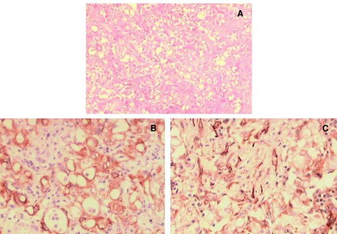
Adrenalectomy and pyelolithotomy were per-formed by open approach, with no intercurrences trans- or post-operatively. The adrenal gland weighted 535 g and upon sectioning it showed predominance of multicystic, yellowish and opaque tissue, with the tumor being predominantly solid. Histological exami-nation with hematoxylin-eosin demonstrated mesothe-lial cells similar to epithemesothe-lial lineage cells arranged as small tubules, cysts or string-shaped, with histo-logical pattern compatible with adenomatoid tumor of adrenal gland (Figure-1). Immunohistochemical study by the immunoperoxidase technique using the markers AE1, AE3, vimentin, CEA and CD31, dem-onstrate that neoplastic cells were positive to AE1,
Figure 1 – Histological and immunohistochemical studies of the adenomatoid tumor of adrenal gland. A) Proliferation of mesothelial cells similar to epithelial lineage cells, arranged as small tubules, cysts, or string-shaped (HE, X80). B) Positivity for cytokeratins in epithelial-like cells. CEA negative. Positive to AE1 and AE3 (Imunolabeling, X200). C) Positivity for vimentin expressed in mesothelial cells (Imunolabeling, X200).
AE3 and vimentin (Figure-1), and negative to the other markers, confirming the diagnosis.
The patient has not shown signs of recurrent lesion after a 3-year follow-up.
COMMENTS
Adenomatoid tumors are benign neoplasias with mesothelial origin, with cases rarely reported in extra-genital sites (2). There are 8 cases describing adenomatoid tumor of adrenal gland, occurring mostly in males, involving more frequently the left adrenal gland (1). With the increasing use of imaging exami-nations for diagnostic of other pathologies, adrenal
A
315
ADENOMATOID TUMOR OF SUPRA-RENAL GLAND
tumors have been more diagnosed, usually in an inci-dental way. Apparently, there are no specific charac-teristics that enable us to radiologically distinguish adenomatoid tumors from other adrenal lesions (4). The differential diagnosis of an adrenal mass includes adrenal adenoma or carcinoma, myelolipoma, pheo-chromocytoma, cysts and metastatic neoplasias (1). There are no reports in the literature of lesion recur-rence following complete surgical removal, and this treatment is definitive.
REFERENCES
1. Raaf HN, Grant LD, Santoscoy C, Levin HS, Abdul-Karim FW: Adenomatoid tumor of the adrenal gland:
a report of four new cases and a review of the litera-ture. Mod Pathol. 1996; 9: 1046-51.
2. Glatz K, Wegmann W: Papillary adenomatoid tumour of the adrenal gland. Histopathology. 2000; 37: 376-7.
3. Isotalo PA, Keeney GL, Sebo TJ, Riehle DL, Cheville JC: Adenomatoid tumor of the adrenal gland: a clini-copathologic study of five cases and review of the lit-erature. Am J Surg Pathol. 2003; 27: 969-77. 4. Rodrigo Gasque C, Marti-Bonmati L, Dosda R,
Gonzalez Martinez A: MR imaging of a case of adenomatoid tumor of the adrenal gland. Eur Radiol. 1999; 9: 552-4.
Received: May 26, 2004 Accepted after revision: July 30, 2004
Correspondence address: Dr. Fabrício Rodrigues Lemos Rua São Marcos 77 / 303
Porto Alegre, RS, 91420-550, Brazil Tel.: + 55 51 9812-4743