pneumoconioses. Environments posing the greatest occu-pational risk include the following: mining and processing of stone; mining of gold and precious stones; well drilling;
Introduction
Silicosis is a fibrosing lung disease caused by inhala-tion and deposiinhala-tion of crystalline silica particles, resulting in a pulmonary response. It is the most prevalent of the
Tuberculosis and silicosis: epidemiology, diagnosis and chemoprophylaxis*
Tuberculose e silicose: epidemiologia, diagnóstico e quimioprofilaxia
Carlos Eduardo Galvão Barboza1, Daniel Hugo Winter2, Márcia Seiscento3,
Ubiratan de Paula Santos4, Mário Terra Filho5
Abstract
Silicosis, the most prevalent of the pneumoconioses, is caused by inhalation of crystalline silica particles. Silica-exposed workers, with or without silicosis, are at increased risk for tuberculosis and nontuberculous mycobacteria-related diseases. The risk of a patient with silicosis developing tuberculosis is higher (2.8 to 39 times higher, depending on the severity of the silicosis) than that found for healthy controls. Various regimens for tuberculosis chemoprophylaxis in patients with silicosis have been studied, all of which present similar efficacy and overall risk reduction to about one half of that obtained with placebo. Long-term regimens have potential side effects (particularly hepato-toxicity). In addition, the use of such regimens can jeopardize adherence to treatment. The current guidelines recommend that tuberculin skin tests be performed, and, if positive, that chemoprophylaxis be instituted. There are several possible regimens, varying in terms of the drugs prescribed, as well as in terms of treatment duration. We recommend the use of isoniazid at 300 mg/day (or 10 mg/kg/day) for six months for patients with silicosis, as well as for healthy patients with periods of exposure to silica longer than 10 years and strongly positive tuberculin skin test results (induration ≥ 10 mm). Nevertheless, further studies are necessary so that indications, drugs, doses and duration of chemoprophylaxis regimens can be more properly defined.
Keywords: Silicosis; Tuberculosis; Diagnostic techniques and procedures; Chemoprevention.
Resumo
A silicose, a mais prevalente das pneumoconioses, é provocada pela inalação de partículas de sílica cristalina. Indivíduos expostos à sílica, com ou sem silicose, apresentam risco aumentado de tuberculose e de micobacterioses não-tuberculosas. O risco de silicóticos desenvolverem tuberculose em relação a controles sadios varia de 2,8 a 39 vezes, em conformidade com a gravidade da doença de base. Têm sido estudados diferentes esquemas de quimioprofilaxia para tuberculose em silicóticos, todos com eficácia semelhante e com redução final de risco para cerca da metade em relação ao uso de placebo. São, no entanto, esquemas de longa duração, o que, acrescido dos possíveis efeitos colate-rais (particularmente hepatotoxicidade), podem prejudicar a aderência. As diretrizes atuais recomendam a realização de prova tuberculínica e, se positiva, a instituição de quimioprofilaxia. São vários os esquemas possíveis, tanto em termos de drogas quanto de duração. Nossa recomendação é de que se use isoniazida na dose de 300 mg/dia (ou 10 mg/kg/dia) por seis meses para os indivíduos com silicose ou sadios com exposição superior a 10 anos, se forem reatores fortes à prova tuberculínica (induração ≥ 10 mm). São necessários, no entanto, novos estudos para que indicações, drogas, doses e duração da profilaxia sejam definidas mais apropriadamente.
Descritores: Silicose; Tuberculose; Técnicas de diagnóstico e procedimentos; Quimioprevenção.
* Study carried out in the Department of Pulmonology of the Instituto do Coração do Hospital das Clínicas da Faculdade de Medicina da Universidade de São Paulo – InCor/HC-FMUSP, Heart Institute of the University of São Paulo School of Medicine Hospital das Clínicas – São Paulo, Brazil.
1. Chief Resident in the Department of Pulmonology of the Instituto do Coração do Hospital das Clínicas da Faculdade de Medicina da Universidade de São Paulo – InCor/HC-FMUSP, Heart Institute of the University of São Paulo School of Medicine Hospital das Clínicas – São Paulo, Brazil.
2. Graduate Student in the Department of Pulmonology of the Instituto do Coração do Hospital das Clínicas da Faculdade de Medicina da Universidade de São Paulo – InCor/HC-FMUSP, Heart Institute of the University of São Paulo School of Medicine Hospital das Clínicas – São Paulo, Brazil.
3. Attending Physician in the Tuberculosis Group of the Department of Pulmonology of the Instituto do Coração do Hospital das Clínicas da Faculdade de Medicina da Universidade de São Paulo – InCor/HC-FMUSP, Heart Institute of the University of São Paulo School of Medicine Hospital das Clínicas – São Paulo, Brazil. 4. Attending Physician in the Occupational Diseases Group of the Department of Pulmonology of the Instituto do Coração do Hospital das Clínicas da Faculdade de Medicina da Universidade de São Paulo – InCor/HC-FMUSP, Heart Institute of the University of São Paulo School of Medicine Hospital das Clínicas – São Paulo, Brazil. 5. Associate Professor in the Department of Pulmonology of the Instituto do Coração do Hospital das Clínicas da Faculdade de Medicina da Universidade de São Paulo – InCor/HC-FMUSP, Heart Institute of the University of São Paulo School of Medicine Hospital das Clínicas – São Paulo, Brazil.
Correspondence to: Carlos Eduardo Galvão Barboza. Rua Luís Góis, 1313, apto. 51, Saúde, CEP 04043-350, São Paulo, SP, Brasil. Tel 55 11 3069-7202. E-mail: [email protected]
Epidemiological aspects
The association between silicosis and tubercu-losis has been studied since the beginning of the twentieth century.(7) The risk of developing
pulmo-nary tuberculosis is reported to be 2.8 to 39 times higher for patients with silicosis than for healthy controls.(4,7-10) The risk of a patient with silicosis
developing extrapulmonary tuberculosis is also as much as 3.7 times higher than in healthy controls . (9,11)
The pleural form is most common, accounting for 61% of the cases,(11) followed by the pericardial form
and the lymph node form.(9) Regarding the
relation-ship between mycobacteria-related diseases and the different forms of silicosis, studies in the inter-national literature have shown that the acute and accelerated forms present the highest incidence.(12)
In Brazil, a 52% prevalence of pulmonary tuber-culosis, in the form of progressive massive fibrosis, has been recently reported in patients with silicosis. Most of those cases were diagnosed by sputum smear microscopy or sputum culture.(13)
In a prospective study evaluating 1,153 gold miners, the annual incidence of tuberculosis was found to be 2.7% in those with silicosis, compared with 0.98% in those without silicosis. This incidence was proportional to the severity of the silicosis, reaching up to 6.3% in the patients whose chest X-rays showed intense nodule profusion.(9) In a
second study, in which the efficacy of chemoproph-ylaxis in patients with silicosis was evaluated (and, therefore, without healthy controls), the annual incidence of tuberculosis was found to be 7% in the group receiving placebo.(11)
Another prospective study, in which more than 2,000 gold miners were monitored for 27 years, showed that the risk of developing pulmonary tuberculosis is proportional to the severity of the silicosis and the intensity of the exposure. The workers with the highest cumulative exposure to dust were 3.22 times more likely to develop tuber-culosis than were those with the lowest loads. The same study observed a mean interval of 6.8 years between the diagnosis of silicosis and the onset of tuberculosis.(7)
Four studies have evaluated the occurrence of tuberculosis in miners exposed to silica but without silicosis. The risk was 1.1 to 4.0 times higher than that found for controls.(4,7,8,10) In three of those
four studies, the development of tuberculosis was sandblasting; ceramic and glass production; and
iron smelting.(1,2) Clinically, silicosis can present
in three different forms: acute; accelerated; and chronic. The acute form is caused by substantial exposure to silica and usually manifests within 2 years after the initial exposure. In the acceler-ated form, symptoms appear after 2 to 10 years. The chronic form develops more than 10 years after exposure, and is typically oligosymptomatic. However, it can evolve to progressive dyspnea on exertion. In patients with the chronic form, the progression of the disease can be rapid, evolving to death within a few months or years. From a histopathological point of view, silicosis is char-acterized by the presence of granulomas, with collagen nuclei surrounded by epithelioid cells, giving rise to silicotic nodules, which are diffusely distributed in the lungs and, with the progression of the disease, can coalesce and form large masses, distorting the parenchyma.(3)
In addition to its importance as an occupa-tional disease, silicosis—or even exposure to silica without established disease—is associated with increased risk of developing various pulmonary and systemic comorbidities. Higher prevalences of chronic obstructive pulmonary disease, lung cancer, tuberculosis, nontuberculous mycobacteria-related diseases, glomerulonephritis, rheumatoid arthritis, scleroderma, and other autoimmune diseases have been documented among patients with silicosis.(1,4)
To date, there is no specific treatment for sili-cosis that is efficient and based on clinical trials. Research in eastern countries has shown improve-ment in pulmonary function and delay in disease progression with the use of tetrandrine,(5) an
alka-loid derived from the plant Stephania tetrandra with antioxidant, antifibrogenic, anti-inflam-matory, and immunomodulatory properties. (6)
Corticosteroids and therapeutic bronchoalveolar lavage (BAL) have also been tested, both of which have produced less than promising results.(2)
Therefore, the appropriate approach is removal of or from the source of exposure. In addition, health care workers involved in the treatment of patients with silicosis must attempt to prevent and detect associated complications early.
Mycobacterium tuberculosis and their controls were evaluated.(8) The risk of tuberculosis was increased
for those who had worked in mining for more than 10 years, with an OR of 1.9. For periods of exposure longer than 15 years, the risk was approximately 4 times higher than that found for controls. Having an occupation considered to involve higher expo-sure to dust (for example, underground work vs. surface work) at diagnosis showed a non-significant tendency toward risk for tuberculosis (OR = 1.3; confidence interval: 0.82-1.94).
The occurrence of diseases caused by other species of the genus Mycobacterium has also been studied. One study compared miners with nontuberculous mycobacteria-related diseases and controls without pulmonary disease.(8) The following risk factors were
associated with the development of mycobacteria-related diseases: having silicosis (OR = 5.0); having worked in mining for more than 10 years (OR = 2.6); having worked in mining for more than 20 years (OR = 7.1); being HIV infected (OR = 3.6); and having a history of tuberculosis (OR = 9.6). In another study, only miners with positive sputum culture for myco-bacteria were included, and those infected with M. tuberculosis were compared with those infected with other mycobacteria. The risk factors for nontu-berculous mycobacteria-related diseases were silicosis and previous treatment for tuberculosis (OR = 12.6 and 3.61, respectively).(14) In both studies, the most
preva-lent species was M. kansasii, which was responsible for approximately 67% of the cases. At our facility, we have observed an increasing presence of M. kansasii in patients with silicosis (unpublished data).
In Brazil, one study investigated the risk factors for tuberculosis in the city of Pelotas (located in the state of Rio Grande do Sul). All cases diagnosed between 1994 and 1995 were paired with controls in the general population. Rock quarrying showed an increased risk (4.7 times higher), whereas living less than 2 km from a rock quarrying site repre-sented no statistically significant difference in risk. The intensity of the exposure to dust was not eval-uated, nor was the presence (and severity) of the silicosis determined.(15)
Pathophysiology of tuberculosis
in silicosis
Little in known about the mechanisms by which the risk of developing pulmonary and extrapul-directly related to the cumulative exposure, similar
to a dose-response effect.(4,7,10) Longer time working
in mining constitutes an additional risk factor in one of the studies but not in another.(8,10) In the largest
of those studies,(4) the authors reviewed more than
4 million death certificates issued between 1982 and 1995 in the United States. For each case possibly related to silica, 5 controls were paired. The death certificates contained information on occupation, based on which a degree of exposure to silica was attributed, although it was not possible to deter-mine the duration of exposure. Among the workers classified as having been exposed to very high levels of silica, there were miners and foundry workers. A total of 6,570 cases of pulmonary tuberculosis were identified, 22% of which presented exposure to silica. The mean odds ratio (OR) found was 1.47, being proportional to the degree of exposure and reaching 2.48 in the group in which exposure was more intense. However, in a second, cross-sectional, study, the cumulative exposure to dust and silica was calculated for each worker, based on environ-mental measurements and occupational history.(10)
In that study, chest X-rays and histories were used in order to evaluate 520 gold miners for the pres-ence of pulmonary tuberculosis. After excluding the miners with silicosis, according to the radiographic classification, the risk for tuberculosis was found to be directly related to the cumulative degree of exposure. The prevalence ranged from 20%, in those with the least exposure, to approximately 35%, in those with the greatest exposure. In isolation, length of time working in mining was not independently related to the prevalence of tuberculosis.
Other authors monitored 2,255 gold miners for 27 years,(7) and, in 115 cases, pulmonary
tubercu-losis was diagnosed by sputum smear microscopy or autopsy (histological finding). Similar to findings from another study,(4) the risk was proportional to
the cumulative degree of exposure, which, in this cohort, was estimated based on the records on the number of work hours of each worker, reaching 4.01 in those with the greatest exposure. The diagnosis of tuberculosis was made, on average, 7.6 years after the end of exposure, which draws attention to the fact that, even after removal of or from the source of exposure, and regardless of the presence of silicosis, this population is still at risk.
resemble alveolar proteinosis, giving rise to the term silicoproteinosis.(18)
High-resolution computed tomography (CT) of the chest, despite its higher sensitivity, should only be used in cases where there is doubt about the clinical or radiological profile.(20)The principal
findings are diffuse nodules (predominantly in the upper and posterior regions), branched centrilobular opacities, subpleural nodules, and lymphadenome-galy (the “eggshell” pattern being seen in only 10% of such cases). Branched opacities, which represent peribronchiolar fibrosis, constitute early signs of sili-cosis.(20) Subpleural nodules, or “candle-wax” lesions,
correspond to visceral pleural thickening and can be confused with asbestos-related pleural plaques, which explains the denomination pseudoplaque.(21)
Pleural alterations have also been observed in sili-cosis, the main ones being pleural effusion (in 12% of the patients evaluated), pleural thickening (in 58% of the cases), and pleural invagination. These findings, including the presence of round atel-ectasis in some cases of progressive massive fibrosis, were more prevalent in the advanced forms of the disease.(22)
Pulmonary tuberculosis associated with
silicosis
In patients with silicosis, it is extremely important to exclude the coexistence of active tuberculosis, a situation in which treatment, rather than chemoproph-ylaxis, would be indicated.(23) However, the diagnosis
of active tuberculosis superimposed on silicosis can be very difficult, particularly in initial profiles, when the clinical manifestations can be benign and the radio-logical alterations can be indistinguishable from those resulting from the preexisting silicosis.(24) Therefore,
in cases of clinical suspicion of concomitant active tuberculosis, an appropriate additional investiga-tion should be performed so that the profile can be correctly managed.
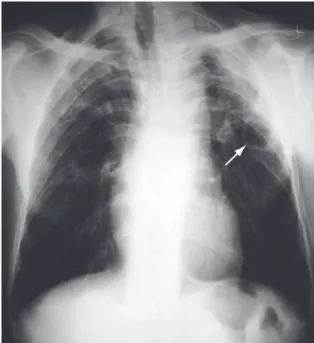
As an initial additional evaluation, it is recom-mended that sputum smear microscopy and sputum culture (induced sputum culture, if necessary, since it has good sensitivity) be performed, as well as chest X-ray,(23,26) as shown in Figure 1. In cases in
which there are still doubts about the presence of active tuberculosis, bronchoscopy with BAL can be used, in conjunction with transbronchial biopsy when possible; biopsies significantly increase the monary tuberculosis in increased in patients with
silicosis or patients exposed to silica. Evidence from experimental studies suggests that silica modi-fies the immune response of the lungs, impairs the metabolism/function of pulmonary macrophages, and, with frequent exposure, causes macrophage apoptosis . (7,11) These findings are consistent with
observations that the incidence of tuberculosis is higher in dust-exposed workers, even in those without established silicosis, than in workers not so exposed.
Another element involved is surfactant protein A, which appears at high levels in the BAL fluid of patients with silicosis. An excess of this protein seems to be associated with higher susceptibility to tuber-culosis, possibly because it allows mycobacteria to enter the alveolar macrophages without triggering cytotoxicity and inhibits the formation of reactive nitrogen species by the activated microphages.(16,17)
It is also believed that the bacilli can remain encapsulated within the silicotic nodules, which would be responsible for the reactivation of tuber-culosis in such patients.(7)
Diagnostic aspects
Silicosis
The diagnosis of silicosis is made based on a history of exposure to silica accompanied by a clinical and radiological profile consistent with the disease. In most cases, routine chest X-ray is sufficient for this purpose.(2,18) The characteristic X-ray finding
is the presence of multiple, diffusedly distributed nodules smaller than 10 mm in diameter, predomi-nantly in the superior and posterior regions of the lungs. The nodules can coalesce and form opacities greater than 10 mm in diameter, which is indicative of progressive massive fibrosis. The type (regular or irregular), size (≤ 1.5 mm; 1.5-3 mm; or > 3 mm), and profusion of these nodules form the basis for the classification established by the International Labour Organization.(19) Hilar and mediastinal
Chemoprophylaxis: efficacy and safety
In the past 20 years, numerous studies have validated the use of chemoprophylaxis against tuber-culosis in HIV-infected patients and have established its efficacy. In immunocompetent patients, protec-tion has been demonstrated in studies involving individuals with tuberculosis-related pulmonary sequelae, as well as contacts of patients with active tuberculosis.(34) Few authors, however, have studied
tuberculosis prevention in silica-exposed workers, with or without silicosis.
A randomized, double-blind, placebo-controlled trial evaluated the effect of three chemoprophy-laxis regimens in 652 patients with silicosis who did not have active tuberculosis and had never been treated for tuberculosis.(11) Tuberculin skin test
reactivity was not an exclusion criterion. However, 94% presented a reaction (induration) > 10 mm at the study outset. The individuals were rand-omized to receive, in an unsupervised manner, one of the following regimens: 300 mg/day of isoni-azid for 24 weeks; 300 mg/day of isoniisoni-azid and 600 mg/ day of rifampicin for 12 weeks; 600 mg/day of rifampicin for 12 weeks; or placebo for 24 weeks. After 5 years, the use of chemoprophylaxis reduced the risk of developing tuberculosis by approximately 50%. The proportion of patients with active tuber-culosis in the placebo group was 27%, compared with 13% in the groups using chemoprophylaxis (combined result of the three different regimens; p < 0.01). The annual incidence was 7% in the placebo group and 4% in the groups using chemo-prophylaxis. Even when the individuals who did not comply with the regimen proposed were included (intention-to-treat analysis), the difference in favor of chemoprophylaxis still remained (proportion of patients with tuberculosis, 27% vs. 17%; p < 0.05). There was no significant difference among the three chemoprophylaxis regimens in term of efficacy.
In that same trial, treatment discontinuation due to adverse reactions occurred in 4% of the individ-uals randomized to receive chemoprophylaxis and in 2% of those in the placebo group. There were two cases of symptomatic hepatitis, one in the group receiving isoniazid and one in the group receiving isoniazid and rifampicin. An isolated increase in the serum levels of alanine aminotransferase (ALT) occurred in up to 30% of those using isoniazid, although those levels returned to baseline values diagnostic yield of the test, even in patients whose
sputum and BAL are negative for mycobacteria.(27)
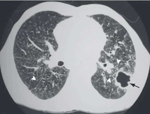
Patterns suggestive of silicotuberculosis have also been recognized on chest CT scans. The prin-cipal findings consistent with active tuberculosis superimposed on silicosis are thick-walled cavi-ties (Figure 2), consolidations, images presenting a tree-in-bud pattern, nodular image asymmetry, and rapid disease progression.(26,28,29).
Positron emission tomography, with or without CT, studies tissue metabolism, based on fluorine-18-deoxyglucose uptake, and is known to identify malignant neoplasms and some benign lesions, such as those secondary to tuberculosis.(30) However,
pulmo-nary lesions and lymphadenomegaly resulting from silicosis also show accelerated metabolism on positron emission tomography scans (particularly in the form of massive fibrosis).(31,32) In addition, both diseases are
accompanied by intense radiotracer uptake, which limits the correct differentiation of the lesions in terms of etiology. Although new radiotracers, possibly with greater specificity for silica-induced lesions, are being studied, they have yet to be validated in terms of their clinical applicability.(33)
Compliance with long-term and potentially toxic regimens represents one more difficulty of chemoprophylaxis. In the group allocated to receive rifampicin plus pyrazinamide for two months, compliance was 55%. Among those randomized to use isoniazid for six months, 63.9% completed the regimen. The difference among the groups in terms of compliance was not found to be statisti-cally significant.(35)
Guideline recommendations
The association between exposure to silica, with or without silicosis, and the risk of developing tuber-culosis and other mycobacteria-related diseases is well documented in the literature. The use of chem-oprophylaxis in this population, however, does not count on equally extensive evidence. Although there have been few studies carefully evaluating indica-tions, efficacy, safety, and duration of the various regimens, national and international guidelines recommend chemoprophylaxis for patients with silicosis.
The II Brazilian Consensus on Tuberculosis recommends that patients with silicosis and strongly positive tuberculin skin test results (indu-ration ≥ 10 mm) receive isoniazid at 300 mg/day (or 10 mg/kg/day) for six months. Considering that chemoprophylaxis for longer periods has similar efficacy, the nine-month regimen should be limited to groups at extremely high risk for tuberculosis.(23)
Similarly, the American Thoracic Society(34)
recommends that tuberculin skin tests be performed in patients with silicosis. In cases of positive results (induration ≥ 10 mm)—and after excluding active tuberculosis—the Society recommends that chem-oprophylaxis be instituted, using one of the four possible regimens: isoniazid for nine months; isoni-azid for six months; rifampicin and pyrazinamide for two to three months; or rifampicin for four months. Although there is no specific recommendation for patients with silicosis, the regimen recommended by this society is isoniazid for nine months, regard-less of the HIV serologic status. The determination of bilirubin and hepatic enzyme levels in the initial evaluation is indicated only for individuals consid-ered to be at high risk for liver disease, such as alcoholics, pregnant women, and HIV-infected indi-viduals.(34)
after discontinuation of the drug. There was no difference between those receiving rifampicin only and those receiving placebo in terms of ALT levels. No induction of resistant strains was associated with the use of the chemoprophylaxis regimens.(11)
Chemoprophylaxis safety was evaluated in a study involving 77 patients with silicosis who had no history of tuberculosis and who had a tuberculin skin test induration ≥ 10 mm.(35) The individuals
were allocated to receive a two-month course of rifampicin at 600 mg/day plus pyrazinamide at 1,500 mg/day or a six-month course of isoniazid at 300 mg/day. The incidence of hepatotoxicity (defined, in the study, as an increase in ALT to at least 1.5 times above the upper limit of normality) was higher in the group receiving rifampicin and pyrazinamide than in the group receiving isoniazid only (47.5% vs. 13.9%; p < 0.01). When the anal-ysis was limited to those presenting an increase of greater than 5 times, the incidence was, respectively, 35% and 2.8% (p < 0.001). Symptoms suggestive of hepatitis were seen in 15% and 2.7% of the patients receiving rifampicin plus pyrazinamide or isoniazid, respectively. However, viral hepatitis was excluded in only 4 of the 7 patients in question. Similarly, the treatment was interrupted due to hepatotoxicity in a greater number of individuals in the group receiving rifampicin plus pyrazinamide (35% vs. 5.6%; p < 0.01).
References
1. Rice FL. Crystalline silica, quartz. Concise international chemical assessment document, 24. Geneva: World Health Organization; 2000.
2. Terra-Filho M, Santos UP. Silicosis. J Bras Pneumol. 2006;32(Suppl 2):S41-7.
3. Castranova V, Vallyathan V. Silicosis and coal workers’ pneumoconiosis. Environ Health Perspect. 2000;108(Suppl 4):675-84.
4. Calvert GM, Rice FL, Boiano JM, Sheehy JW, Sanderson WT. Occupational silica exposure and risk of various diseases: an analysis using death certificates from 27 states of the United States. Occup Environ Med. 2003;60(2):122-9.
5. Xie QM, Tang HF, Chen JQ, Bian RL. Pharmacological actions of tetrandrine in inflammatory pulmonary diseases. Acta Pharmacol Sin. 2002;23(12):1107-13.
6. Lai JH. Immunomodulatory effects and mechanisms of plant alkaloid tetrandrine in autoimmune diseases. Acta Pharmacol Sin. 2002;23(12):1093-101.
7. Hnizdo E, Murray J. Risk of pulmonary tuberculosis relative to silicosis and exposure to silica dust in South African gold miners. Occup Environ Med. 1998;55(7):496-502. Erratum in: Occup Environ Med 1999;56(3):215-6.
8. Corbett EL, Churchyard GJ, Clayton T, Herselman P, Williams B, Hayes R, et al. Risk factors for pulmonary mycobacterial disease in South African gold miners. A case-control study. Am J Respir Crit Care Med. 1999;159(1):94-9.
9. Cowie RL. The epidemiology of tuberculosis in gold miners with silicosis. Am J Respir Crit Care Med. 1994;150(5 Pt 1):1460-2.
10. teWaternaude JM, Ehrlich RI, Churchyard GJ, Pemba L, Dekker K, Vermeis M, et al. Tuberculosis and silica exposure in South African gold miners. Occup Environ Med. 2006;63(3):187-92.
11. A double-blind placebo-controlled clinical trial of three antituberculosis chemoprophylaxis regimens in patients with silicosis in Hong Kong. Hong Kong Chest Service/Tuberculosis Research Centre, Madras/British Medical Research Council. Am Rev Respir Dis. 1992;145(1):36-41.
12. Adverse effects of crystalline silica exposure. American Thoracic Society Committee of the Scientific Assembly on Environmental and Occupational Health. Am J Respir Crit Care Med. 1997;155(2):761-8.
13. Ferreira AS, Moreira VB, Ricardo HM, Coutinho R, Gabetto JM, Marchiori E. Progressive massive fibrosis in silica-exposed workers. High-resolution computed tomography findings. J Bras Pneumol. 2006; 32(6):523-8.
14. Sonnenberg P, Murray J, Glynn JR, Thomas RG, Godfrey-Faussett P, Shearer S. Risk factors for pulmonary disease due to culture-positive M. tuberculosis or nontuberculous mycobacteria in South African gold miners. Eur Respir J. 2000;15(2):291-6.
15. Menezes AMB, Costa JD, Gonçalves H, Saul M, Menezes M, Soila L et al. Incidência e Fatores de Risco para Tuberculose em Pelotas, Uma Cidade do Sul do Brasil. Rev Bras Epidemiol. 1998;1(1):50-60.
16. Pasula R, Wright JR, Kachel DL, Martin WJ 2nd. Surfactant protein A suppresses reactive nitrogen intermediates by alveolar macrophages in response to Mycobacterium tuberculosis. J Clin Invest. 1999;103(4):483-90.
Our opinion is that patients with silicosis, as well as those with periods of exposure to silica longer than 10 years, even without the disease, should be submitted to tuberculin skin tests in the initial evaluation, since it is recognized that this popu-lation is at increased risk for tuberculosis.(8) If the
result is negative, it is recommended that the test be performed again within two weeks for confirmation, considering that exposure to the tuberculin skin test can stimulate the immunological memory against the bacillus. If the negative result persists, the test should thereafter be performed annually.(36) If
posi-tive (induration ≥ 10 mm), a six-month course of chemoprophylaxis with isoniazid at 300 mg/day (or 10 mg/kg/day) should be instituted. Despite the lack of confirmation regarding its benefits, this position is supported by specialists in the interna-tional literature.(37)
Final considerations
Silicosis is a prevalent disease for which there is currently no specific treatment. Its management should center on removing workers from the source of exposure and preventing worsening, by, for example, ensuring vaccination against influenza and pneumococci. Chief among the potential complica-tions is tuberculosis, for which patients with silicosis are at an up to 40 times higher risk than is the general population. The efficacy of chemoprophy-laxis is well established for HIV-infected individuals and, to a lesser degree, for patients with silicosis. Therefore, in the follow-up evaluation of such patients, the individual risk of developing tubercu-losis should be estimated using tuberculin skin tests, and, for those with strongly positive tuberculin skin test results (induration ≥ 10 mm), chemoprophy-laxis should be prescribed. However, further studies are needed in order to consolidate this indication, as well as to identify the best regimen in terms of drug of choice, dose, and treatment duration.
smear negative pulmonary tuberculosis. Respir Med. 1995;89(9):621-3.
28. Lee KS, Hwang JW, Chung MP, Kim H, Kwon OJ. Utility of CT in the evaluation of pulmonary tuberculosis in patients without AIDS. Chest. 1996;110(4):977-84.
29. Chong S, Lee KS, Chung MJ, Han J, Kwon OJ, Kim TS. Pneumoconiosis: comparison of imaging and pathologic findings. Radiographics. 2006;26(1):59-77.
30. Bombarda S, Soares Jr J, Terra-Filho M. Estudo do metabolismo da glicose na tuberculose pulmonar ativa utilizando a tomografia por emissão de pósitrons (18F-FDG PET). J Pneumol. 2002;28(5):270-6.
31. Chang JM, Lee HJ, Goo JM, Lee HY, Lee JJ, Chung JK, et al. False positive and false negative FDG-PET scans in various thoracic diseases. Korean J Radiol. 2006;7(1):57-69. 32. O’Connell M, Kennedy M. Progressive massive fibrosis
secondary to pulmonary silicosis appearance on F-18 fluorodeoxyglucose PET/CT. Clin Nucl Med. 2004;29(11):754-5.
33. Wallace WE, Gupta NC, Hubbs AF, Mazza SM, Bishop HA, Keane MJ, et al. Cis-4-[(18)F]fluoro-L-proline PET imaging of pulmonary fibrosis in a rabbit model. J Nucl Med. 2002;43(3):413-20.
34. American Thoracic Society. Targeted Tuberculin Testing and Treatment of Latent Tuberculosis Infection. Am J Respir Crit Care Med. 2000; 161(4): S221-S247.
35. Leung CC, Law WS, Chang KC, Tam CM, Yew WW, Chan CK, et al. Initial experience on rifampin and pyrazinamide vs isoniazid in the treatment of latent tuberculosis infection among patients with silicosis in Hong Kong. Chest. 2003;124(6):2112-8.
36. Balaan DR, Banks DE. Silicosis. In: Rom WN, editor. Environmental and occupational medicine. Boston: Little, Brown; 1992. p. 345-58.
37. Rees D, Murray J. Silica, silicosis and tuberculosis. Int J Tuberc Lung Dis. 2007;11(5):474-84.
17. Gold JA, Hoshino Y, Tanaka N, Rom WN, Raju B, Condos R, et al. Surfactant protein A modulates the inflammatory response in macrophages during tuberculosis. Infect Immun. 2004;72(2):645-50.
18. Davis GS. Silicosis. In: Hendick DJ, Burge PS, Beckett WS, Churg A, editors. Occupational disorders of the lung: recognition, management and prevention. London: W. B. Saunders; 2002. p. 105-127.
19. Organização Internacional do Trabalho. Diretrizes para Utilização da Classificação Internacional da OIT de Radiografias de Pneumoconioses. São Paulo: OIT; 2005. 20. Antao VC, Pinheiro GA, Terra-Filho M, Kavakama J,
Müller NL. High-resolution CT in silicosis: correlation with radiographic findings and functional impairment. J Comput Assist Tomogr. 2005;29(3):350-6.
21. Müller NL, Fraser RS, Colman NC, Paré PD. Doença Pulmonar Causada por Inalação de ou Aspiração de Partículas, de Sólidos ou de Líquidos. In: Muller NL, editors. Diagnóstico radiológico das doenças do tórax. Rio de Janeiro: Guanabara Koogan; 2003. p. 511-553.
22. Arakawa H, Honma K, Saito Y, Shida H, Morikubo H, Suganuma N, et al. Pleural disease in silicosis: pleural thickening, effusion, and invagination. Radiology. 2005;236(2):685-93. 23. Sociedade Brasileira de Pneumologia e Tisiologia. II
Consenso Brasileiro de Tuberculose: Diretrizes Brasileiras para Tuberculose 2004. J Pneumol. 2004;30(Supl 1):S1-86. 24. Snider DE Jr. The relationship between tuberculosis and
silicosis. Am Rev Respir Dis. 1978;118(3):455-60
25. Conde MB, Soares SL, Mello FC, Rezende VM, Almeida LL, Reingold AL, et al. Comparison of sputum induction with fiberoptic bronchoscopy in the diagnosis of tuberculosis: experience at an acquired immune deficiency syndrome reference center in Rio de Janeiro, Brazil. Am J Respir Crit Care Med. 2000;162(6):2238-40.
26. Bombarda S, Figueiredo CM, Funari MB, Soares Jr J, Seiscento M, Terra-Filho M. Imagem em tuberculose pulmonar. J Pneumol. 2001;27(6):329-40.