Accuracy of Percutaneous Core Biopsy in the Diagnosis of
Small Renal Masses (
d
4.0 cm): A Meta-analysis
_______________________________________________
Qiqi He1,2, Hanzhang Wang3, Jonathan Kenyon2, Guiming Liu2, Li Yang1, Junqiang Tian1, Zhongjin Yue1, Zhiping Wang1
1Department of Urology, Key laboratory of disease of Urological systems, Gansu Nepho-Urological clinical Center, Second hospital of Lanzhou University, Lanzhou, Gansu, China; 2Department of Urology, University Hospitals Case Medicine Center, Case Western Reserve University, Cleveland, OH, USA; 3Tulane School of Public Health and Tropical Medicine, New Orleans, USA
ABSTRACT ARTICLE INFO
______________________________________________________________ ______________________
Objective: To use meta-analysis to determine the accuracy of percutaneous core needle biopsy in the diagnosis of small renal masses (SMRsd4.0 cm).
Materials and Methods: Studies were identified by searching PubMed, Embase, and the Cochrane Library database up to March 2013. Two of the authors independently asses-sed the study quality using QUADAS-2 tool and extracted data that met the inclusion criteria. The sensitivity, specificity, likelihood ratios, diagnostic odds ratio (DOR) and also summary receiver operating characteristic (SROC) curve were investigated and draw. Deek’s funnel plot was used to evaluate the publication bias.
Result: A total of 9 studies with 788 patients (803 biopsies) were included. Failed biopsies without repeated or aborted from follow-up/surgery result were excluded (232 patients and 353 biopsies). For all cases, the pooled sensitivity was 94.0% (95% CI: 91.0%, 95.0%), the pooled positive likelihood was 22.57 (95% CI: 9.20-55.34), the poo-led negative likelihood was 0.09 (95% CI: 0.06-0.13), the poopoo-led DOR was 296.52(95% CI: 99. 42-884.38). The area under the curve of SROC analysis was 0.959±0.0254.
Conclusion: Imaging-guided percutaneous core needle biopsy of small renal masses (SMRsd4.0 cm) is highly accurate to malignant tumor diagnosis with unknown metas-tatic status and could be offered to some patients after clinic judgment prior to surgical intervention consideration.
Key words:
Nephrostomy, Percutaneous; Biopsy; Carcinoma, Renal Cell
Int Braz J Urol. 2015; 41: 15-25
_____________________
Submitted for publication: September 16, 2013
_____________________
Accepted after revision: February 07, 2014
INTRODUCTION
Renal cell carcinoma (RCC) is in the top 15 most common malignancies of both men and women and incidence has steadily increased since 1975 (1). Increasingly, these malignant tumors are recognized more frequently as small masses (2). CT-guided and sonographically guided percuta-neous biopsy of small renal masses seemed to be effective in early reports. Several studies of renal mass biopsy have demonstrated high degrees of
IBJU| ACCURACY OF PERCUTANEOUS CORE BIOPSY IN SMALL RENAL MASSES
16 No large randomized controlled trials comparing percutaneous to other methods of metastatic detection are available; we set out to review the available literatures on percutaneous renal mass biopsies. Therefore, this meta-analysis included only well-designed, comparative stu-dies in order to mainly evaluate the safety and accuracy of percutaneous core needle biopsy in diagnosis of patients presenting with small renal masses (SMRsd4.0 cm). The purpose of this meta--analysis is to determine the diagnosis accuracy of images-guided percutaneous needle biopsies of small renal masses in adult patients.
MATERIALS AND METHODS
Search strategy
A Medline search of the English-language Literature searches were performed to identify re-views of well-designed, comparative studies on the accuracy of percutaneous core needle biopsies in diagnosis of RCC in patients presenting with small renal mass. The search included words iden-tified in the whole text as well as in the Medical Subjects Heading (MeSH) terms: ‘kidney’, ‘renal mass’, ‘renal cell carcinoma’, ‘ percutaneous’, ‘nee-dle’, ‘diagnosis’, ‘biopsy’, ‘accuracy’. The following databases were used: Pubmed (1966-March 2013), Embase (1974-March 2013), the Cochrane Library (2011 issue 5). No language restrictions were used. Publications addressing evaluated renal masses or recurrent disease after radiofrequency ablation or nephrectomy were excluded.
Eligibility criteria
Publications were included in the meta--analysis if the pre-set inclusion of below were met: (1) the renal lesions had to be limited in size (d4cm) and location (kidney mass); (2) all his-tological diagnoses of large-core needle biopsy specimens had to be confirmed by either surgical pathology or follow-up (defined as a minimum of 12 months in at least 90% of the patients). (3) The absolute number of benign and malignant diag-noses had to be derivable; (4) Renal core biopsy was performed under ultrasonography or CT gui-dance using local anesthesia and an 18-G core biopsy gun. Exclusion criteria included masses
>4.0 cm in any dimension and biopsy of tumor masses outside the kidney, lacking of confirmed by surgical pathology or adequate follow-up for the mass diagnosis, vague patients counting and different biopsy tools and methods, others which could not meet our eligibility criteria.
Data Extraction and Quality Assessment
A total of 744 papers were obtained in our initial search, 690 of which failed to meet our in-clusion criteria. Of the 84 studies remaining, 75 publications were excluded for lacking follow-up data (22 papers), unsatisfactorily confirmed his-tological diagnoses on needle biopsy (16 papers), failed to allow absolute patient small mass num-ber to be derived (d4 cm)(37 papers). Only 9 stu-dies (7-15) were included (Figure-1).
The aim of our study was to evaluate the ac-curacy of percutaneous core needle biopsy of small renal masses (d4 cm), especially for the malignancy. The results of the percutaneous renal mass biopsy were defined as positive if the pathologic examina-tion presented a RCC, a metastasis, or a specific ex-trarenal malignancy invading the kidney. In all other cases, the biopsy results were considered negative, including those specimens in which cytopathology revealed malignancy not otherwise specified (i.e. the malignant cells were found, but the type of tumor could not be specified).
All positive percutaneous renal mass biop-sy results (i.e. identification of a specific malig-nancy) which were confirmed by surgical proce-dure, surgical pathology or follow-up information (individuals refusing surgery however proved malignancy in a follow-up) were considered true--positive. The false-positive rate was defined as zero because there usually no positive biopsy re-sults would outcome benign mass. Studies were included if patients with negative percutaneous renal mass biopsies were followed up to confirm negative or metastasis.
biopsy result as true-negative. If the final diagno-sis and the diagnodiagno-sis based on the initial results of the percutaneous renal mass biopsy were discor-dant, also including the malignancy not specified initially, the results of the biopsy were defined as false-negative. Our use of these methodological definitions is in accordance with a similar large prospective study (16).
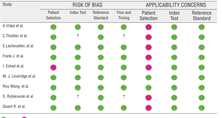
Quality assessment of the study was asses-sed by using the Quality Assessment of Diagnos-tic Accuracy Studies (QUADAS-2) checklist (17). Briefly, QUADAS-2 is a 4-domain tool, the last domain of which assists authors of systematic re-views in rating: 1) bias and 2) applicability. The risk of bias is assessed in four key areas: patient selection, index test, reference standard, and flow and timing. Concern for applicability is assessed in three key areas: patient selection, index test, and reference standard. For both categories, risk of bias and concern for applicability, the
indivi-dual criteria were classified as low risk, high risk, or unclear and the results were presented using tables from the QUADAS web site (www.quadas. org) (Table-1 and Figure-2).
Data analysis
One author extracted the data from in-cluded studies and entered them into the data extraction form. A second reviewer checked the extracted data to ensure data quality. Disagree-ments were resolved by discussion between the two review authors; if no agreement could be re-ached, it was planned that corresponding author would decide.
The 2x2 data were summarized in forest plots of sensitivity and specificity for each stu-dy. We calculated and assessed the true positive rate (TPR, sensitivity), specificity, likelihood ratios, diagnostic odds ratio along with 95 % confidence intervals (CI). Since the false positive rate was
IBJU| ACCURACY OF PERCUTANEOUS CORE BIOPSY IN SMALL RENAL MASSES
18 fined as 0, a correction was required to make 2x2 tables rational. Thus, a correction value of ½ was added to all cells in the 2x2 matrix. Heterogenei-ty was assessed by means of the Cochran Q and I2 test. Heterogeneity was classified as not
like-ly contributory (I2=0%-40%), moderate (I2 =30%-60%), substantial (I2=50%-90%), or considerable (I2>75%) (18). Pooled summary of sensitivity and specificity was calculated by using the DerSimo-nian and Laird random-effects models for weighted
Figure 2 - Graphical Display of 9 studies results (QUADAS-2). Table 1 - Quality Assessment for 9 studies (QUADAS 2).
Study RISK OF BIAS APPLICABILITY CONCERNS Patient
Selection
Index Test Reference Standard
Flow and Timing
Patient Selection
Index Test
Reference Standard
A Volpe et al.
C.Thuillier et al. ? ?
E.Lechevallier, et al. Frank J. et al. I. Eshed et al. M. J. Leveridge et al. Rou Wang, et al.
S. Rybikowski et al. ? ?
Stuart R. et al.
Low Risk; High Risk; ? Unclear Risk
Flow and Timing
Reference Standard
Index Test
Patients Selection
0% 20% 40% 60% 80% 100%
if present. To assess the diagnostic accuracy of the percutaneous core needle biopsies of small renal masses, we used the Rutter and Gastonis version of formulas for constructing summary receiver operating characteristic (SROC) curve. To identi-fy if publication bias was present in our study, we calculated the diagnostic odds ratio inverse of the square root of the effective sample size from Deek’s funnel plots for all included studies.
Meta-analysis in this study was conduc-ted with the Meta-Disc software package (Clinical Biostatistics Unit, Ramony Cajal Hospital, Madrid, Spain) (version 1.40) and Stata (version 11, Col-lege Station, TX, USA); probability values of less than 5% were considered significant.
RESULTS
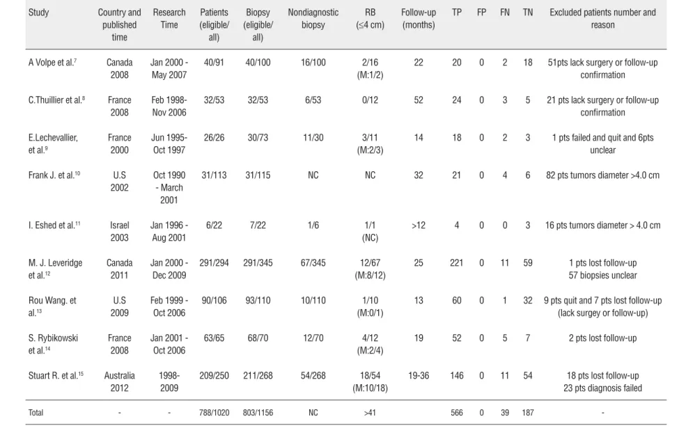
Search results and characteristics of studies Our initial search yielded 744 literatures and the process of study selection is summari-zed in Figure-1. In total, there were 1020 patients and 1156 percutaneous core needle biopsies in 9 studies. All failed biopsies without repeat biop-sies or quit from follow-up/surgery result were excluded (232 patients and 353 biopsies). Finally, 788 patients and 803 percutaneous core needle biopsies were included in our meta-analysis and Table 2 summarizes the main characteristics of included studies.
Quality assessment
Table-2 and Figure-2 summarized the me-thodological quality of our nine studies assessment by QUADAS-2 tool. If the answers to all questions of a domain are judged as ‘yes’ indicating low risk of bias, then this domain will be judged to be at low risk of bias. In advert, if one judged as ‘no’ would indicate ‘high risk’, the potential bias might exist. ‘Unclear’ indicated insufficient information to deter-mine whether partial verification was present.
Of the 9 studies’ risk bias, 8/9 provided clear definition of an exclusion criteria, one stu-dy just described the basic information about subjects without exclusion criteria and thus sco-red “high risk”. Two studies (8, 14) did not show enough information about the methods in details clearly, and also did not described an exact time
of interval between the biopsies and follow-up of patients. Studies where language differences were thought to exist were scored ‘unclear’ in the index tests bias and patient flow and timing domains which we were not sure about in these studies. The remaining studies presented a clear interpretation in all the questions of domains and scored well. In relation to applicability, patient selection criteria in 2/9 studies were in accordance to our analysis inclusion criteria and scored well, the other 7 stu-dies might have some different aims, neither some patients were not applicable for our inclusion cri-teria and were excluded from our analysis. So we considered it might have high risk bias in patient selection domain. The other two domains scored well for all studies.
Diagnostic accuracy
Because of our definition about the true positive, which is the specific malignancy biop-sies could be confirmed by surgery procedure or follow-up final diagnosis, the false positive could not exist, that’s to say, if the cytopathology report of the biopsies defined a malignancy, the correc-ted benign results conduccorrec-ted from surgery proce-dure or follow-up would mostly never happened. So the specificity would be calculated as 100%.
The pooled sensitivity was 94.0% (95% CI: 91.0%, 95.0%, p=0.28), with I2 of 17.7% (no likely contributory), specificity was 100% (95% CI: 98.4%, 100%, p=1), with I2 of 0% (no likely contributory). The pooled positive likelihood was 22.57 (95% CI: 9.20-55.34, p=0.54), the pooled negative likelihood was 0.09 (95% CI: 0.06-0.13, p=0.13) (Figure-3).
The pooled DOR was 296.52 (95% CI: 99. 42-884.38, p=0.36). The overall diagnostic accu-racy according to the results of SROC curve analy-sis was 0.959±0.0254 and the overall diagnostic accuracy (Q*) was 0.903±0.037 (Figure-4). Thus, percutaneous core needle biopsies to diagnose malignancy in small renal masses (d4 cm) is a hi-ghly accurate method.
Publication bias
IBJU
|
A
CCURA
CY OF PERCUT
ANEOUS CORE BIOPSY IN SMALL RENAL MASSES
20
Table 2 - Basic characteristics of the included studies of using 18 gauge needles biopsy on d4 cm renal mass.
Study Country and published time Research Time Patients (eligible/ all) Biopsy (eligible/ all) Nondiagnostic biopsy RB (d4 cm)
Follow-up (months)
TP FP FN TN Excluded patients number and reason
A Volpe et al.7 Canada
2008
Jan 2000 - May 2007
40/91 40/100 16/100 2/16 (M:1/2)
22 20 0 2 18 51pts lack surgery or follow-up confirmation
C.Thuillier et al.8 France
2008
Feb 1998-Nov 2006
32/53 32/53 6/53 0/12 52 24 0 3 5 21 pts lack surgery or follow-up confirmation
E.Lechevallier, et al.9
France 2000
Jun 1995-Oct 1997
26/26 30/73 11/30 3/11 (M:2/3)
14 18 0 2 3 1 pts failed and quit and 6pts unclear
Frank J. et al.10 U.S
2002
Oct 1990 - March
2001
31/113 31/115 NC NC 32 21 0 4 6 82 pts tumors diameter >4.0 cm
I. Eshed et al.11 Israel
2003
Jan 1996 - Aug 2001
6/22 7/22 1/6 1/1
(NC)
>12 4 0 0 3 16 pts tumors diameter > 4.0 cm
M. J. Leveridge et al.12
Canada 2011
Jan 2000 - Dec 2009
291/294 291/345 67/345 12/67 (M:8/12)
25 221 0 11 59 1 pts lost follow-up 57 biopsies unclear
Rou Wang. et al.13
U.S 2009
Feb 1999 - Oct 2006
90/106 93/110 10/110 1/10 (M:0/1)
13 60 0 1 32 9 pts quit and 7 pts lost follow-up (lack surgey or follow-up)
S. Rybikowski et al.14
France 2008
Jan 2001 -Oct 2006
63/65 68/70 12/70 4/12 (M:2/4)
19 52 0 5 7 2 pts lost follow-up
Stuart R. et al.15 Australia
2012
1998-2009
209/250 211/268 54/268 18/54 (M:10/18)
19-36 146 0 11 54 18 pts lost follow-up 23 pts diagnosis failed
Total - - 788/1020 803/1156 NC >41 566 0 39 187
Figure 3 - Forest plots of index.
A
B
C
IBJU| ACCURACY OF PERCUTANEOUS CORE BIOPSY IN SMALL RENAL MASSES
22 bias of literature. The shape of the funnel plots revealed a bit asymmetry (p=0.00, Figure-5). An existing publication bias were indicated; the re-ason for this might be related to the presence of a potential publication bias, a language bias, in-flated estimates in smaller studies, and the lack of publication of small trials with opposite results.
COMMENTS
Recent advances in radiological examina-tions have improved small renal mass malignancy diagnosis accuracy. In many cases, if an enhanced
Figure 4 - SROC curve.
E
effect in radiographic appeared, renal lesions were assumed to be malignant and patients were direc-ted toward surgery intervention mostly. However, some solid and complex cystic lesions cannot be well defined and diagnosed by images; and urolo-gists still encounter some masses that cannot be de-finitively categorized as either benign or malignant, especially for small renal masses (d4cm). Therefore, percutaneous needle core biopsy is becoming an efficient tool in the characterization of incidentally discovered SRMs in the diagnosis for some uncer-tain renal lesions. In our analysis, data of the po-oled sensitivity, popo-oled positive likelihood, popo-oled
1-specificity
Sensitivity SROC Curve
Figure 5 - Deeks’ Funnel plots of publications bias. As observed in many meta-analysis of diagnostic test, the concept of the false negati-ve remains a complex issue. Some patients recei-ved an indeterminate biopsy on their mass, which does not precisely determine subtype even it was a malignancy. Moreover, some patients’ mass may be compounded by some existence of hybrid tu-mors containing elements of benign and malig-nant histology after surgical intervention, such as oncocytoma related to chromophobe RCC (21). In all such cases the event was defined as false nega-tive. Only truly identified subtype of the benign / malignancy could be recognized as true negative/ positive. This definition, while strict, is much in accordance with similar studies (10, 13).
In our analysis of 9 included studies, the-re wethe-re 71.5% biopsies (561/788) that wethe-re true positive malignancy and 29.7(233/788) truly be-nign, the ratio between malignancy and benign was 2.4:1 which agrees nicely to others’ reports (5, 12, 13, 20). As the high morbidity of malignancy in SRMs suggests a probably trend toward over treatment of these mass with surgery intervention, some reports indicate that only one third of SRMs would become significant if managed conservati-vely (22). Some studies showed that biopsy could alter management in 41%-61% of cases (5, 6, 16).
If the patient receives an initially non--diagnostic biopsy, repeat biopsy is recommended. In our analysis, repeat biopsy was carried out in 41 patients and 23 patients were malignancy after repeat biopsies in 9 studies. Laguna and collea-gues (23) analyzed published renal-mass biopsy series and determined that repeat biopsy or sur-gical resection of tumors followed indeterminate biopsy in 46.4% of cases, and that repeat biopsy identified cancer in 71% of cases. That means that if normal, insufficient tissue or necrosis obtained from the initial biopsy is caused by technical fai-lure, a repeat biopsy is necessary with respect of 70% malignancy morbidity in SRMs.
Like other meta-analyses of diagnostic tests, our work has limitations. We have tried to identify and calculate all the studies about diagnosis accu-racy of the percutaneous core needle biopsies of the small renal mass (d4.0 cm). By means of an exten-sive Medline-based search, we aimed to retrieve and extract all published data available regarding SRM negative likelihood was similar to many literature
reviews demonstrating that renal mass biopsy has an overall success rate above 90% (19, 20).
Accuracy might be defined by several va-riables, including the diagnostic ability to subtype/ grade tissue, as well as the ability to obtain an in-terpretable tissue specimen to conduct a consistent diagnosis. The RCC subtype was not routinely pro-vided; however available, the accuracy for RCC sub-type is estimated at nearly 96.6% (13). Tissue grade, another diagnosis index, was not reported as high accuracy as the subtype reports. Some studies sho-wed that the biopsies accuracy for the Fuhrman nu-clear grade was lower at 46%-85% (4, 5).
In respect to the complication rates of the percutaneous biopsy, numerous studies (4-6) have already presented enough evidences that there is a very low complication rate. The most common complications observed are ble-eding, syncope, flank pain, pneumothorax, etc. The most feared potential complication is tumor seeding, however, similar to several other analy-ses (4-7) no contemporary study has observed this complication. In the 9 included studies of our meta-analysis, the major complications and mortality reports were extremely low.
Deek’s Funnel Plot Asymmetry Test pvalue = 0.00
1/root(ESS)
Diagnostic Odds Ratio
Study Regression Line
0
1
2
3
4
IBJU| ACCURACY OF PERCUTANEOUS CORE BIOPSY IN SMALL RENAL MASSES
24 percutaneous core needle biopsy diagnosis of ma-lignancy. Some published studies may have been overlooked due to our strict exclusion criteria. Gi-ven diagnostic accuracy studies require large sample numbers and long follow-up periods with which to facilitate metastatic events, it seems likely studies of this magnitude would miss follow-up data and ge-nerate bias. Furthermore, publication bias (a lower probability of the publication of negative results) was more difficult to avoid than it would have been in a meta-analysis of a randomized controlled trial. Future studies should follow the Standards for Re-porting of Diagnostic Accuracy recommendations and also include enough subjects.
CONCLUSIONS
Our meta-analysis identified that imaging--guided percutaneous core needle biopsy of small renal masses (SMRsd4.0 cm) is highly accurate in differentiating benign from malignant tumors. We propose percutaneous core needle biopsies be offered to adapted patients prior to consideration of surgical intervention. Accordingly, a number of unnecessary nephrectomies might be avoided by tissues biopsy pathology.
CONFLICT OF INTEREST
None declared.
REFERENCES
1. Chow WH, Devesa SS, Warren JL, Fraumeni JF Jr: Rising incidence of renal cell cancer in the United States. JAMA. 1999; 281: 1628-31.
2. Edwards BK, Brown ML, Wingo PA, Howe HL, Ward E, Ries LA, et al. Annual report to the nation on the status of cancer, 1975-2002, featuring population-based trends in cancer treatment. J Natl Cancer Inst. 2005; 97: 1407-27.
3. Caoili EM, Bude RO, Higgins EJ, Hoff DL, Nghiem HV. Evaluation of sonographically guided percutaneous core biopsy of renal masses. AJR Am J Roentgenol. 2002; 179: 373-8.
4. Lebret T, Poulain JE, Molinie V, Herve JM, Denoux Y, Guth A, et al. Percutaneous core biopsy for renal masses: indications, accuracy and results. J Urol. 2007; 178: 1184-8; discussion 1188.
5. Neuzillet Y, Lechevallier E, Andre M, Daniel L, Coulange C. Accuracy and clinical role of fine needle percutaneous biopsy with computerized tomography guidance of small (less than 4.0 cm) renal masses. J Urol. 2004; 171: 1802-5.
6. Wood BJ, Khan MA, McGovern F, Harisinghani M, Hahn PF, Mueller PR. Imaging guided biopsy of renal masses: indications, accuracy and impact on clinical management. J Urol. 1999; 161: 1470-4.
7. Volpe A, Mattar K, Finelli A, Kachura JR, Evans AJ, Geddie WR, et al. Contemporary results of percutaneous biopsy of 100 small renal masses: a single center experience. J Urol. 2008; 180: 2333-7.
8. Thuillier C, Long JA, Lapouge O, Pasquier D, Terrier N, Bocqueraz F, et al. Value of percutaneous biopsy for solid renal tumours less than 4 cm in diameter based on a series of 53 cases. Prog Urol. 2008; 18: 435-9.
9. Lechevallier E, André M, Barriol D, Daniel L, Eghazarian C, De Fromont M, et al. Fine-needle percutaneous biopsy of renal masses with helical CT guidance. Radiology. 2000; 216: 506-10.
10. Rybicki FJ, Shu KM, Cibas ES, Fielding JR, vanSonnenberg E, Silverman SG. Percutaneous biopsy of renal masses: sensitivity and negative predictive value stratified by clinical setting and size of masses. AJR Am J Roentgenol. 2003; 180: 1281-7.
11. Eshed I, Elias S, Sidi AA. Diagnostic value of CT-guided biopsy of indeterminate renal masses.Clin Radiol. 2004; 59: 262-7.
12. Leveridge MJ, Finelli A, Kachura JR, Evans A, Chung H, Shiff DA, et al. Outcomes of small renal mass needle core biopsy, nondiagnostic percutaneous biopsy, and the role of repeat biopsy. Eur Urol. 2011; 60: 578-84.
13. Wang R, Wolf JS Jr, Wood DP Jr, Higgins EJ, Hafez KS. Accuracy of percutaneous core biopsy in management of small renal masses. Urology. 2009; 73: 586-90; discussion 590-1.
14. Rybikowski S, Tomatis L, Arroua F, Ragni E, Rossi D, Bastide C. Value of percutaneous kidney biopsy in the management of solid renal tumours less or equal to 4 cm. Prog Urol. 2008; 18: 337-43.
15. Menogue SR, O’Brien BA, Brown AL, Cohen RJ. Percutaneous core biopsy of small renal mass lesions: a diagnostic tool to better stratify patients for surgical intervention. BJU Int. 2013; 111: E146-51.
16. Maturen KE, Nghiem HV, Caoili EM, Higgins EG, Wolf JS Jr, Wood DP Jr. Renal mass core biopsy: accuracy and impact on clinical management. AJR Am J Roentgenol. 2007; 188: 563-70.
18. Deeks JJ HJ, Altman DG. Analysing data and undertaking meta-analyses. In:Higgins JPT , Green S , eds. Cochrane hand book for systematic reviews of interventions. Vol 5.0.1. 2008 278.
19. Lane BR, Samplaski MK, Herts BR, Zhou M, Novick AC and Campbell SC. Renal mass biopsy--a renaissance? The Journal of urology. 2008; 179: 20.
20. Volpe A, Kachura JR, Geddie WR, Evans AJ, Gharajeh A, Saravanan A, et al. Techniques, safety and accuracy of sampling of renal tumors by fine needle aspiration and core biopsy. J Urol. 2007; 178: 379-86.
21. Waldert M, Klatte T, Haitel A, Ozsoy M, Schmidbauer J, Marberger M, et al. Hybrid renal cell carcinomas containing histopathologic features of chromophobe renal cell carcinomas and oncocytomas have excellent oncologic outcomes. Eur Urol. 2010; 57: 661-5.
22. Volpe A, Panzarella T, Rendon RA, Haider MA, Kondylis FI, Jewett MA. The natural history of incidentally detected small renal masses. Cancer. 2004;100:738-45.
23. Laguna MP, Kümmerlin I, Rioja J, de la Rosette JJ. Biopsy of a renal mass: where are we now? Curr Opin Urol. 2009; 19: 447-53.