Exposure to Ionizing Radiation during Dental
X-Rays Is Not Associated with Risk of
Developing Meningioma: A Meta-Analysis
Based on Seven Case-Control Studies
Ping Xu, Hong Luo, Guang-Lei Huang, Xin-Hai Yin, Si-Yang Luo, Ju-Kun Song*
Gui Zhou provincial people’s hospital, Guiyang 550002, PR China
Abstract
Background
Many observational studies have found that exposure to dental X-rays is associated with the risk of development of meningioma. However, these findings are inconsistent. We con-ducted a meta-analysis to assess the relationship between exposure to dental X-rays and the risk of development of meningioma.
Methods
The PubMed and EMBASE databases were searched to identify eligible studies. Summary odds ratio (OR) estimates and 95% confidence intervals (95% CIs) were used to compute the risk of meningioma development according to heterogeneity. Subgroup and sensitivity analyses were performed to further explore the potential heterogeneity. Finally, publication bias was assessed.
Results
Seven case-control studies involving 6,174 patients and 19,459 controls were included in the meta-analysis. Neither exposure to dental X-rays nor performance of full-mouth panorex X-rays was associated with an increased risk of development of meningioma (overall: OR, 0.97; 95% CI, 0.70–1.32; dental X-rays: OR, 1.05; 95% CI, 0.89–1.25; panorex X-rays: OR, 1.01; 95% CI, 0.76–1.34). However, exposure to bitewing X-rays was associated with a slightly increased risk of development of meningioma (OR, 1.73; 95% CI, 1.28–2.34). Simi-lar results were obtained in the subgroup and sensitivity analyses. Little evidence of publica-tion bias was observed.
Conclusion
Based on the currently limited data, there is no association between exposure to dental X-rays and the risk of development of meningioma. However, these results should be
a11111
OPEN ACCESS
Citation:Xu P, Luo H, Huang G-L, Yin X-H, Luo S-Y, Song J-K (2015) Exposure to Ionizing Radiation dur-ing Dental X-Rays Is Not Associated with Risk of De-veloping Meningioma: A Meta-Analysis Based on Seven Case-Control Studies. PLoS ONE 10(2): e0113210. doi:10.1371/journal.pone.0113210
Academic Editor:Michael Scheurer, Baylor College of Medicine, UNITED STATES
Received:June 19, 2014
Accepted:October 20, 2014
Published:February 6, 2015
Copyright:© 2015 Xu et al. This is an open access article distributed under the terms of theCreative Commons Attribution License, which permits unre-stricted use, distribution, and reproduction in any me-dium, provided the original author and source are credited.
Data Availability Statement:All relevant data are within the paper and its Supporting Information files.
Funding:The authors have no support or funding to report.
cautiously interpreted because of the heterogeneity among studies. Additional large, high-quality clinical trials are needed to evaluate the association between exposure to dental X-rays and the risk of development of meningioma.
Introduction
Intracranial meningioma is a solid tumor that arises from the meninges, which protect the cen-tral nervous system. Meningioma, one of the most clinically serious tumors of the cencen-tral ner-vous system, accounts for approximately 20% of all intracranial tumors in male patients and 38% in female patients [1–4]. The estimated prevalence of pathologically confirmed meningio-ma is 3.5 in 100,000 cases per year worldwide [4]. According to a report of the National Cancer Data Base, the overall 2- and 5-year survival rates for patients with meningioma are 81% and 69%, respectively [5]. Furthermore, patients with meningioma may show neurological symp-toms of increased intracranial pressure (e.g., headaches, nausea, vomiting, lethargy, and papil-loedema) or focal brain dysfunction (e.g., limb weakness/numbness and seizures). Pain, disability, and mortality are patient burdens that result in high costs to society. The above data highlight the importance of screening patients at highest risk and identifying potential risk fac-tors for the development of meningioma.
Previous studies have shown that biological risk factors of ionizing radiation (IR) are associ-ated with a high incidence of atomic bomb-induced health issues, including various types of cancer [6,7]. Exposure to IR is considered to be strongly associated with the risk of developing meningioma. Patients who undergo routine dental examination are those most frequently af-fected by X-ray exposure. In 1980, Longstreth et al. first investigated the association between dental X-ray exposure and the risk of development of meningioma. They found that exposure to dental X-rays can increase the incidence of meningioma and is a strong risk factor for me-ningioma [8–11]. Since then, a number of observational studies have been published [12–16]. However, the findings of these studies are varied or even conflicting. Given the widespread use of dental X-rays and poor prognosis of meningioma, any risk factors for the development of meningioma would have a substantial impact on public health. Therefore, we conducted a meta-analysis of case-control studies to evaluate the association between exposure to dental X-rays and the risk of development of meningioma.
Methods
Search strategy
We searched the PubMed and EMBASE databases up to March 2014 to identify relevant stud-ies that evaluated the association between exposure to dental X-ray and the risk of development of meningioma. The following search terms were employed: (1)“meningioma(s),” “brain neo-plasm(s),” “brain tumor(s),” “brain Neoplasm(s),” “meningeal Neoplasm(s),”and (2)“dental x-ray(s),” “tooth radiography,” “teeth radiography,”and“dental radiography.”Furthermore, we reviewed the reference lists of all eligible articles.
Eligibility criteria
available; and they provided the adjusted and/or unadjusted odds ratio (OR), relative risk (RR) with corresponding 95% confidence interval (95%CI), or raw data with which to estimate the crude OR or RR. Studies were excluded if they met the following criteria: they were letters, comments, correspondence, conference reports, or laboratory studies or they did not contain enough data with which to calculate the OR. When multiple publications covered the same study population, only the study with the larger sample was included. Two authors (J.K.S. and X.H.Y.) independently assessed the inclusion of all retrieved studies and resolved any disagree-ments through discussion or consultation with a third author (G.L.H.).
Data extraction
Two authors (J.K.S. and X.H.Y.) independently collected the following basic information using a standardized data extraction form: first author’s name, year of publication, study design, source of control, study location, number of participants (cases/controls), crude and/or adjust-ed point estimates and corresponding 95%CIs, and covariate features includadjust-ed in the multivari-able model. Disputes were resolved by discussion and consensus with a third author (G.L.H.).
Quality assessment
We evaluated the methodological quality of each study using the Newcastle-Ottawa scale (NOS) [17]. Three major components were judged as follows: representativeness of the study groups (0–4 points), determination for interested exposure in the studies (0–3 points), and comparability of groups (0–2 points). A higher score indicated better methodological quality. The quality of each study was graded as either low-level (0–4 points) or high-level (5–9 points).
Statistical analysis
We used the OR with 95%CI as a common measure across all eligible studies. Because meningi-oma is a relatively rare disease, differences among the estimates of relative risk were ignored and the RR was directly converted to the OR. We used the Cochrane Q test to evaluate statisti-cal heterogeneity. A P value of<0.05 and/or an I2statistic of>50% was considered statistically
significant. A random-effects model was used if heterogeneity was observed, while a fixed-effects model was used if the P value was>0.05 and/or the I2statistic was<50%. We further
performed sensitivity analyses to evaluate robustness and stability by sequential omission of one study in each turn. Moreover, subgroup analyses were performed to explore the potential presence of heterogeneity and assess the influence of different inclusion criteria on the overall estimate. Publication bias was evaluated using the Begg and Egger tests [18,19]. All statistical analyses were carried out using STATA version 12.0 (Stata Corporation, College Station, TX, USA).
Results
Study selection
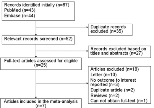
Fig. 1shows a flow chart of the inclusion criteria. In total, 87 studies were screened in the initial
Study characteristics
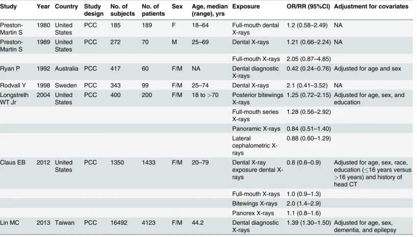
The characteristics of all included articles are presented inTable 1. Seven case-control studies including 19,459 individuals and 6,174 incident cases were identified. These studies were pub-lished from 1980 to 2013. Among all studies, the sample size ranged from 342 to 20,615. Four studies were conducted in the United States [8,9,13,16], one in Sweden [14], one in Taiwan [12], and one in Australia [15].
All seven studies reported exposure to dental X-rays; four reported exposure to full-mouth series [8,9,13,16], one reported exposure to bitewing X-rays, and one reported exposure to panorex X-rays [13,16]. Five studies evaluated the OR of developing meningioma [8,9,12,
13,16], and two evaluated the RR of developing meningioma [14,15]. One study investigated only men [8], one investigated only women [9], and five investigated both men and women
[12–16]. The association between exposure to dental X-rays and the risk of development of
me-ningioma was the primary outcome in two studies [13,16], while it was a secondary outcome in five studies [8,9,12,14,15]. Three studies did not adjust for confounding factors [8,9,14], whereas the others controlled for various risk factors for meningioma including age, sex, edu-cation, and others [12,13,15,16].
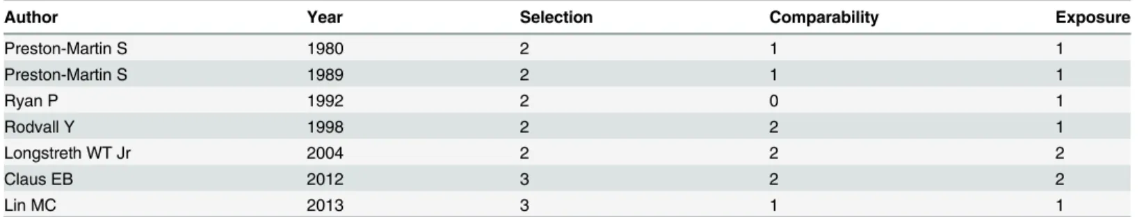
We used the NOS to evaluate the quality of the included studies (Table 2). The median NOS score of the eligible studies was 5.0 (range, 3–7).
Exposure to dental X-rays and risk of developing meningioma
The overall ORs were pooled to obtain the total estimate of risk using a random-effects model (OR, 0.97; 95%CI, 0.70–1.32; P = 0.82) with significant heterogeneity (P<0.001, I2= 86.5%).
The results suggested that exposure to dental X-rays has no important effects on the risk of de-velopment of meningioma, and substantial heterogeneity was observed (Fig. 2). We
Figure 1. Flow chart of identification of eligible studies to final inclusion.
subsequently conducted sensitivity analyses to test the stability and robustness of these results. The exclusion of any single study did not materially affect the overall combined OR, which ran-ged from 0.93 (95%CI, 0.66–1.32) to 1.08 (95%CI, 0.81–1.45); substantial heterogeneity was ob-served. When we excluded the study by Lin et al. [12], who reported an association between exposure to dental X-rays and the risk of development of benign brain tumors, we obtained anal-ogous results (OR, 0.86; 95%CI, 0.67–1.11) with moderate heterogeneity (P = 0.086, I2= 48.2%). The subgroup analyses based on different exclusion criteria yielded similar results (seeTable 3).
Exposure to full-mouth X-rays and risk of developing meningioma
Four studies examined the association between exposure to full-mouth dental X-rays and the risk of development of meningioma [8,9,13,16]. The overall OR estimates for each study were pooled to obtain the total estimate of risk using a fixed-effects model (OR, 1.05; 95%CI, 0.89–1.25; P = 0.57), and the test for heterogeneity revealed no statistical significance (P = 0.40, I2= 0.0%). These results suggested that exposure to dental X-rays has no important effects on the risk of development of meningioma. No significant heterogeneity was observed (Fig. 3).
Exposure to dental bitewing X-rays and risk of developing meningioma
Two studies contained data on exposure to dental bitewings X-rays [13,16]. The overall OR es-timates for each study were pooled to obtain the total estimate of risk using a fixed-effects Table 1. Characteristics of studies included in the meta-analysis.
Study Year Country Study design
No. of subjects
No. of patients
Sex Age, median (range), yrs
Exposure OR/RR (95%CI) Adjustment for covariates
Preston-Martin S
1980 United States
PCC 185 189 F 18–64 Full-mouth dental
X-rays
1.2 (0.58–2.49) NA
Preston-Martin S
1989 United States
PCC 272 70 M 25–69 Dental X-rays 1.21 (0.66–2.24) NA
Full-mouth X-rays 2.05 (0.87–4.85)
Ryan P 1992 Australia PCC 417 60 F/M NA Dental diagnostic
X-rays
0.42 (0.24–0.76) Adjusted for age and sex
Rodvall Y 1998 Sweden PCC 343 99 F/M 25–74 Dental X-rays 2.1 (0.41–3.52) NA Longstreth
WT Jr
2004 United States
PCC 400 200 F/M 18 to>70 Posterior bitewings
X-rays
1.25 (0.72–2.15) Adjusted for age, sex, and education
Full-mouth series X-rays
1.28 (0.56–2.92)
Panoramic X-rays 0.84 (0.51–1.40) Lateral
cephalometric X-rays
0.88 (0.60–1.29)
Claus EB 2012 United States
PCC 1350 1433 F/M 20–79 Dental X-ray
exposure dental X-rays
0.8 (0.6–0.9) Adjusted for age, sex, race, education (16 years versus
>16 years) and history of
head CT Full-mouth X-rays 1.0 (0.9–1.3)
Bitewings X-rays 2.0 (1.4–2.9) Panorex X-rays 1.1 (0.8–1.6) Lin MC 2013 Taiwan PCC 16492 4123 F/M 44.2 Dental diagnostic
X-rays
1.39 (1.30–1.50) Adjusted for age, sex, dementia, and epilepsy
NA, not available; M, male; F, female; PCC, population-based case-control
model (OR, 1.73; 95%CI, 1.28–2.34; P = 0.00) with low heterogeneity (P = 0.16, I2= 49.1%). These results suggest that exposure to dental bitewing X-rays is associated with a slightly in-creased risk of development of meningioma. Substantial heterogeneity was observed (Fig. 4). However, because only two studies reported the association, these results should be interpreted with caution.
Exposure to dental panorex X-rays and risk of developing meningioma
A pooled analysis of two studies [13,16] showed that exposure to dental panorex X-rays does not increase the risk of development of meningioma (OR, 1.01; 95%CI, 0.76–1.34; P = 0.95). No significant heterogeneity was detected (P = 0.39, I2= 0.0%) (Fig. 5).
Table 2. Quality assessment of included studies based on Newcastle–Ottawa scale.
Author Year Selection Comparability Exposure
Preston-Martin S 1980 2 1 1
Preston-Martin S 1989 2 1 1
Ryan P 1992 2 0 1
Rodvall Y 1998 2 2 1
Longstreth WT Jr 2004 2 2 2
Claus EB 2012 3 2 2
Lin MC 2013 3 1 1
doi:10.1371/journal.pone.0113210.t002
Figure 2. Forest plot of exposure to dental X-rays and risk of meningioma.Studies are pooled with a random-effects model.
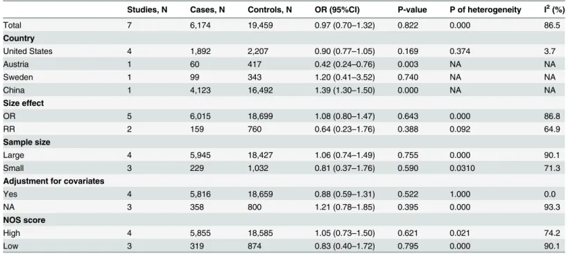
Table 3. Summary of results.
Studies, N Cases, N Controls, N OR (95%CI) P-value P of heterogeneity I2(%)
Total 7 6,174 19,459 0.97 (0.70–1.32) 0.822 0.000 86.5
Country
United States 4 1,892 2,207 0.90 (0.77–1.05) 0.169 0.374 3.7
Austria 1 60 417 0.42 (0.24–0.76) 0.003 NA NA
Sweden 1 99 343 1.20 (0.41–3.52) 0.740 NA NA
China 1 4,123 16,492 1.39 (1.30–1.50) 0.000 NA NA
Size effect
OR 5 6,015 18,699 1.08 (0.80–1.47) 0.643 0.000 86.8
RR 2 159 760 0.64 (0.23–1.76) 0.388 0.092 64.9
Sample size
Large 4 5,945 18,427 1.06 (0.74–1.49) 0.755 0.000 90.1
Small 3 229 1,032 0.81 (0.37–1.76) 0.590 0.0310 71.3
Adjustment for covariates
Yes 4 5,816 18,659 0.88 (0.59–1.31) 0.522 1.000 0.0
NA 3 358 800 1.21 (0.78–1.85) 0.395 0.000 93.3
NOS score
High 4 5,855 18,585 1.05 (0.73–1.50) 0.621 0.021 74.2
Low 3 319 874 0.83 (0.40–1.72) 0.795 0.000 90.1
OR, odds ratio; CI, confidence interval; NA, not available; Large,100 cases; Small,<100 cases; High, NOS score of5; Low, NOS score of<5
doi:10.1371/journal.pone.0113210.t003
Figure 3. Forest plot of exposure to dental full-mouth X-rays and risk of meningioma.Studies are pooled with a fixed -effects model.
Figure 4. Forest plot of exposure to dental bitewing X-rays and risk of meningioma.Studies are pooled with a fixed -effects model.
doi:10.1371/journal.pone.0113210.g004
Figure 5. Forest plot of exposure to dental panorex X-rays and risk of meningioma.Studies are pooled with a fixed-effects model.
Publication bias
Both the Begg rank correlation test and the Egger funnel plot asymmetry test (regression meth-od) in the meta-analysis indicated no significant publication bias (exposure to dental X-rays: Begg test, P = 0.37; Egger test, P = 0.14) (Fig. 6). Because of the limited number of articles, we did not assess the publication bias for the other outcomes.
Discussion
The pathogenetic mechanism of meningioma remains unknown. Many conditions have been identified as risk factors, including brain injury, smoking, chronic virus infection, and occupa-tional exposure. The most threatening risk factor for the development of meningioma is expo-sure to IR. Notably, a moderate to high dose of IR could increase the risk of developing various cancers as confirmed by studies of atomic bomb survivors and children irradiated for benign medical conditions and primary tumors. However, most of the general population receives lower-dose exposure during procedures such as diagnostic radiography, computed tomography (CT), or other types of radiation. The precise nature of the relationship between exposure to IR (especially low-dose IR) and the development of meningioma is not well characterized. With the progression of medical technology, medical diagnostic X-ray instruments have become widely used among the general population. Dental X-rays have been widely employed since 1919 [35]. Four radiographic techniques are commonly applied during dental examinations: periapical films, panoramic films, lateral skull or cephalometric (temporomandibular joint) films, and Figure 6. Funnel plots of exposure to dental X-rays and risk of meningioma for assessment of publication bias.
dental CT. The dose to which the patient is exposed is 0.018 to 1.200 mSv for periapical films, 0.135 to 0.900 mSv for panoramic films, and 0.030 to 0.200 mSv for diagnostic X-ray films [16,36]. Although dental X-ray equipment emit a very low dose of X-rays to which the patient is exposed, there is a long-standing dispute about the association between exposure to dental X-rays and the risk of development of meningioma. Several observational studies have evaluated the association between the risk of development of meningioma and exposure to lower doses of X-rays among the general population; however, the findings are limited due to the relatively small sample sizes [8–10,14,15]. In their reviews of rodent studies, Claus et al. [16] and Lin et al. [12] reported that exposure to some dental X-rays appears to be associated with an increased risk of intracranial meningioma. However, some discrepancies were noted in the evaluated studies
[20–29]. Therefore, a meta-analysis is required to merge and assess these findings.
The meta-analysis design serves as a valuable tool with which to study the rare effects of an intervention or treatment, permitting data synthesis and providing more convincing estimates of effect. To the best of our knowledge, this is the first meta-analysis to explore the role of expo-sure to dental X-rays in patients with meningioma. The overall results of the present meta-analysis of seven case-control studies using a random-effects model provide evidence that ex-posure to dental X-rays is not likely to have any important effects on the risk of development of meningioma. The pooled estimates were robust across the sensitivity analyses, and no publica-tion bias was observed. Exposure to dental full-mouth and panorex X-rays may not increase the risk of development of meningioma, while exposure to dental bitewing X-rays may slightly increase the risk. These findings can partly explained as follows. First, only two studies reported an association between exposure to dental bitewing X-rays and the risk of development of me-ningioma. Second, patients who undergo dental bitewing examination are exposed to a rela-tively higher dose of X-rays. However, because of the few studies that reported this association, the present meta-analysis does not have enough power for a decisive conclusion. Further stud-ies should focus on this association. Only three studstud-ies reported the frequency of dental X-rays, and all showed negative associations. Because the classification of exposure differed among the studies, it is difficult to merge the results using a meta-analysis.
Some limitations should be considered in the present meta-analysis. First, this meta-analysis was based on case-control studies. Although the case-control study is the most appropriate de-sign for exposure causing rare event, this dede-sign has inherent limitations such as selective bias and recall or memory bias. Additionally, some confounding factors (e.g., race, sex, head trau-ma, history of head CT) are difficult to control in case-control studies. Second, substantial het-erogeneity was a potential problem when interpreting the results of our analysis. This
The following points should be considered for further studies. First, it is necessary to estab-lish a standardized protocol with respect to exposure dose, method of examination, and dura-tion of exposure to dental X-rays because great variadura-tion exists in the literature. Second, more large-scale studies should be performed on the relationship between various types of dental X-ray exposure and the risk of development of meningioma. Finally, the average follow-up pe-riod was not reported in this meta-analysis; longer-term studies are needed.
In conclusion, the currently available evidence indicates that exposure to dental X-rays is unlikely to have any important effects on the risk of development of meningioma. Although these findings are encouraging, the results of this meta-analysis should be interpret with cau-tion because of the heterogeneity among the studies and the relatively limited number of stud-ies, Further large-scale, well-designed trials on this topic are needed.
Supporting Information
S1 PRISMA Checklist.
(DOC)
Author Contributions
Conceived and designed the experiments: PX. Performed the experiments: JKS. Analyzed the data: JKS XHY. Contributed reagents/materials/analysis tools: HL. Wrote the paper: JKS PX. Assisted with the full text and revised the grammar: GLH SYL.
References
1. Bondy M, Ligon BL (1996) Epidemiology and etiology of intracranial meningiomas: a review. J Neu-rooncol 29: 197–205. doi:10.1007/BF00165649PMID:8858525
2. Surawicz TS, McCarthy BJ, Kupelian V, Jukich PJ, Bruner JM, et al. (1999) Descriptive epidemiology of primary brain and CNS tumors: results from the Central Brain Tumor Registry of the United States, 1990–1994. Neuro Oncol 1: 14–25. doi:10.1093/neuonc/1.1.14PMID:11554386
3. Longstreth WT Jr., Dennis LK, McGuire VM, Drangsholt MT, Koepsell TD (1993) Epidemiology of intra-cranial meningioma. Cancer 72: 639–648. doi: 10.1002/1097-0142(19930801)72:3%3C639::AID-CNCR2820720304%3E3.0.CO;2-PPMID:8334619
4. Claus EB, Bondy ML, Schildkraut JM, Wiemels JL, Wrensch M, et al. (2005) Epidemiology of intracrani-al meningioma. Neurosurgery 57: 1088–1095; discussion 1088–1095. doi:10.1227/01.NEU.
0000188281.91351.B9PMID:16331155
5. McCarthy BJ, Davis FG, Freels S, Surawicz TS, Damek DM, et al. (1998) Factors associated with sur-vival in patients with meningioma. J Neurosurg 88: 831–839. doi:10.3171/jns.1998.88.5.0831PMID: 9576250
6. Sadamori N, Shibata S, Mine M, Miyazaki H, Miyake H, et al. (1996) Incidence of intracranial meningio-mas in Nagasaki atomic-bomb survivors. Int J Cancer 67: 318–322. doi:10.1002/(SICI)1097-0215 (19960729)67:3%3C318::AID-IJC2%3E3.3.CO;2-NPMID:8707402
7. Shintani T, Hayakawa N, Hoshi M, Sumida M, Kurisu K, et al. (1999) High incidence of meningioma among Hiroshima atomic bomb survivors. J Radiat Res 40: 49–57. doi:10.1269/jrr.40.49PMID: 10408177
8. Preston-Martin S, Mack W, Henderson BE (1989) Risk factors for gliomas and meningiomas in males in Los Angeles County. Cancer Res 49: 6137–6143. PMID:2790826
9. Preston-Martin S, Paganini-Hill A, Henderson BE, Pike MC, Wood C (1980) Case-control study of intra-cranial meningiomas in women in Los Angeles County, California. J Natl Cancer Inst 65: 67–73. PMID: 6930521
10. Preston-Martin S, Henderson BE, Yu MC (1983) Epidemiology of intracranial meningiomas: Los Ange-les County, California. Neuroepidemiology 2: 164–178. doi:10.1159/000110522
12. Lin MC, Lee CF, Lin CL, Wu YC, Wang HE, et al. (2013) Dental diagnostic X-ray exposure and risk of benign and malignant brain tumors. Ann Oncol 24: 1675–1679. doi:10.1093/annonc/mdt016PMID: 23406732
13. Longstreth WT Jr., Phillips LE, Drangsholt M, Koepsell TD, Custer BS, et al. (2004) Dental X-rays and the risk of intracranial meningioma: a population-based case-control study. Cancer 100: 1026–1034. doi:10.1002/cncr.20036PMID:14983499
14. Rodvall Y, Ahlbom A, Pershagen G, Nylander M, Spannare B (1998) Dental radiography after age 25 years, amalgam fillings and tumours of the central nervous system. Oral Oncology 34: 265–269. doi: 10.1016/S1368-8375(97)00096-1PMID:9813721
15. Ryan P, Lee MW, North B, McMichael AJ (1992) Amalgam fillings, diagnostic dental X-rays and tu-mours of the brain and meninges. European Journal of Cancer Part B: Oral Oncology 28: 91–95. doi: 10.1016/0964-1955(92)90034-X
16. Claus EB, Calvocoressi L, Bondy ML, Schildkraut JM, Wiemels JL, et al. (2012) Dental x-rays and risk of meningioma. Cancer 118: 4530–4537. doi:10.1002/cncr.26625PMID:22492363
17. Hartling L, Milne A, Hamm MP, Vandermeer B, Ansari M, et al. (2013) Testing the Newcastle Ottawa Scale showed low reliability between individual reviewers. J Clin Epidemiol 66: 982–993. doi:10.1016/ j.jclinepi.2013.03.003PMID:23683848
18. Begg CB, Mazumdar M (1994) Operating characteristics of a rank correlation test for publication bias. Biometrics 50: 1088–1101. doi:10.2307/2533446PMID:7786990
19. Egger M, Davey Smith G, Schneider M, Minder C (1997) Bias in meta-analysis detected by a simple, graphical test. BMJ 315: 629–634. doi:10.1136/bmj.315.7109.629PMID:9310563
20. Jorgensen TJ (2013) Dental x-rays and risk of meningioma. Cancer 119: 463. doi:10.1002/cncr.27710 PMID:23254822
21. White SC, Hildebolt CF, Lurie AG (2013) Dental x-rays and risk of meningioma. Cancer 119: 464. doi: 10.1002/cncr.27709PMID:23254766
22. Dirksen D, Runte C, Berghoff L, Scheutzel P, Figgener L (2013) Dental X-rays and risk of meningioma: anatomy of a case-control study. J Dent Res 92: 397–398. doi:10.1177/0022034513484338PMID: 23539560
23. Claus EB, Wiemels J, Wrensch M (2013) Dental x-rays and risk of meningioma: response to Drs. Cal-non, Jorgensen, and White. Cancer 119: 465–466. doi:10.1002/cncr.27708PMID:23254687
24. Calnon WR (2013) Shortcomings of study on dental x-rays and risk of meningioma. Cancer 119: 464–465. doi:10.1002/cncr.27709PMID:23254924
25. (2013) Dental X-rays: meningiomas? Prescrire Int 22: 16–17. PMID:23383406
26. Tetradis S, White SC, Service SK (2012) Dental x-rays and risk of meningioma; the jury is still out. J Evid Based Dent Pract 12: 174–177. doi:10.1016/j.jebdp.2012.06.012PMID:22935292
27. Potera C (2012) Older dental X-rays linked to meningioma. Am J Nurs 112: 18. doi:10.1097/01.NAJ. 0000415943.02290.eaPMID:22739596
28. Fonzar F (2012) Are dental x-rays associated with an increased risk of meningioma? Eur J Oral Implan-tol 5: 107; discussion 108. PMID:22866287
29. Abt E (2012) Can dental x-rays increase the risk of meningioma? Evid Based Dent 13: 37–38. doi:10. 1038/sj.ebd.6400852PMID:22722407
30. Maillie HD, Gilda JE (1993) Radiation-induced cancer risk in radiographic cephalometry. Oral Surg Oral Med Oral Pathol 75: 631–637. doi:10.1016/0030-4220(93)90239-ZPMID:8488031
31. Neuberger JS, Brownson RC, Morantz RA, Chin TD (1991) Association of brain cancer with dental X-rays and occupation in Missouri. Cancer Detect Prev 15: 31–34. PMID:1646072
32. Preston-Martin S, White SC (1990) Brain and salivary gland tumors related to prior dental radiography: implications for current practice. J Am Dent Assoc 120: 151–158. PMID:2405031
33. Umansky F, Shoshan Y, Rosenthal G, Fraifeld S, Spektor S (2008) Radiation-induced meningioma. Neurosurg Focus 24: E7. doi:10.3171/FOC/2008/24/5/E7PMID:18447746
34. Preston-Martin S, Henderson BE, Bernstein L (1985) Medical and dental x rays as risk factors for re-cently diagnosed tumors of the head. Natl Cancer Inst Monogr 69: 175–179. PMID:3834328
35. Richards AG, Colquitt WN (1981) Reduction in dental X-ray exposures during the past 60 years. J Am Dent Assoc 103: 713–718. PMID:6946136