Clinical and psychosocial challenges in the treatment of a
patient with Proteus syndrome
Desafios clínicos e psicossociais no tratamento de um paciente com
síndrome de Proteus
Matheus Bertanha1
*
, Regina Moura1, Marcone Lima Sobreira1, Lied Martins Santiago Pereira2, Rodrigo Gibin Jaldin1,
Manuella Pacíico de Freitas Segredo2, Hamilton Almeida Rollo1, Winston Bonetti Yoshida1
Abstract
Proteus syndrome is a rare combination of malformations that can afect several tissues and organs. It is characterized by bilateral macrodactyly, cranial hypertrophy, bone anomalies, scoliosis, soft-tissue hamartomas, verrucous pigmented nevus, visceral abnormalities and other forms of hypertrophy. Just over 200 cases have been reported worldwide. his article reports on the clinical course of a pediatric patient with this syndrome. he child had severe malnutrition associated with extreme gigantism of the lower limbs and also psychosocial problems related to social exclusion. As the disease progressed it exacerbated and evolved into a wasting syndrome. After several years, the parents agreed to amputation of the hypertrophic lower limbs. One year after the amputations the child had been rehabilitated and had adapted to prostheses, with nutritional improvement and notable psychological recovery and social reintegration, which represented a signiicant improvement in his quality of life.
Keywords: amputation; congenital abnormalities; musculoskeletal abnormalities.
Resumo
A Síndrome de Proteus é uma rara associação de malformações que podem afetar vários tecidos e órgãos. É caracterizada por macrodactilia bilateral, hipertroia craniana, anomalias ósseas, escoliose, hamartomas de tecidos moles, nevo verrucoso pigmentar, anormalidades viscerais e outras hipertroias. Há pouco mais de 200 casos notiicados em todo o mundo. O presente artigo relata a evolução clínica de um paciente pediátrico com essa síndrome. A criança apresentou desnutrição grave associada ao extremo gigantismo de membros inferiores. Além disso, apresentou repercussões psicossociais relacionadas à exclusão social. A doença tornou-se mais grave e progrediu como síndrome consumptiva. Finalmente, os pais concordaram com a amputação dos membros inferiores hipertróicos. Um ano após as amputações, a criança estava totalmente reabilitada, protetizada dos membros amputados, com melhora nutricional, além de apresentar notória recuperação psicológica e reinserção social, o que representou melhora signiicativa da qualidade de vida para o paciente.
Palavras-chave: amputação; anormalidades congênitas; anormalidades musculosqueléticas.
1 Universidade Estadual Paulista – UNESP, Faculdade de Medicina de Botucatu, Department of Surgery and Orthopedics, Botucatu, SP, Brazil. 2 Universidade Estadual Paulista – UNESP, Faculdade de Medicina de Botucatu, Department of Pediatrics, Botucatu, SP, Brazil.
Financial support: None.
Conlicts of interest: No conlicts of interest declared concerning the publication of this article. Submitted: August 04, 2015. Accepted: September 14, 2015.
INTRODUCTION
Proteus syndrome was irst described by Cohen and Hayden in 19791 as a new disorder characterized
by uncontrolled growth of tissues and organs. The disease was labeled Proteus syndrome (PS) in 1983 by Wiedemann et al.2 as a way of describing the
great variation in clinical expression, which include gigantism of hands, feet or both extremities, craniofacial hypertrophy,3 bone abnormalities,4 scoliosis,5 soft
tissue hamartomas, pigmented verrucous nevus, visceral anomalies and accelerated growth during the patient’s irst years of life.6-8 Only approximately
200 cases have been described worldwide.9
Other conditions were also linked to this syndrome later, such as: partial or complete hemi-hypertrophy, macrodactylia, exocytosis, palmar or plantar gyriform mass, linear epidermal nevus and subcutaneous tumors with a blood and lymph vascular component.7,10,11
Some of these abnormalities are also seen in other syndromes, such as Klippel-Trenaunay, Maffucci and neuroibromatosis, but PS can be differentiated from other syndromes because it characteristically includes mesodermal and asymmetrical malformations.9,12
The objective of this report is to describe the clinical case of a patient with PS who had severe malnutrition
and psychosocial problems, which were reversed after amputation of the heavily involved lower limbs.
PART I - CLINICAL SITUATION
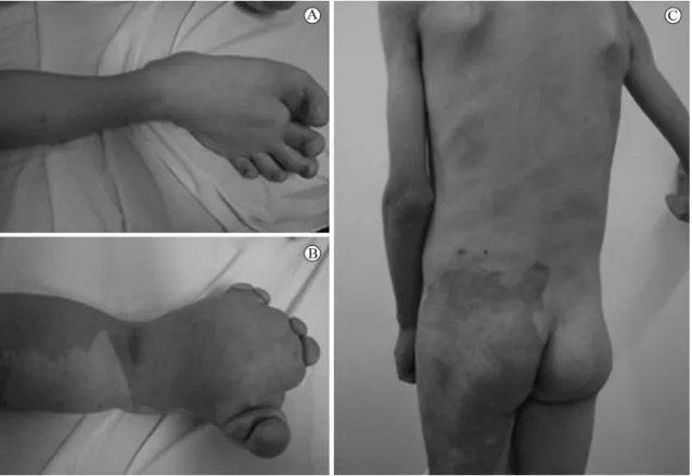
A 3-year-old male child who was being managed by a vascular surgery team exhibited gigantism of both feet, gigantism of the left leg and thigh, verrucous and hyperpigmented nevus on the thigh, varicose veins in the left lower limb and lat hemangioma on the left side, including left arm and leg, initially thought to be Klippel-Trenaunay syndrome (Figure 1). However, this patient also had scoliosis of the cervical and thoracic spine and hamartoma of the right hemithorax, leading to a re-designation of the case as PS (Figure 1).
For 4 years management was limited to clinical monitoring of the child, whose family did not authorize any type of intervention during this period. A large number of supplementary examinations were conducted during this period of clinical observation. Of these, vascular duplex ultrasound (USD) demonstrated that the deep vein system in the lower limbs was normal, but there was a dilated posterior sciatic vein in the left lower limb and supericial arteriovenous malformations including communicating istulas in the same limb. Angiotomography and magnetic resonance
angiography did not add to the USD indings. X-rays, computed tomography, magnetic resonance imaging, electrocardiogram and hematological and urinary tests all showed no signiicant abnormalities beyond the orthopedic conditions already described.
During the period of observation, the child exhibited progressive worsening of the hypertrophy of the feet and began to become extremely underweight. In addition to malnutrition, ambulation worsened, primarily because of the hypertrophy of the feet (Figure 2) and because of his deformities the child developed an introverted psychological proile, making few friends and with weak social relationships.
Faced with this situation, after 4 years, the patient’s family inally agreed to the proposal to perform a bilateral amputation of the lower limbs.
The treatment options for PS are as follows: 1) Clinical observation (watchful waiting); 2) Embolization of vascular malformations with
microspheres;
3) Reconstructive plastic surgery; 4) Amputation of limbs.
PART II - WHAT WAS DONE
A multidisciplinary team comprising orthopedists, physiotherapists, vascular surgeons, plastic surgeons, pediatricians and psychiatrists arrived at a consensus that the best option would be to perform a transfemoral amputation of the left lower limb and a transtibial amputation of the right lower limb. This option was presented to the child’s parents and they accepted the suggestion.
The procedures were conducted during a single operation: left transfemoral amputation and right transtibial amputation. The intraoperative appearance of the skin and deeper tissues of the right lower limb were normal, but the left lower limb exhibited vascular malformations, with tangles of vessels with ibrosis and thrombosis, with no active bleeding, and supericial arteriovenous istulas. The procedures were accomplished with no intercurrent conditions. During the postoperative period, healing of the right side was satisfactory and fast. However, on the left side, which was the side with vascular malformations, dehiscence set in and healing was slow and by secondary intention. Infection of the dehiscent operating wound was treated with antibiotics and complete healing took 2 months.
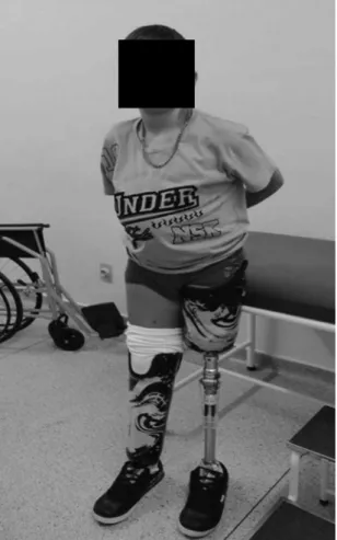
One year after the amputations, the child had adapted to and undergone rehabilitation with prostheses for both lower limbs and had recovered from malnutrition
(Figure 3). He also exhibited psychosocial improvement, recovered his self-conidence and had resocialized, with increased enthusiasm for the games that are normal for his age. He achieved good intellectual performance at school, with no dificulty keeping up with the activities appropriate for his age.
Figure 2. Patient with hypertrophy of both lower limbs, hamartomas, hemangiomas and malnutrition.
DISCUSSION
Diagnosing PS remains dificult today13,14 and the
criteria for its diagnosis are controversial.8 Clinically,
the disease involves hypertrophy of skin, connective tissue, brain, bones and other tissues. Genetic mapping of PS identiied a somatic activation mutation of AKT1. This enzyme is called v-akt murine thymoma viral oncogene homolog 1 (AKT1), or protein kinase B-alpha (PKB-ALPHA), and belongs to the protein kinase superfamily. AKT1 is one of the most important mediators of the responses to insulin, to insulin-like growth factor (IGF1) and to glucose. Somatic activation of the AKT1 mutation provokes disproportional growth of tissues and also makes tumors more likely.15
Treatment of PS tends to be complex.16 Lesions
involving the ovaries and testicles should be treated aggressively because of the risk of neoplasms.17
Gastrointestinal and renal lesions can be treated conservatively, with rigorous follow-up using serial imaging exams.18 The lungs may exhibit
emphysematous abnormalities, sometimes requiring pneumonectomies.19 Involvement of the bladder is
rare.20 The airways and tonsils should be assessed
before surgery, because malformations are common21
and could make oral endotracheal intubation dificult. Craniofacial abnormalities affect around 30% of patients and reconstructive treatment should be initiated early because the condition is progressive.3,22,23 Since the
incidence of neoplasms is high, systematic screening is fundamental.24
The extremities tend to exhibit large scale deformities, involving soft tissues, bones and blood vessels. Treatment with reconstructive plastic surgery is challenging and often unfeasible,25 because of
concurrent malformations of deep vascular systems. Consequently, in some cases, the option that remains is amputation of the affected limb, but family members are often resistant to this course of action.
In the case described here, when amputation of the lower limbs was proposed, because restorative treatment was not possible, the parents were reluctant to accept the suggestion, which delayed the deinitive treatment, during which time the nutritional and psychological status of the patient deteriorated. However, as the case worsened, the parents accepted surgery. This was performed with a good outcome, allowing rehabilitation with prostheses to be initiated; to which the patient adapted easily. The patient reintegrated well socially and gained weight, with consequent improvement in quality of life.
In conclusion, the deformities seen in PS have a wide variety of clinical presentations and include complex deformities that can have a severe negative impact on patients’ self-esteem. Treatment should be managed by a multidisciplinary team and will very often be of a palliative nature. In some cases, in which reconstructive surgery is impossible, major amputations will be needed to save the lives of the most severely affected patients. Although amputations are an extreme solution, they can have good results because they open the way to physical rehabilitation with prostheses, allowing for reintegration into society and nutritional recovery. Proteus syndrome is a disease that is little understood and is rare, but PS patients suffer from serious limitations and so presentation of these rare cases can provide useful guidance for treatment in similar situations.
REFERENCES
1. Cohen MM Jr, Hayden P. A newly recognized hamartomatous syndrome. Birth Defects Orig Artic Ser. 1979;15(5B):291-6. PMid:118782.
2. Wiedemann HR, Burgio GR, Aldenhoff P, Kunze J, Kaufmann HJ, Schirg E. The proteus syndrome: partial gigantism of the hands and/or feet, nevi, hemihypertrophy, subcutaneous tumors, macrocephaly or other skull anomalies and possible accelerated growth and visceral affections. Eur J Pediatr. 1983;140(1):5-12. http://dx.doi.org/10.1007/BF00661895. PMid:6873112.
3. Sakamoto Y, Nakajima H, Kishi K, Shimizu R, Nakajima T. Management of craniofacial hyperostosis in Proteus syndrome. J Craniofac Surg. 2010;21(2):414-8. http://dx.doi.org/10.1097/ SCS.0b013e3181cfa7f0. PMid:20216456.
4. Pazzaglia UE, Beluffi G, Bonaspetti G, Ranchetti F. Bone malformations in Proteus syndrome: an analysis of bone structural changes and their evolution during growth. Pediatr Radiol. 2007;37(8):829-35. http://dx.doi.org/10.1007/s00247-007-0486-1. PMid:17569038.
5. White NJ, Cochrane DD, Beauchamp R. Paraparesis caused by an angiolipomatous hamartoma in an adolescent with Proteus syndrome and scoliosis. J Neurosurg. 2005;103(3, Supl):282-4. PMid:16238085.
6. Samlaska CP, Levin SW, James WD, Benson PM, Walker JC, Perlik PC. Proteus syndrome. Arch Dermatol. 1989;125(8):1109-14. http://dx.doi.org/10.1001/archderm.1989.01670200085015. PMid:2667470.
7. Barmakian JT, Posner MA, Silver L, Lehman W, Vine DT. Proteus syndrome. J Hand Surg Am. 1992;17(1):32-4. http://dx.doi. org/10.1016/0363-5023(92)90109-3. PMid:1538109.
8. Biesecker L. The challenges of Proteus syndrome: diagnosis and management. Eur J Hum Genet. 2006;14(11):1151-7. http://dx.doi. org/10.1038/sj.ejhg.5201638. PMid:16883308.
9. Turner JT, Cohen MM Jr, Biesecker LG. Reassessment of the Proteus syndrome literature: application of diagnostic criteria to published cases. Am J Med Genet A. 2004;130A(2):111-22. http://dx.doi. org/10.1002/ajmg.a.30327. PMid:15372514.
11. Thomason JL, Abramowsky CR, Rickets RR, Culbertson JH, Clifton MS, Shehata BM. Proteus syndrome: three case reports with a review of the literature. Fetal Pediatr Pathol. 2012;31(3):145-53. http://dx.doi.org/10.3109/15513815.2012.656830. PMid:22413928.
12. Gontijo B, Pereira LB, Silva CMR. Malformações vasculares. An Bras Dermatol. 2004;79(1):7-25. http://dx.doi.org/10.1590/ S0365-05962004000100002.
13. Satter E. Proteus syndrome: 2 case reports and a review of the literature. Cutis. 2007;80(4):297-302. PMid:18038691.
14. Cohen MM Jr. Proteus syndrome: clinical evidence for somatic mosaicism and selective review. Am J Med Genet. 1993;47(5):645-52. http://dx.doi.org/10.1002/ajmg.1320470514. PMid:8266991.
15. Lindhurst MJ, Sapp JC, Teer JK, et al. A mosaic activating mutation in AKT1 associated with the Proteus syndrome. N Engl J Med. 2011;365(7):611-9. http://dx.doi.org/10.1056/NEJMoa1104017. PMid:21793738.
16. Vieira NRN, Pereira LB, Silva CMR, Gontijo B. Síndrome de Proteus: relato de caso. An Bras Dermatol. 2001;76:201-208.
17. Raju RR, Hart WR, Magnuson DK, Reid JR, Rogers DG. Genital tract tumors in Proteus syndrome: report of a case of bilateral paraovarian endometrioid cystic tumors of borderline malignancy and review of the literature. Mod Pathol. 2002;15(2):172-80. http:// dx.doi.org/10.1038/modpathol.3880510. PMid:11850547.
18. Kaduthodil MJ, Prasad DS, Lowe AS, Punekar AS, Yeung S, Kay CL. Imaging manifestations in Proteus syndrome: an unusual multisystem developmental disorder. Br J Radiol. 2012;85(1017):e793-9. http:// dx.doi.org/10.1259/bjr/92343528. PMid:22514103.
19. Li CY, Chang YL, Chen WC, Lee YC. Pulmonary manifestations and management of proteus syndrome. J Formos Med Assoc. 2010;109(5):397-400. http://dx.doi.org/10.1016/S0929-6646(10)60069-1. PMid:20497874.
20. Abbo O, Bouali O, Galinier P, Moscovici J. Proteus syndrome: case report of bladder vascular malformation causing massive hematuria. Prog Urol. 2012;22(2):132-5. http://dx.doi.org/10.1016/j. purol.2011.07.011. PMid:22284599.
21. Lublin M, Schwartzentruber DJ, Lukish J, Chester C, Biesecker LG, Newman KD. Principles for the surgical management of patients with Proteus syndrome and patients with overgrowth not meeting Proteus criteria. J Pediatr Surg. 2002;37(7):1013-20. http://dx.doi. org/10.1053/jpsu.2002.33832. PMid:12077761.
22. Adolphs N, Tinschert S, Bier J, Klein M. Craniofacial hyperostoses in Proteus syndrome: a case report. J Craniomaxillofac Surg. 2004;32(6):391-4. http://dx.doi.org/10.1016/j.jcms.2004.06.007. PMid:15555524.
23. Becktor KB, Becktor JP, Karnes PS, Keller EE. Craniofacial and dental manifestations of Proteus syndrome: a case report. Cleft Palate Craniofac J. 2002;39(2):233-45. http://dx.doi.
org/10.1597/1545-1569(2002)039<0233:CADMOP>2.0.CO;2. PMid:11879083.
24. Nazario APM, Alves MR, Ellis C, Morais FC, Panico MB, Vieira GM. Síndrome de Proteus: hamartose desde o nascimento. J Vasc Bras. 2011;10:124.
25. Havard S, Enjolras O, Lessana-Leibowitch M, Escande JP. Proteus syndrome. 8 cases. Ann Dermatol Venereol. 1994;121(4):303-8. PMid:7702248.
*
Correspondence
Matheus Bertanha Faculdade de Medicina de Botucatu, Universidade Estadual Paulista – UNESP Campus de Botucatu Rubião Júnior, s/n CEP 18618-970 - Botucatu (SP), Brazil Tel.: +55 (14) 3815-7428 E-mail: [email protected]
Author information
MB - Assistant professor at the Department of Surgery and Orthopedics, Faculdade de Medicina de Botucatu, Universidade Estadual Paulista (UNESP). RM and MLS - PhD’s assistant professors at the Department of Surgery and Orthopedics, Faculdade de Medicina de Botucatu, Universidade Estadual Paulista (UNESP). LMSP and MPFS - Primary physicians at the Department of Pediatrics, Faculdade de Medicina de Botucatu, Universidade Estadual Paulista (UNESP). RGJ - Primary physician at the Department of Surgery and Orthopedics, Faculdade de Medicina de Botucatu, Universidade Estadual Paulista (UNESP). HAR - Adjunct professor at the Department of Surgery and Orthopedics, Faculdade de Medicina de Botucatu, Universidade Estadual Paulista (UNESP). WBY - Full professor at the Department of Surgery and Orthopedics, Faculdade de Medicina de Botucatu, Universidade Estadual Paulista (UNESP).
Author contributions
Conception and design: MB, RM Analysis and interpretation: MB, RM, MLS, RGJ Data collection: LMSP, MPFS Writing the article: MB, MLS, RGJ, WBY Critical revision of the article: MB, HAR, MLS Final approval of the article*: MB, RM, MLS, LMSP, RGJ, MPFS, HAR, WBY Statistical analysis: N/A. Overall responsibility: MB, WBY