Clinical and echocardiographic characteristics associated
with the evolution of the ductus arteriosus in the
neonate with birth weight lower than 1,500g
Características clínicas e ecocardiográficas associadas à evolução do canal arterial
em recém-nascidos com peso de nascimento inferior a 1.500g
Luiza Fortunato Visconti1, Samira Saady Morhy1, Alice D’Agostini Deutsch1, Gláucia Maria Penha Tavares1,
Tatiana Jardim Mussi Wilberg1, Felipe de Souza Rossi1
ABSTRACT
Objective: To identify clinical and echocardiographic parameters associated with the evolution of the ductus arteriosus in neonates with birth weight lower than 1,500g. Methods: Retrospective study of 119 neonates in which clinical parameters (Prenatal: maternal age, risk of infection and chorioamnionitis, use of corticosteroid, mode of delivery and gestational age. Perinatal: weight, Apgar score, gender and birth weight/gestational age classification; Postnatal: use of surfactant, sepsis, fluid intake, heart murmur, heart rate, precordial movement and pulses, use of diuretics, oxygenation index, desaturation/apnea, ventilatory support, food intolerance, chest radiography, renal function, hemodynamic instability, and metabolic changes) and echocardiographic parameters (ductus arteriosus diameter, ductus arteriosus/weight ratio, left atrium/ aorta ratio, left ventricular diastolic diameter, and transductal flow direction, pattern and velocity) were analyzed. The clinical and echocardiographic parameters analyzed were considered statistically significant when p<0.05. Results: In the 119 neonates, the incidence of patent ductus arteriosus was 61.3%; 56 received treatment (46 pharmacological and 10 surgical treatment), 11 had spontaneous closure, 4 died, and 2 were discharged with patent ductus arteriosus. A higher incidence of chorioamnionitis, use of surfactant, lower weight and gestational age, sepsis, heart murmur, ventilatory support and worse oxygenation indices were observed in the neonates receiving treatment. The group with spontaneous closure had a smaller ductus arteriosus diameter, lower ductus arteriosus/weight ratio, and higher transductal flow velocity. Conclusion: Based on clinical and echocardiographic parameters, the neonates with spontaneous closure of the ductus arteriosus could be differentiated from those who required treatment.
Keywords: Ductus arteriosus patency; Neonate; Premature
RESUMO
Objetivo: Identificar parâmetros clínicos e ecocardiográficos associados à evolução do canal arterial em recém-nascidos com peso de nascimento <1.500g. Métodos: Estudo retrospectivo de 119 recém-nascidos, no qual foram analisados parâmetros clínicos (pré-natais: idade materna, risco infeccioso e corioamnionite, uso de corticoide, tipo de parto e idade gestacional; perinatais: peso, Apgar, gênero e classificação peso/idade gestacional; pós-natais: surfactante, sepse, oferta hídrica, sopro cardíaco, frequência cardíaca, movimento precordial e pulsos, diurético, índice de oxigenação, queda de saturação/apneia, suporte ventilatório, intolerância alimentar, radiografia de tórax, função renal, instabilidade hemodinâmica e alterações metabólicas); parâmetros ecocardiográficos (diâmetro do canal arterial, relação canal arterial/peso, relação átrio esquerdo/ aorta, diâmetro diastólico ventrículo esquerdo, direção, padrão e velocidade de fluxo pelo canal arterial). Os parâmetros clínicos e ecocardiográficos analisados foram considerados estatisticamente significantes quando p<0,05. Resultados: Nos 119 recém-nascidos, a incidência de canal arterial foi de 61,3%, 56 receberam tratamento (46 medicamentoso e 10 cirúrgico), 11 tiveram fechamento espontâneo, 4 foram a óbito e 2 receberam alta com persistência do canal arterial. Houve maior incidência de corioamnionite, uso de surfactante, menor peso e idade gestacional, sepse, sopro cardíaco, ventilação e piores índices de oxigenação nos recém-nascidos tratados. O grupo com fechamento espontâneo apresentou menor diâmetro do canal arterial, menor relação canal arterial/peso e maior velocidade do fluxo pelo canal arterial. Conclusão: Com base em parâmetros clínicos e ecocardiográficos, foi possível diferenciar os recém-nascidos com fechamento espontâneo do canal arterial daqueles com necessidade de tratamento.
Descritores: Permeabilidade do canal arterial; Recém-nascido; Prematuro
Study carried out at Hospital Israelita Albert Einstein, São Paulo, SP, Brazil.
1 Hospital Israelita Albert Einstein, São Paulo, SP, Brazil.
Corresponding author: Samira Saady Morhy – Avenida Albert Einstein, 627/701 – Morumbi – Zip code: 05652-900 – São Paulo, SP, Brazil – Phone: (55 11) 2151-1233 – E-mail: [email protected]
Received on: Feb, 5, 2013 – Accepted on: Aug 13, 2013
INTRODUCTION
The ductus arteriosus (DA) is a structure that is present in the fetus and connects the aorta and the pulmonary artery, diverting the right ventricular flow
into the systemic circulation(1,2).
Soon after birth, the process of functional DA
closure starts(1). In preterm neonates (PTN), patent
ductus arteriosus (PDA) is common due to increased sensitivity to prostaglandins, a higher incidence of hypoxia and acidosis, and defective migration of the
smooth muscle that leads to DA vasoconstriction(3-5).
With PDA, there is high pulmonary flow and low systemic flow, which may be associated with morbidities in the PTN, such as chronic pulmonary disease, ventricular hemorrhage, leukomalacia, necrotizing enterocolitis, renal failure, congestive heart failure, retinopathy of prematurity, and increased mortality in patients with
respiratory distress syndrome(6,7).
For the assessment of DA, in addition to the clinical characteristics resulting from the hemodynamic changes caused by PDA, echocardiographic data that describe the cardiac morphology (DA and cardiac
chamber sizes)(8,9), and the flow through the DA
(flow direction, pattern and velocity)(10-13) are also
considered.
The procedures currently available for the treatment of PDA are the use of non-steroidal anti-inflammatory drugs and surgical closure. However, although these treatments have been proven effective, their use remains controversial due to their side effects (risk of transient change in brain perfusion and transient decrease in renal function when anti-inflammatory drugs are used; and pneumothorax, infection, hemorrhage, chylothorax and vocal cord paralysis when surgery is used), and to the lack of evidence that they improve the patients’ long-term
outcome(7).
Additionally, data from the literature have shown spontaneous closure occurring in the neonatal period
in 31% to 34% of the neonates weighing ≤1,000g(3,14)
and in 67% of those weighing between 1,000 and
1,500g(14), within 7 days after birth.
Thus, further studies are required to identify the groups which would really benefit from the treatment
of PDA(15,16).
OBJECTIVE
To identify clinical and echocardiographic parameters associated with the evolution of the ductus arteriosus in neonates weighing <1,500g.
METHODS
Single-center, cohort, longitudinal and retrospective study conducted in the Neonatal Intensive Care Unit of
Hospital Israelita Albert Einstein (HIAE), in São Paulo. The study was approved by the HIAE Research Ethics Committee no. 314/2011.
The study population comprised neonates with birth weight (BW) <1,500g, in the period from January 2008 to December 2010.
The diagnosis of PDA was made using bedside echocardiography, requested by the neonatologist.
Patients with complex congenital heart defects or those who did not undergo echocardiography were excluded from the study.
For the analysis of variables, the patients were divided into three groups: patients who had spontaneous closure of the DA (Group I); patients who received pharmaceutical treatment (Group II); and patients who received surgical treatment (Group III).
Clinical parameters
Prenatal, perinatal and postnatal clinical parameters were analyzed. The following prenatal clinical parameters were studied: maternal age, presence of risk of infection (ruptured membranes for longer than 18 hours, maternal urinary infection, and maternal fever), presence of chorioamnionitis, antenatal use of corticosteroid, mode of delivery (vaginal or cesarean), gestational age (GA – the GA analyzed was that considered by the service, based on the last menstrual period, on the first-trimester ultrasonography, or on the Capurro’s
method(17). The perinatal parameters analyzed were:
BW, 1 and 5-minute Apgar score, gender, classification as small for GA (SGA) or appropriate for GA (AGA),
based on the Babson-Benda growth charts(18). The
postnatal parameters observed were: use of surfactant, presence of early or late neonatal sepsis, and fluid intake in the first two and four days of life.
On the day the patients underwent echocardiographic study, in which either spontaneous closure of DA was observed or pharmaceutical or surgical treatment were indicated, the following parameters were also collected: age, presence of heart murmur, presence of persistent tachycardia (heart rate – HR>160), hyperdynamic
precordium (visible ictus), wide pulses, use of diuretics,
Echocardiographic parameters
The echocardiographic variables were obtained by reviewing the reports and echocardiographic images stored. Data from all echocardiograms performed were collected, from hospitalization until the test in which DA closure was verified. On the first echocardiographic study performed, the parameters described below were analyzed. In the other echocardiographic studies, only DA persistence/closure was observed.
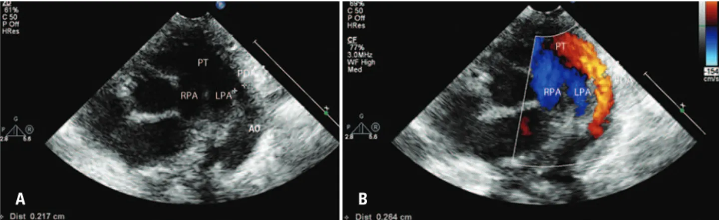
The following variables were analyzed: DA diameter (mm) (Figure 1), DA diameter corrected for the patient’s weight (mm/kg), on two-dimensional echocardiography; left atrial to aorta diameter ratio, left ventricular diastolic diameter (mm) on M-mode echocardiography; and transductal flow direction,
Figure 2. Transductal flow pattern on pulsed Doppler(13). (A) Hypertension: bidirectional flow from the pulmonary to the aorta, on early systole (below baseline), followed by a small flow from the aorta to the pulmonary (above baseline). (B) Growing: bidirectional flow, however with decreased flow from the pulmonary to the aorta and increased flow from the aorta to the pulmonary (C) Pulsatile: flow only from the aorta to the pulmonary, pulsatile, with peak velocity of approximately 1.5m/s. (D) Closing: flow directed from the aorta to the pulmonary, not pulsatile but rather continuous, with a higher peak velocity
A B
PT: pulmonary trunk; PDA: persistent ductus arteriosus; AO: aorta; LPA: left pulmonary artery; RPA: right pulmonary artery.
Figure 1. (A) Two-dimensional image showing the presence of the ductus arteriosus, with its diameter measurement. (B) Two-dimensional image of the ductus arteriosus, with color flow mapping, showing the transductal flow in red, going from the aorta to the pulmonary artery
A B
peak velocity (m/s) and patterns on color pulsed Doppler.
The transductal flow patterns were classified as: a) pulmonary hypertension pattern, if bidirectional flow, from the pulmonary to the aorta, in early systole (below baseline), followed by a small flow from the aorta to the pulmonary (above baseline), suggesting increased pulmonary vascular resistance; b) growing pattern, if bidirectional flow, with decreased flow from the pulmonary to the aorta, and increased flow from the aorta to the pulmonary, suggesting decreased pulmonary vascular resistance; c) pulsatile pattern, if only flow from the aorta to the pulmonary, pulsatile, with peak velocity of approximately 1.5m/s; d) closing pattern, if flow directed from the aorta to the pulmonary, not pulsatile but rather
Statistical analysis
The SAS 9.0 software program was used for the statistical analyses.
Continuous variables were described as mean and standard deviation or median and interquartile range. Categorical variables were described as absolute and relative frequencies. The association between categorical
variables was tested using the χ2 test. The Student
t test was used for comparisons between numerical
variables.
Results were considered statistically significant when p<0.05.
RESULTS
In the study period, 138 patients with BW<1,500g were born. Of these, 19 were excluded, 2 for congenital heart defect (truncus arteriosus type II and coarctation of the aorta), and 17 for not having undergone echocardiography (5 died in the first day of life and the test was not requested by the medical team for the other 12). Thus 119 patients were included in the study.
The incidence of DA was 61.3% (73/119). Among neonates weighing <1,000g, the incidence was 75% (30/40), and, among those with less than 30 weeks, the incidence was 75% (57/76).
Mortality was 10.1% (12/119), of whom only one patient did not have PDA.
Of the 73 patients with PDA, 4 (5.5%) died early,
before the 5th day of life, with no possibility of treatment,
and 2 (2.8%) were discharged with PDA, untreated. Spontaneous closure (Group I) was observed in 11 patients (15%). The mean time for closure was approximately 13.5 days; one of these patients had spontaneous closure at 18 days, but died at 162 days of life.
A total of 46 patients (63%) underwent pharmacological treatment (Group II), with a mean closure time of 8 days
of life. Of these, 3 died between the 12th and 17th day of life,
after DA closure.
Ten patients (13.7%) underwent surgical closure (Group III), four as the first treatment choice and six after unsuccessful pharmacological treatment (mean closure time 12.7 days of life). Of these, three died at 14, 35 and 41 days of life (the time elapsed between surgery and death was longer than 1 week in the three cases).
Clinical parameters
We observed a higher incidence of the use of surfactant in Group II in relation to Group I; a higher incidence of risk of infection, chorioamnionitis, late sepsis, and use of surfactant, as well as of lower BW, GA, and 1-minute Apgar score in Group III in relation to Group I, with statistically significant differences (p<0.05) (Table 1).
A higher incidence of heart murmur and need for ventilatory support was observed in Groups II and III.
Table 1. Differences of pre- and perinatal variables among patients with spontaneous ductus arteriosus closure (Group I), patients with pharmacological closure (Group II) and those with surgical closure (Group III)
Patient characteristics Group I (n=11) Group II (n=46) Group III (n=10)
Maternal age (years) 33.9 34.7* 38.8***
Risk of infection, n (%) 1 (9.1) 15 (34.09)* 6 (60.0%)****
Chorioamnionitis, n (%) 1 (9.1) 9 (20.0)* 6 (66.7)****
Antenatal use of corticosteroid, n (%) 9 (81.8) 29 (63.0)* 10 (100)***
Cesarean delivery, n (%) 9 (81.8) 38 (82.61)* 8 (80.0)***
Gestational age (weeks) 29.6 28.4* 25.9****
Birth weight (grams) 1.154.1 1.092.7* 828.5****
1-minute Apgar score 7.6 6.6* 4.8****
5-minute Apgar score 8.5 8.6* 8.2***
Male gender, n (%) 5 (45.5) 29 (63.04)* 8 (80)***
Classification as SGA, n (%) 3 (27.3) 8 (17.39)* 1 (10)***
Use of surfactant, n (%) 4 (36.4) 34 (73.9)** 8 (80)****
Early sepsis, n (%) 5 (45.5) 27 (58.7)* 8 (80)***
Late sepsis, n (%) 4 (36.4) 16 (34.8)* 8 (80)****
FI at 48 hours 96.5 93.6* 102.6***
FI at 96 hours 112.2 108.4* 126.4***
* No statistically significant difference was observed between Groups I and II; ** statistically significant difference was observed between Groups I and II, p<0.05; *** no statistically significant difference was observed between Groups I and III; **** statistically significant difference was observed between Groups I and III, p<0.05.
Values of oxygenation index and metabolic changes were only assessed in patients with blood gas analysis on the day the treatment was indicated or spontaneous DA closure occurred; few Group I patients had their blood gas collected – this explains the difference between the two groups (Table 2).
Echocardiographic parameters
The echocardiographic measurements studied were compared among the three groups. The mean DA diameter and the mean weight-corrected DA diameter were greater in Groups II and III in relation to Group I. The transductal flow velocity was significantly lower in Group III in relation to Group I (Table 3).
In relation to the transductal flow patterns assessed in the first echocardiogram, 57.2% of patients with PDA with spontaneous closure had a hypertensive (14.3%) or closing pattern (42.9%), whereas 67.5% of patients undergoing pharmacological closure had a growing pattern (17.5%) or pulsatile pattern (50%), and 66.6% of patients undergoing surgical closure had a growing pattern (22.2%) or pulsatile pattern (44.4%). However, no statistically significant difference was observed between the groups (Table 4).
Table 2. Differences in clinical signs among patients with spontaneous ductus arteriosus closure (Group I), patients with pharmacological closure (Group II) and those with surgical closure (Group III)
Clinical signs Group I
(n=11)
Group II (n=46)
Group III (n=10)
Patient age (days) 2.5 2.2* 2.1***
Presence of heart murmur, n (%) 2 (18.2) 22 (47.8)** 9 (90)****
HR >160, n (%) 0 (0) 1 (2.2)* 1 (10)***
Hyperdynamic precordium, n (%) 0 (0) 4 (40)* 2 (66.7)***
Wide pulse, n (%) 1 (50) 8 (61.5)* 2 (100)***
Use of diuretics, n (%) 0 (0) 1 (2.2)* 1 (10)***
Abnormal renal function, n (%) 1 (9.1) 0 (0)** 4 (40)***
Oxygenation index ** ****
≤6, n (%) 0 (0) 27 (96.4) 9 (90)
7-14, n (%) 1 (50) 1 (3.6) 1 (10)
≥15, n (%) 1 (50) 0 (0) 0 (0)
Desaturation/apnea * ***
Absent, n (%) 8 (72.7) 23 (52.3) 5 (50)
Up to 6 episodes, n (%) 2 (18.2) 19 (43.1) 2 (20)
Frequent, n (%) 1 (9.1) 2 (4.6) 3 (30)
Ventilatory support ** ****
Room air, n (%) 6 (54.6) 5 (10.9) 0 (0)
Inhaled O2, n (%) 0 (0) 0 (0) 1 (10)
CPAP ou MAP ≤8, n (%) 2 (18.2) 31 (67.4) 5 (50)
MAP 9-12, n (%) 0 (0) 4 (8.7) 1 (10)
MAP ≥12 HFV, n (%) 3 (27.3) 6 (13) 3 (30)
Food intolerance * ***
Absent, n (%) 8 (72.7) 16 (34.8) 2 (20)
Fasting, n (%) 2 (18.2) 18 (39.1) 4 (40)
Minimal enteral nutrition, n (%) 0 (0) 6 (13) 0 (0)
Residual gastric content, n (%) 1 (9.1) 5 (10.9) 2 (20)
Abdominal distention/vomiting, n (%) 0 (0) 1 (2.1) 2 (20)
Chest radiography * ***
Normal, n (%) 2 (50) 5 (17.2) 1 (14.3)
Increased pulmonary vasculature, n (%) 2 (50) 20 (69) 4 (57.1)
Cardiomegaly/pulmonary edema, n (%) 0 (0) 4 (13.8) 2 (28.6)
Hemodynamic instability * ***
Absent, n (%) 8 (72.7) 39 (84.8) 5 (50)
Hypotension (1 VAD), n (%) 1 (9.1) 6 (13.0) 4 (40)
Hypotension (more than 1 VAD), n (%) 2 (18.2) 1 (2.2) 1 (10)
Metabolic change ** ***
Absent, n (%) 1 (33.3) 26 (92.9) 8 (80)
Mild metabolic acidosis, n (%) (pH 7.1-7.25 and/or BE -7 to -12), n (%)
2 (66.7) 2 (7.1) 1 (10)
Moderate to severe metabolic acidosis, n (%)
(pH <7.1 or BE <-12), n(%)
0 (0) 0 (0) 1 (10)
* No statistically significant difference was observed between Groups I and II; ** statistically significant difference was observed between Groups I and II, p<0.05; *** No statistically significant difference was observed between Groups I and III; **** statistically significant difference was observed between Groups I and III, p<0.05.
HR: heart rate; CPAP: constant positive airway pressure; MAP: medium airway pressure; HFV: high-frequency ventila-tion; VAD: vasoactive drug; BE: base excess.
Table 3. Differences of the echocardiographic measurements among patients with spontaneous ductus arteriosus closure (Group I), patients with pharmacological closure (Group II) and those with surgical closure (Group III)
Echocardiographic data Group I (n=11)
Group II (n=46)
Group III (n=10)
DA diameter (mm) 1.63 2.24** 2.39****
DA/W (mm/kg) 1.55 2.22** 3.44****
LA/Ao 1.10 1.20* 1.26***
LV diastolic diameter (mm) 12.73 12.98* 11.78***
Flow direction (L-R), n (%) 6 (66.7) 33 (71.7)* 5 (50)***
Transductal flow velocity (m/s) 2.45 1.92* 1.62****
DA: ductus arteriosus diameter; DA/W: ductus arteriosus diameter/patient weight ratio; LA/Ao: Left atrium size/aorta size ratio; LV: left ventricle; L-R: from the left to the right.
* No statistically significant difference was observed between Groups I and II; ** statistically significant difference was observed between Groups I and II, p<0.05; *** no statistically significant difference was observed between Groups I and III; **** statistically significant difference was observed between Groups I and III, p<0.05.
Table 4. Differences of the transductal flow patterns among patients with spontaneous ductus arteriosus closure (Group I), patients with pharmacological closure (Group II) and those with surgical closure (Group III)
Flow patterns on the first echo-cardiogram
Group I (n=11)
Group II (n=46)
Group III (n=10)
Hypertension or closing, n (%) 4 (57.2) 13 (32.5)* 3 (33.3)***
Growing or pulsatile, n (%) 3 (42.8) 27 (67.5)* 6 (66.7)***
DISCUSSION
PDA is an anomaly that affects a large number of premature neonates and may lead to significant clinical consequences, with an impact in the morbidity and mortality of these patients.
In this study, we observed an incidence of 61.3% of PDA, of which 75% in neonates weighing <1,000g or with less than 30 weeks. This results is in agreement with data from the literature that describe incidences between 20% and 60%, depending on the diagnostic criteria and population studied, with increasing incidences with the
decrease in BW and GA(3).
Currently, the core issue in the clinical management of patients with PDA is to identify, by means of epidemiological, clinical and echocardiographic data, the group that would benefit from treatment indication, given the adverse consequences of the pharmacological and surgical treatments, of the incidence of spontaneous closure, and of the lack of evidence that the prophylactic pharmacological treatment is beneficial in the long term(19-21).
Disagreement is common between clinical and echocardiographic parameters in the assessment of the hemodynamic consequences of PDA. In practice, DA size on echocardiogram, the presence of chronic pulmonary disease, and the increase in ventilatory support parameters are taken into consideration.
Echocardiogram can provide an early diagnosis and evaluate the hemodynamic consequences. However, there is still no consensus on which echocardiographic parameters should be used to guide therapy.
In the present study, among pre- and perinatal parameters, the use of surfactant was significantly more frequent in the groups with pharmacological and surgical closure in relation to the group with spontaneous closure, so this population could be more prone to require treatment. Surfactants have no effect on the contractile behavior of DA, but they change the pulmonary vascular
resistance, increasing the left to right transductal flow(22).
The presence of risk of infection, chorioamnionitis and late sepsis was more frequent in the group requiring surgery, and this could be explained by the release of inflammatory mediators in these processes; these mediators would impair the spontaneous DA closure, as
described by Shannon et al.(1). BW and GA were lower
in the group with surgical closure, with a statistically significant difference. Jhaveri et al. also found failure in the treatment with indomethacin in 70% of neonates
<28 weeks of GA, thus requiring surgical treatment(23).
In relation to the echocardiographic parameters analyzed, the comparison between the three groups
with PDA showed higher mean DA diameter and DA diameter/weight ratio in the groups with pharmacological and surgical closure. These results are similar to
those described by Hajjar et al.(8), who observed an
inverse relation between DA size and the probability of spontaneous closure. Mc Namara and Sehgal also related the DA size to the hemodynamic consequences of PDA, categorizing them as small, when DA diameter is <1.5mm; moderate, when DA diameter is between 1.5
and 3mm; and large, when DA diameter >3mm(15).
On the other hand, Jhaveri et al. considered small DA those with a diameter <1.5mm. However, diameters >1.5mm were considered as DA with moderate hemodynamic consequences. The difference between moderate and severe hemodynamic consequences was defined by the mean transductal flow gradient, which
was <8mmHg in cases with severe consequences(23).
In this study, the transductal flow velocity was higher in the cases of PDA in closing process, and lower in the group with surgical closure; these findings are
similar to those observed by Su et al.(13). Transductal
flow acceleration in the neonate is an indirect sign of its closing process and should be taken into consideration for the decision-making concerning therapy.
However, in this study, unlike in Su et al’s study(13),
no statistically significant difference in the transductal flow pattern was observed between the groups. In our opinion, this difference can be explained by the fact that, in this study, the flow patterns analyzed were only those from the first echocardiogram, which was performed early (mean time of 2.3 days). It would interesting to analyze follow-up echocardiographic studies to observe whether a significant difference would occur that could help decide therapy.
The major limitation of this study is that it is a retrospective study, with the patients being divided into groups according to the absence, presence or treatment of PDA, based on criteria of the neonatal intensive care unit, and data collected by means of review of medical charts and echocardiograms previously performed. Addittionally, it was not possible to evaluate all the echocardiographic parameters used to quantify the hemodynamic consequences of PDA, as reported in studies of the literature, such as: mean flow velocity in the left pulmonary artery, end-diastolic velocity in the left pulmonary artery, E/A wave ratio through the diastolic tricuspid valve flow, and reverse diastolic flow
in the descending aorta and mesenteric artery(8,15,23).
CONCLUSION
ventilatory support, DA diameter, and DA diameter/ weight ratio could differentiate patients who progressed with spontaneous DA closure (Group I) from those requiring pharmacological treatment (Group II) and/or surgical treatment (Group III).
Other parameters, such as risk of infection, chorioamnionitis, birth weight, gestational age, 1-minute Apgar score, presence of late sepsis, and transductal flow velocity on Doppler, could differentiate patients who progressed with spontaneous DA closure (Group I) from those requiring surgical treatment (Group III).
REFERENCES
1. Hamrick SE, Hansmann G. Patent ductus arteriosus of the preterm infant. Pediatrics. 2010;125(5):1020-30.
2. Aguiar CR, Costa HP, Rugolo LM, Sadeck LS, Costa MZ, Pachi PR, et al. O recém-nascido de muito baixo peso. Atheneu, 2010. 2a ed. Seção 6, cap.29: 375-85. 3. Koch J, Hensley G, Roy L, Brown S, Ramaciotti C, Rosenfeld CR. Prevalence
of spontaneous closure of the ductus arteriosus in neonates at a birth weight of 1000 grams or less. Pediatrics. 2006;117(4):1113-21.
4. Afiune JY, Singer JM, Leone CR. Evolução Ecocardiográfica de recém-nascidos com persistência do canal arterial. J Pediatr. 2005;81(6):454-60. 5. Agarwal R, Deorari AK, Paul VK. Patent ductus arteriosus in preterm
neonates. Indian J Pediatr. 2008;75(3):277-80.
6. Thébaud B, Lacaze-Mazmonteil T. Patent ductus arteriosus in premature infants: A never-closing act. Paediatr Child Health. 2010;15(5):267-70. 7. Beniz WE. Treatment of persistent patent ductus arteriosus in preterm
infants: time to accept the null hypothesis. J Perinatol. 2010;30(4):241-52. 8. El Hajjar M, Vaksmann G, Rakza T, Kongolo G, Storme L. Severity of the ductal
shunt: a comparison of different markers. Arch Dis Child Fetal Neonatal. 2005;90(5):419-22.
9. Brissaud O, Guichoux J. Patent ductus arteriosus in the preterm infant: A survey of clinical practices in French neonatal intensive care units. Pediatr Cariol. 2011;32(5):607-14.
10. Kiuckow M, Seri I, Evans N. Functional echocardiography: an emerging clinical tool for the neonatologist. J Pediatr. 2007;150(2):125-30.
11. Silverman NH, Lewis AB, Heymann MA, Rudolph AM. Echocardiographic assessment of ductus arteriosus shunt in premature infants. Circulation. 1974;50(4):821-5.
12. Sehgal A, McNamara PJ. Does echocardiography facilitate determination of hemodynamic significance attributable to the ductus arteriosus? Eur J Pediatr. 2009;168(8):907-14.
13. Su BH, Watanabe T, Shimizu M, Yanagisawa M. Echocardiographic assessment of patent ductus arteriosus shunt flow pattern in premature infants. Arch Dis Child. 1997;77(1):36-40.
14. Nemerofsky SL, Parravicini E, Bateman D, Kleinman C, Polin RA, Lorenz JM. The ductus arteriosus rarely requires treatment in infants > 1000 grams. Am J Perinatol. 2008;25(10):661-6.
15. McNamara PJ, Sehgal A. Towards rational management of the patent ductus arteriosus: the need for disease staging. Arch Dis Child Fetal Neonatal Ed. 2007;92(6):424-7. Erratum in: Arch Dis Child Fetal Neonatal Ed. 2008;93(1):F78.
16. Knight DB. The treatment of patent ductus arteriosus in preterm infants. A review and overview of randomized trials. Semin Neonatol. 2001;6(1):63-73. 17. Capurro H, Korichzky S, Fonseca O, Caldeyro-Barcia R. A simplified method for
diagnosis of gestational age in the newborn infant. J Pediatr. 1978;93(1):120-2. 18. Babson SG, Benda GI. Growth graphs for the assessment of infants of varying
gestational age. J Pediatr. 1976;89(5):814-20.
19. Sehgal A, McNamara PJ. The ductus arteriosus: a refined approach! Semin Perinatol. 2012;36(2):105-13.
20. Clyman RI, Couto J, Murphy GM. Patent ductus arteriosus: are current neonatal treatment options better or worse than no treatment at all? Semin Perinatol. 2012;36(2):123-9.
21. Hammerman C, Bin-Nun A, Kaplan M. Managing the patent ductus arteriosus in premature neonate: a new look at what we thought we knew. Semin Perinatol. 2012;36(2):130-8.
22. Reller MD, Rice MJ, McDonald RW. Review of studies evaluating ductal patency in the premature infant. J Pediatr. 1993;122(6):S59-62.