Abstract
Submitted: September 5, 2016
Modiied: November 16, 2016
Accepted: November 27, 2016
Effect of vegetable oils applied over
acquired enamel pellicle on initial
erosion
Objective: The prevalence of dental erosion has been recently increasing, requiring new preventive and therapeutic approaches. Vegetable oils have been studied in preventive dentistry because they come from a natural, edible, low-cost, and worldwide accessible source. This study aimed to evaluate the protective effect of different vegetable oils, applied in two concentrations, on initial enamel erosion. Material and Methods: Initially, the acquired pellicle was formed in situ for 2 hours. Subsequently, the enamel blocks were treated in vitro according to the study group (n=12/per group): GP5 and GP100 – 5% and pure palm oil, respectively; GC5 and GC100 – 5% and pure coconut oil; GSa5 and GSa100 – 5% and pure saflower oil; GSu5 and GSu100 – 5% and pure sunlower oil; GO5 and GO100 – 5% and pure olive oil; CON− – Deionized Water (negative control) and CON+ – Commercial Mouthwash (Elmex® Erosion Protection Dental Rinse, GABA/ positive control). Then, the enamel blocks were immersed in artiicial saliva for 2 minutes and subjected to short-term acid exposure in 0.5% citric acid, pH 2.4, for 30 seconds, to promote enamel surface softening. The response variable was the percentage of surface hardness loss [((SHi - SHf) / SHf )×100]. Data were analyzed by one-way ANOVA and Tukey’s test (p<0.05). Results: Enamel blocks of GP100 presented similar hardness loss to GSu100 (p>0.05) and less than the other groups (p<0.05). There was no difference between GP5, GC5, GC100, GSa5, GSu100, GSa100, GSu5, GO5, GO100, CON− and CON+. Conclusion: Palm oil seems to be a promising alternative for preventing enamel erosion. However, further studies are necessary to evaluate a long-term erosive cycling.
Keywords: Tooth erosion. Plant oils. Primary prevention. Dental enamel. Franciny Querobim IONTA1
Catarina Ribeiro Barros de ALENCAR2 Poliana Paciico VAL1 Ana Paula BOTEON3
Maisa Camillo JORDÃO1
Heitor Marques HONÓRIO1
Marília Afonso Rabelo BUZALAF4
Daniela RIOS1
http://dx.doi.org/10.1590/1678-7757-2016-0436
1Universidade de São Paulo, Faculdade de Odontologia de Bauru, Departamento de Odontopediatria,
Ortodontia e Saúde Coletiva, Bauru, SP, Brasil.
2Universidade Estadual da Paraíba, Faculdade de Odontologia, Centro de Ciência e Tecnologia em
Saúde, Campina Grande, PB, Brasil.
3Universidade de São Paulo, Faculdade de Odontologia de Bauru, Departamento de Dentística,
Endodontia e Materiais Odontológicos, Bauru, SP, Brasil.
4Universidade de São Paulo, Faculdade de Odontologia de Bauru, Departamento de Ciências
Biológicas, Bauru, SP, Brasil. Corresponding address:
Daniela Rios Faculdade de Odontologia de Bauru -
Universidade de São Paulo. Alameda Dr. Octávio Pinheiro Brisolla, 9-75 - CP 73 -
Introduction
The prevalence of dental erosion has been increasing in recent years17. Dental erosion is deined as a chemical process that involves gradual loss
of dental hard tissue by intrinsic or extrinsic acids
of non-bacterial origin22. Advanced stages of this condition may impair esthetics and function, affecting
the patient’s quality of life16. Therefore, establishing
effective preventive and therapeutic approaches
focused on the etiopathogenesis of the lesion is required. Preventive measures should start as early
as possible and involve causal measures, such as
dietary advice, to reduce the erosive challenges. In
addition, the development of strategies to enhance biological protective factors may help preventing
dental erosion. Saliva has been considered the most
important biological factor on the pathogenesis of
dental erosion3,14. The protective mechanism of saliva includes the formation of the acquired enamel pellicle
(AEP)9,25, a non-bacterial organic ilm formed over the enamel surface by the adsorption of proteins,
peptides, lipids, and other macromolecules available in saliva6,9. The AEP plays an important role on the
prevention of dental erosion, working as a mediator
that diminishes the direct contact of acids with the
enamel surface9. The protective potential of the AEP depends on its physical properties, including
thickness and maturation time9. Studies have shown
that pellicles formed during two hours or less offer
maximum protection against erosive demineralization, without any increase in enamel erosion prevention with
longer periods of maturation10,26. One possible strategy
to increase AEP protection may be the modiication
of its composition, to improve the protective effect during an erosive challenge by the maintenance of
the AEP on the enamel. Lipids consist of about 25%
of the dry weight of acquired pellicle10, and it is known
that lipophilic components are able to modulate the composition and ultrastructure of the AEP12. Therefore,
it is believed that lipid-rich AEPs are more resistant to
acid challenges, protecting against enamel erosion12.
The preventive potential of vegetable oils has been widely studied, since they are a natural, edible,
low-cost, and worldwide accessible source2,8,12. A
previous study showed that 2% olive oil and 2% olive
oil associated to luoride mouthwash were able to
prevent erosion, but to a lower extent when compared
with the positive control (acidulated luoride solution,
250 ppm, pH 3.88)27. Various types of vegetable oils
are available and their anti-erosive potential might be different according to their composition, including the
types of fatty acids and other components. This study
aimed to evaluate the in vitro effect of different types
of vegetable oils, in pure form or as emulsions, applied on AEP formed in situ, on the protection of enamel
against initial erosive demineralization.
Material and methods
Experimental design
This study was conducted according to the
Declaration of Helsinki. The protocol was approved by the local Research Ethics Committee (Protocol
1.173.522/2015). All individuals signed an informed
consent form before the conirmation of their eligibility
for the study.
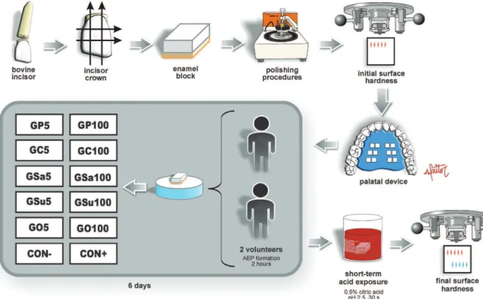
This study evaluated the in vitro potential of distinct
vegetable oils, in different concentrations, to inhibit
enamel erosive demineralization. The experimental
design is shown in Figure 1. Before applying the oils, the AEP was formed in situ on 24 enamel blocks
worn by two volunteers (1 block for each group per
volunteer) for 2 hours. Subsequently, the enamel
blocks were treated in vitro according to the groups (n=12/per group): GP5 – 5% palm oil; GP100 –
pure palm oil; GC5 – 5% coconut oil; GC100 – pure
coconut oil; GSa5 – 5% saflower oil; GSa100 – pure saflower oil; GSu5 – 5% sunlower oil; GSu100 – pure sunlower oil; GO5 – 5% olive oil; GO100 – pure olive oil; Control− – negative control, deionized water;
Control + – positive control, mouthwash commercial
solution containing 125 ppm F− as AmF, 375 ppm
F− as NaF, 800 ppm Sn2+ as SnCl
2; pH 4.5 (Elmex® Erosion Protection Dental Rinse/EP – CP GABA GmbH;
Hamburg, Germany). After application of the oils (5
drops, 30 seconds), the blocks were immersed in 0.5% citric acid (nascent pH 2.4) during 30 seconds
to promote the softening of enamel surface. The
percentage of surface hardness change was assessed
(response variable). The mentioned procedures were repeated for 6 days, in which one sample per group
was ixed in each volunteer intraoral appliance per day.
Sample size
water) per group. Thus, a standard deviation of 8.5%
was obtained. Twelve samples per group were set, considering 12 groups with a minimally detectable
difference of 15% in hardness loss and 8.5% of
standard deviation, with alpha and beta errors of 5%
and 20%, respectively.
Enamel blocks preparation
Enamel blocks (4×4×3 mm3, n=160) were
prepared from the labial surfaces of bovine incisor
crowns. The blocks were cut using a IsoMet® low speed saw cutting machine (Buehler Ltd.; Lake Bluff,
Illinois, United States) with two diamond disks (Extec
Corp.; Enield, Connecticut, United States), which were separated by a 4-mm thickness spacer. The blocks’ surfaces were smoothed with water-cooled silicon
carbide discs (320, 600, and 1200 grade papers;
Buehler Ltd.; Lake Bluff, Illinois, United States), and
wet polished with felt paper and diamond spray (1 µm; Buehler Ltd.; Lake Bluff, Illinois, United States).
The blocks were cleaned using an ultrasonic device
for 2 min and veriied regarding the presence of
white spots and cracks using a microscope (40×). Knoop surface hardness (SHi) was determined by
the mean values of ive indentations performed 100
µm away from each other, with 25 g for 10 seconds
(Micromet® 5114 hardness tester; Buehler Ltd., Lake
Bluff, Illinois, United States). One hundred and forty
four enamel blocks were selected by excluding values 10% higher or lower than the mean microhardness
of all specimens (interblock variability), to avoid bias
regarding initial enamel condition. The blocks were
allocated using Microsoft Excel to distribute blocks with lower and higher initial hardness values equally
into all groups. The randomization was done to divide
the enamel blocks between the groups and the two
volunteers (position of the block in the intraoral device and 6 days of experiment).
Before the in situ phase for AEP, the blocks were
sterilized with ethylene oxide23.
In situ
phase – acquired enamel pellicle
formation
Two healthy adult volunteers with the same
age (22 years old), residing in the same luoridated
area (0.70 mg F/l), participated in the study,
after satisfying the following inclusion criteria: physiological salivary parameters (stimulated >1 ml/
min; unstimulated >0.1 ml/min; neutral pH 7.0-7.5);
absence of erosive tooth wear, untreated carious
lesions, or periodontitis. The exclusion criteria were: presence of systemic diseases; use of medicines that
affect the salivary characteristics (antidepressants,
narcotics, diuretics, or antihistamines); undergoing
radiation or chemotherapy; gastro-esophageal relux;
frequent regurgitation and/or vomiting; pregnancy or breastfeeding; smoking; practicing pool activities
(exposure to pH treated water); working in
low-pH environment (e.g., batteries industry); or luoride
topical application in the past two months. The intraoral palatal appliances were made with acrylic
resin containing six sites (9×6×3 mm) for two blocks
ixation in each (n=12).
The position of the blocks in the intraoral appliance was randomly determined for each volunteer.
Seven days prior to and during the experiment,
the volunteers brushed their teeth with commercial
luoride toothpaste containing 1,450 ppm F (Tripla
Ação® – Colgate-Palmolive Comercial Ltda; São
Paulo, São Paulo, Brazil). The volunteers were
also warned not to use any other luoride product. Toothbrushing with luoride toothpaste was performed
by the volunteers one hour prior to the insertion of
the intraoral appliance. During 6 days, at the same
time (8-10 AM), two volunteers used the intraoral
appliances containing one block for each studied group during 2 hours to allow the formation of the AEP. The
volunteers did not eat or drink in this period.
In vitro
phase – treatment and acid
exposure
Immediately after the formation of the AEP, the enamel blocks were removed from the intraoral
appliance and ixed in an acrylic disk to receive the
treatment. The commercial brands of the vegetable
oils used in this study were: GP5 and GP100: palm oil (Kidendê - Dendê Light Indústria de Produtos
Alimentícios Ltda; Valença, Bahia, Brazil); GC5 and
GC100: extra virgin coconut oil (COPRA - COPRA
Indústria Alimentícia Ltda; Maceió, Alagoas, Brazil);
GSa5 and GSa100: extra virgin saflower oil (Giroil -
Giroil Agroindústria Ltda, Entre-Ijuís, Rio Grande do
Sul, Brazil); GSu5 and GSu100: extra virgin sunlower
oil (Pazze - Pazze Indústria de Alimentos Ltda; Panambi, Rio Grande do Sul, Brazil); GO5 and GO100:
extra virgin olive oil (Cirio - Sandeleh Alimentos;
Paranaguá, Paraná, Brazil).
The 5% emulsions of the vegetable oils in deionized water were daily prepared prior to the application by
using a high-speed household mixer, resulting in a
inely dispersed emulsion27.
The treatment consisted in applying ive drops
on each enamel block of the respective group during
30 seconds. Then, the enamel block was separately
immersed in 17.6 ml of artiicial saliva (0.33 g KH2PO4, 0.34 g Na2HPO4, 1.27 g KCl, 0.16 g NaSCN, 0.58 g
NaCl, 0.17 g CaCl2, 0.16 g NH4Cl, 0.2 g urea, 0.03
g glucose, 0.002 g ascorbic acid, pH 713 – without
mucin) for 2 minutes, under constant agitation, to simulate a natural rinsing process occurring in the oral
cavity. After that, the enamel blocks were subjected
to short-term erosive demineralization by immersing
each block in 17.6 ml of 0.5% citric acid pH 2.4, under constant agitation, for 30 seconds. Then, the blocks
were washed with deionized water for 30 seconds.
Surface hardness assessment
After the short-term acid exposure, the surface hardness determination was performed again (SHf)
with ive indentations performed at 100 µm distance
in relation to initial indentations (Micromet® 5114
hardness tester; Buehler Ltd., Lake Bluff, Illinois, United States). The percentage of hardness loss was
calculated [((SHi - SHf) / (SHi)) × 100] for each block
and averaged to represent the studied groups.
Statistical analysis
Statistical analysis was performed with SigmaPlot™
version 12.3 (Systat Software GmbH; Erkrath,
Germany). Assumptions of equality of variances
and normal distribution of errors were veriied by
Bartlett’s and Shapiro–Wilk tests, respectively. Once
the assumptions were satisied, two-way ANOVA (for
the factors “volunteers” on two levels and “treatments”
on 12 levels) and Tukey’s post hoc test were applied.
The signiicance level was set at 5%.
Results
We found no statistically signiicant difference for
the factor “volunteers” (p=0.911), and no interaction
between “volunteers” and “treatments” (p=0.634), but
we found signiicant difference between “treatments”
(p=0.002). The percentage of hardness loss of the
evaluated groups is shown in Table 1. Only GP100
(pure palm oil) was statistically different from the control group, showing the lowest surface hardness
loss (p<0.05). All the other studied oils presented
surface hardness loss similar to the control groups
Discussion
Lipids consist of about 25% of the pellicle’s dry
weight19, and lipophilic components are able to
modulate the pellicle composition and ultrastructure12. Therefore, authors have suggested that lipid-rich
pellicles might be more resistant to acids23, thus
reducing erosion12.
This study aimed to elucidate the protective effect of different vegetable oils, applied after in situ formation
of AEP, against initial erosion demineralization.
Two of the vegetable oils assessed here (coconut
oil and palm oil) have not been previously studied regarding their anti-erosive potential, requiring an
initial in vitro evaluation of their effect. However, in
vitro studies are not able to accurately replicate the biological characteristics of the oral cavity, such as the presence of human saliva and the formation of
AEP, which could interfere with the development of
erosion. Some limitations occur in protocols using
human saliva in vitro, such as fast extraoral protein decomposition18. Natural and in vitro formed AEPs
also present differences in their characteristics, e.g.,
the natural pellicle is more hydrophobic than the one
formed in vitro24. Therefore, a combined in situ/in
vitro protocol was chosen in this study to allow the physiological formation of the AEP in situ prior to the
in vitro application of the vegetable oils and short-term exposure to citric acid. A single short-term erosive challenge was performed to more precisely evaluate
the protective ability of the AEP modiied by the
studied oils against initial enamel erosion. Studies have
shown that the hardness test is an adequate method
to evaluate the initial softening of enamel surface11,20.
Vegetable oils are extracted from oil plants and
seeds and are commonly used in foods, cosmetics, and medical products12. Studies have shown that,
when hard tooth tissue is exposed to vegetable oils,
the supericial layer of the AEP gets rich in lipids
micelles4,7. However, the protective effect of these oils against caries and erosion demineralization processes
remains unclear, because only a few evidence-based
researches are available in the literature2,8,27.
Only one study evaluated the direct effect of 5 and 50% olive oil emulsions compared to distilled water
and luoride solution on dentin caries demineralization2. Dentin samples were subjected to three cycles per day
of 5 min treatment application followed by 8 hours of immersion in demineralization solution (pH 5.0) during
9 days. The olive oil emulsions showed a decrease in
mineral loss in comparison with deionized water, and
the luoride solution presented better results2. Our results showed that pure palm oil was
capable to protect enamel against initial erosion
demineralization, but the same was not found for
the 5% palm oil emulsion. No protective effect was observed to 5% emulsion and pure form of coconut,
saflower, sunlower, and olive oils. The effect of olive
oil-based emulsions (100%, 2%, and 2% associated
with mouthwash) on enamel subjected to 10 erosive
cycles was previously assessed using proilometry
analysis27. Each cycle consisted of samples treatment
with oil-based emulsions during 5 min, immersion in
artiicial saliva for 30 min, demineralization in 1% citric
Groups SHi SHf % Hardness Loss
GP5 – 5% palm oil 329.92 (±35.81) 253.90 (±45.15) 23.24 (±8.436)a GP100 – pure palm oil 337.58 (±28.41) 310.80 (±34.58) 7.89 (±7.5)b GC5 – 5% coconut oil 334.16 (±26.88) 250.92 (±37.49) 24.65 (±11.50)a GC100 – pure coconut oil 336.24 (±30.50) 240.26 (±48.45) 28.47 (±13.37)a GSa5 – 5% saflower oil 341.19 (±31.87) 241.90 (±37.23) 28.74 (±11.53)a GSa100 – pure saflower oil 337.85 (±29.91) 248.89 (±43.01) 26.56 (±9.51)a
GSu5 – 5% sunlower oil 335.76 (±26.05) 259.23 (±50.07) 22.92 (±12.94)a GSu100 – pure sunlower oil 338.51 (±27.63) 265.02 (±55.67) 21.78 (±14.83)ab GO5 – 5% olive oil 337.27(±32.29) 252.81 (±57.71) 25.35 (±12.76)a GO100 – pure olive oil 337.36 (±29.49) 249.86 (±46.95) 25.91(±12.51)a CON− – deionized water
(negative control)
335.45 (±29.71) 240.90 (±38.02) 28.09 (±9.95)a
CON+ – luoride mouthrinse (positive control)
337.19 (±28.92) 256.44 (±22.04) 23.74 (±6.15)a
In the fourth column, different letters show signiicant differences between the groups (two-way ANOVA and Tukey’s test, p<0.05)
acid for 3 min, and remineralization in artiicial saliva
for 60 min. The researchers found that 2% emulsion or 2% olive-oil containing mouthrinse offered protection
against tooth erosion, but in a lesser extent than the
positive control (250 ppm acidic luoride solution),
and that pure olive oil did not offer protection27. The authors hypothesized that the adhesion properties of
olive oil might increase when applied as emulsion27. In
contrast, our study did not ind any protective potential
for 5% and pure olive oil. The different results between the studies might be explained by the different
methodologies used. We adopted a short-term erosive
demineralization model and the abovementioned study
used an erosive cycling model.
The effect of safflower oil on the protective
properties of the AEP formed in situ against the
exposure to hydrochloric acid for 2 min was previously
described8. Enamel mineral loss was determined by measurement of calcium and phosphate release,
and the ultrastructure of the AEP was evaluated by
transmission electron microscopy. The results showed
that the surface of AEP was rich in lipids, but no substantial lipids integration was found in the pellicle’s
basal layer. The in situ AEP modiied by saflower oil
rinsing was more susceptible to acid degradation than
the in situ physiological AEP8. In contrast, our study
showed that saflower oil (GSa5 and GSa100) did not
present a negative impact on enamel demineralization
when compared to the control groups.
To our knowledge, palm oil has never been investigated for the prevention of erosion. Palm
oil is the second largest produced and consumed
vegetable oil in the world, due to its high productivity,
low production cost, and rich nutritional content21. It is rich in tocotrienols that have presented health
beneits1. Tocotrienols allow an eficient penetration into tissues that have saturated fatty layers and exhibit
antioxidant protection of cellular membranes against oxidative damage1. The antioxidant property has been
attributed to its ability of distribution in lipid layers of
the cell membrane1.
In previous studies, the outer layer of the AEP
was modiied by the increase of lipids micelles4,7. However, the outer layers of the AEP are supposed
to be easily removed after an erosive challenge, in
contrast to the basal layer that might not be affected6. In this study, despite the ultrastructure of the AEP not
being analyzed, it is hypothesized that palm oil might
have modiied the basal layer of the acquired pellicle,
increasing its protective potential. The tocotrienols
contained in the palm oil might have allowed its penetration and distribution into the basal layers of
the acquired pellicle, increasing its protective role1. We
also highlight that we found no differences between the
protective effect of pure palm oil and pure sunlower
oil. This result can be explained by the tocotrienols
content of the sunlower oil, but in a lesser extent when
compared to palm oil1, which enables a borderline
behavior between palm oil and the other tested oils
(coconut, saflower, and olive oil).
In this study, the commercial mouthwash solution
– Elmex® Erosion Protection Dental Rinse/EP, 125 ppm
F− as AmF, 375 ppm F− as NaF, 800 ppm Sn2+ as SnCl 2; pH 4.5 (CP GABA GmbH; Hamburg, Germany) – did
not present any effect on the inhibition of initial enamel
erosion, being similar to deionized water (negative
control). The role of luoride on tooth erosion is not
fully evidenced14. Differing from our result, some
studies have shown a preventive capacity of luoride
solution containing AmF/NaF (500 ppm F) and SnCl2
(800 ppm Sn) against enamel erosion5,15.
Although palm oil has shown superior protective
capacity against tooth erosion, its effect to prevent
the enamel erosive wear needs to be further evaluated
under long-term erosive challenges. Moreover, the effect of palm oil on the physical properties, quality,
and composition of the acquired pellicle should also
be assessed.
Conclusion
Considering our study design, palm oil seems to
be a promising alternative for the prevention of initial enamel erosion.
Acknowledgements
The authors would like to gratefully acknowledge
the volunteers who participated in this study and FAPESP – São Paulo Research Foundation for the
inancial support (process numbers 2014/00102-0,
References
1- Ahsan H, Ahad A, Siddiqui WA. A review of characterization of tocotrienols from plant oils and foods. J Chem Biol. 2015;8(2):45-59.
2- Buchalla W, Attin T, Roth P, Hellwig E. Inluence of olive oil emulsions
on dentin demineralization in vitro. Caries Res. 2003;37(2):100-7.
3- Buzalaf MA, Hannas AR, Kato MT. Saliva and dental erosion. J Appl Oral Sci. 2012;20(5):493-502.
4- Das SK, Adhikary PK, Bhattacharyya DK. Effects of dietary fats on the fatty acid composition of enamel and dentinal lipids of rabbit
molars. J Dent Res. 1976;55(6):1061-6.
5- Esteves-Oliveira M, Witulski N, Hilgers RD, Apel C, Meyer-Lueckel
H, de Paula Eduardo C. Combined tin-containing luoride solution and
CO2 laser treatment reduces enamel erosion in vitro. Caries Res.
2015;49(6):565-74.
6- Hannig C, Hannig M. The oral cavity – a key system to understand
substratum- dependent bioadhesion on solid surfaces in man. Clin Oral Investig. 2009;13(2):123-39.
7- Hannig C, Kirsch J, Al-Ahmad A, Kensche A, Hannig M, Kümmerer
K. Do edible oils reduce bacterial colonization of enamel in situ. Clin
Oral Investig. 2013;17:649-58.
8- Hannig C, Wagenschwanz C, Pötschke S, Kümmerer K, Kensche A, Hoth-Hannig W, et al. Effect of saflower oil on the protective properties
of the in situ formed salivary pellicle. Caries Res. 2012;46:496-506.
9- Hannig M, Hannig C. The pellicle and erosion. Monogr Oral Sci. 2014;25:206-14.
10- Hannig M, Hess NJ, Hoth-Hannig W, De Vrese M. Inluence of
salivary pellicle formation time on enamel demineralization - an in situ
pilot study. Clin Oral Investig. 2003;7:158-61.
11- Hara AT, Zero DT. Analysis of the erosive potential of
calcium-containing acidic beverages. Eur J Oral Sci. 2008;116:60-5.
12- Kensche A, Reich M, Kümmerer K, Hannig M, Hannig C. Lipids in
preventive dentistry. Clin Oral Investig. 2013;17(3):669-85.
13- Klimek J, Hellwig E, Ahrens G. Effect of plaque on luoride stability in the enamel after amine luoride application in the artiicial mouth.
Dtsch Zahnarztl Z. 1982;37(10):836-40.
14- Magalhães AC, Wiegand A, Rios D, Honório HM, Buzalaf MA. Insights into preventive measures for dental erosion. J Appl Oral Sci.
2009;17(2):75-86.
15- Oliveira TA, Scaramucci T, Nogueira FN, Simões A, Sobral MA. Effect of mouthrinses with different active agents in the prevention of initial
dental erosion. Indian J Dent Res. 2015;26(5):508-13.
16- Papagianni CE, van der Meulen MJ, Naeije M, Lobbezoo F. Oral
health-related quality of life in patients with tooth wear. J Oral Rehabil. 2013;40:185-90.
17- Salas MM, Nascimento GG, Huysmans MC, Demarco FF. Estimated prevalence of erosive tooth wear in permanent teeth of children and
adolescents: an epidemiological systematic review and meta-regression analysis. J Dent. 2015;43:42-50.
18- Schipper RG, Silletti E, Vingerhoeds MH. Saliva as research material: biochemical, physicochemical and practical aspects. Arch
Oral Biol. 2007;52(12):1114-35.
19- Slomiany BL, Murty VL, Zdebska E, Slomiany A, Gwozdzinski K,
Mandel ID. Tooth surface-pellicle lipids and their role in the protection of dental enamel against lactic-acid diffusion in man. Arch Oral Biol.
1986;31(3):187-91.
20- Stenhagen KR, Hove LH, Holme B, Taxt-Lamolle S, Tveit AB.
Comparing different methods to assess erosive lesion depths and progression in vitro. Caries Res. 2010;44(6):555-61.
21- Sundram K, Sambanthamurthi R, Tan YA. Palm fruit chemistry and nutrition. Asia Pac J Clin Nutr. 2003;12:355-62.
22- Ten Cate JM, Imfeld T. Dental erosions summary. Eur J Oral Sci. 1996;104(2 Pt 2):241-4.
23- Toro MJ, Lukantsova LL, Williamson M, Hinesley R, Eckert GJ, Dunipace AJ. In vitro luoride dose-response study of sterilized enamel
lesions. Caries Res. 2000;34(3):246-53.
24- Van der Mei HC, White DJ, Kamminga-Rasker HJ, Knight J, Baig
AA, Smit J, et al. Inluence of dentifrices and dietary components in
saliva on wettability of pellicle-coated enamel in vitro and in vivo. Eur
J Oral Sci. 2002;110(6):434-8.
25- Vukosavljevic D, Custodio W, Buzalaf MA, Hara AT, Siqueira WL.
Acquired pellicle as a modulator for dental erosion. Arch Oral Biol. 2014;59(6):631-8.
26- Wetton S, Hughes J, West N, Addy M. Exposure time of enamel and dentine to saliva for protection against erosion: a study in vitro.
Caries Res. 2006;40:213-7.
27- Wiegand A, Gutsche M, Attin T. Effect of olive oil and an