Effect of adhesive remnant removal on enamel
topography after bracket debonding
Larissa Adrian Meira Cardoso1, Heloísa Cristina Valdrighi2, Mario Vedovello Filho3, Américo Bortolazzo Correr4
How to cite this article: Cardoso LAM, Valdrighi HC, Vedovello Filho M,
Correr AB. Effect of adhesive remnant removal on enamel topography after brack-et debonding. Dental Press J Orthod. 2014 Nov-Dec;19(6):105-12. DOI: http:// dx.doi.org/10.1590/2176-9451.19.6.105-112.oar
» The authors report no commercial, proprietary or financial interest in the prod-ucts or companies described in this article.
Contact address: Larissa Adrian Meira Cardoso
Av. Dr. Maximiliano Baruto, 500 - Jd. Universitário / Araras — Brazil CEP: 13607-339 – E-mail: [email protected] / [email protected] » Patients displayed in this article previously approved the use of their facial and
intraoral photographs.
1 DDS, Hermínio Ometto Foundation, UNIARARAS.
2 Professor, Graduate Dental Program, Hermínio Ometto Foundation,
UNIARARAS.
3 Professor, Masters Program in Dentistry, São Leopoldo Mandic. 4 Professor, Masters Program in Dentistry and at the Department of Dental
Material, Hermínio Ometto Foundation, UNIARARAS.
Submitted: December 10, 2013 - Revised and accepted: May 26, 2014
Introduction: At orthodontic treatment completion, knowledge about the effects of adhesive remnant removal on enamel is paramount. Objective: This study aimed at assessing the effect of different adhesive remnant removal methods on enamel topography (ESI) and surface roughness (Ra) after bracket debonding and polishing. Methods: A total of 50 human premolars were selected and divided into five groups according to the method used for adhesive remnant removal: high speed tungsten carbide bur (TCB), Sof-Lex discs (SL), adhesive removing plier (PL), ultrasound (US) and Fiberglass burs (FB). Metal brackets were bonded with Transbond XT, stored at 37oC for 24 hours before debonding with adhesive
removing plier. Subsequently, removal methods were carried out followed by polishing with pumice paste. Qualitative and quantitative analyses were conducted with pre-bonding, post-debonding and post-polishing analyses. Results were submitted to statistical analysis with F test (ANOVA) and Tukey’s (Ra) as well as with Kruskal-Wallis and Bonferroni tests (ESI) (P < 0.05). Results: US Ra and ESI were significantly greater than TCB, SL, PL and FB. Polishing minimized Ra and ESI in the SL and FB groups. Conclusion: Adhesive remnant removal with SL and FB associated with polishing are recommended due to causing little damage to the enamel.
Keywords: Orthodontic brackets. Dental enamel. Device removal.
DOI: http://dx.doi.org/10.1590/2176-9451.19.6.105-112.oar
Introdução: na finalização do tratamento ortodôntico, torna-se relevante o conhecimento da ação dos métodos de remoção do remanescente resinoso sobre o esmalte. Objetivo: o objetivo do estudo foi avaliar o efeito de métodos de remoção do remanescente de resina após a descolagem do braquete e do polimento na rugosidade (Ra) e topografia (ESI) do esmalte. Métodos: foram selecionados 50 pré-molares humanos, divididos em cinco grupos, de acordo com o método empregado para a remoção da resina residual: broca carbide tungstênio em alta rotação (BCT), discos Sof-Lex (SL), alicate removedor de resina (AL), ultrassom (US) e pontas Fiberglass (PF). Braquetes metálicos foram colados com Transbond XT, armazenados a 37° por 24 horas antes da descolagem com alicate removedor de braquete, sendo aplicados posteriormente os meios de remoção e executado o polimento com pasta de pedra-pomes. Realizou-se análises quali-quantitativas, com avaliações antes da colagem dos braquetes, após a descolagem e após o polimento, sendo os valores ob-tidos submeob-tidos à análise estatística com teste F (ANOVA), de Tukey (Ra) e testes de Kruskal-Wallis e Bonferroni (ESI) (p < 0,05). Resultados: a Ra e o ESI do US foram significativamente maiores do que BCT, SL, AL e PF. O polimento reduziu a Ra e ESI dos grupos SL e PF. Conclusão: a remoção do adesivo resinoso com SL e PF associados ao polimento são os métodos mais indicados por ocasionarem as menores alterações do esmalte.
INTRODUCTION
Orthodontic accessories were, for many years,
fea-tured by band welding systems1. In 1955, Buonocore
enabled orthodontic therapy to be conducted with
restorative material bonded over enamel surface.2
Later on, Newman allowed metallic material to be
bonded over enamel surface,1,3 thereby ofering many
beneits provided by direct bonding: improved esthet-ics and performance, better hygiene, low costs, caries
risk reduction, and accurate bracket positioning.1,4,5,6
The bonding process is no longer an issue. The greatest challenges are with regard to accurate
remov-al of adhesive remnant,3,4,7-10 so as to avoid not only
ir-reversible iatrogenic injuries, such as rough surfaces, vertical cracks, pulp necrosis, loss of the external sur-face rich in fluorine (20 μm), but also the presence of
adhesive remnant at the adhesion area.2,4,11,12,13 These
injuries can be caused by inappropriate removal tech-niques, prophylaxis with abrasives, bonding material, acid conditioning and color similarity between
re-sidual adhesive and enamel.11,12,14,15
The literature presents a great variety of mechani-cal removal methods, namely: adhesive removing
plier,1,3,5,8,12,16 high and low speed tungsten carbide
bur,1,3,5,6,8,9,10,14-19 laser application,15,16 Shofu bur,1,2,3,8,12,16
Sof-Lex® discs,5,6,15,16,18,20 iberglass burs12,19 and
ultra-sound.5,10,14,17 For polishing, rubber cup with pumice
and water5,6,7,9,18 as well as diamond paste5 are used.
Nevertheless, no consensus has yet been reached in the literature regarding the most eicient and safe
technique to this end.3 Considering that the
aforemen-tioned tools are largely used by orthodontists, scien-tiic knowledge about these techniques as well as their biological cost to tooth structure is essential. As a re-sult, there is a great need for choosing efective removal techniques in order to cause the least damages to the patient at the end of treatment and, whenever possible,
preserve the tooth original condition.1,2,3,6,7,9,11,12,13,16-19
This study aimed to conduct an in vitro assess-ment so as to investigate the effect of adhesive rem-nant removal after bracket debonding and polishing on roughness (Ra) and enamel topography (ESI).
MATERIAL AND METHODS
A total of 50 premolars obtained from a teeth bank were submitted to local Institutional Review Board. The study was approved under protocol #162.677.
The teeth were standardized with minimal pre-vious lesions, based on exclusion criteria (fracture, caries, restorations or coronal cracks, orthodon-tic and endodonorthodon-tic treatment). Subsequently, they were washed in tap water and cleaned with
peri-odontal curettes,2,8 submerged in distilled water
(ISO 3696:1987)21 and stored at -8°C.3 The
immer-sion means was weekly changed in order to prevent dehydration and bacterial growth, and to improve
adhesive strength.3,21
The sample was divided into ive groups (n = 10) according to the adhesive remnant removal method used.
Specimens were manufactured with the roots cut in the cement-enamel junction using a double-sided diamond disc (KG-Sorensen, Rio de Janeiro, RJ, Brazil). The crowns were embedded in PVC and fixed with polystyrene adhesive (Resina Cris-tal, Piraglass, Piracicaba, SP, Brazil) with the buc-cal surface exposed. That was where the bracket bonding area was delimited with #0 brush (Mar-ta-Tigrei bristle) and nail polish (color: red - Col-orama). This procedure allowed both rugosimetric analysis and digital photographs to be taken in the enamel region.
Roughness (Ra) quantitative analysis was carried out by means of a rugosimeter (Surfcorder, mod. 1700, Japan), with horizontal readings towards the center of the delimited surface (distance = 2.5 mm; speed = 0.1 mm; s-Cut Off = 0.25 mm). Qualita-tive analysis was conducted with digital photographs (Sony Cyber-shot Digital Camera /12.1 Mp/zoom 1.5 x) under magnification of 40 x and 100 x obtained by stereomicroscopic imaging (Carl Zeiss-Citoval, mod. 2, Germany).
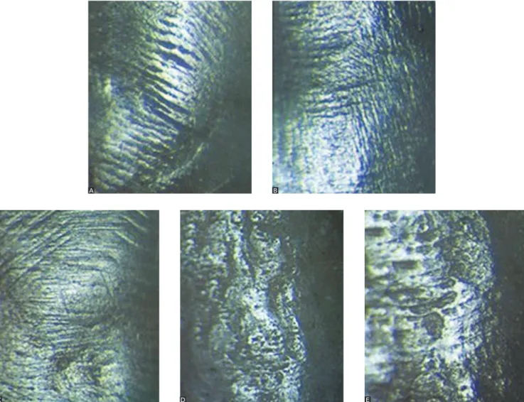
Data were transferred to a computer (JPG format with 12-MB resolution), and ESI classified
accord-ing to Zachrisson and Arthun’s17 criteria (Fig 1):
» Score 0: perfect surface (with no scratches, intact enamel).
» Score 1: regular surface (minor scratches and some healthy enamel).
» Score 2: acceptable surface (many deep scratches, absent healthy enamel).
» Score 3: defective surface (many large, deep scratches, absent healthy enamel).
Figure 1 - Digital photographs (100 x) for ESI classification: A) Score 0; B) Score 1; C) Score 2; D) Score 3; and E) Score 4.
Before bracket bonding, prophylaxis was carried out with pumice paste (Extra-fine/SS White, Rio de Janeiro, RJ, Brazil) and rubber cup (Microdont, São Paulo, SP, Brazil) during 10 seconds. Conditioning was carried out with 37% phosphoric acid (FGM, Joinville, SC, Brazil) for 30 seconds, washed for oth-er 30 seconds. Transbond XT Light Cure Adhesive Primer (3M/ Unitek, Monrovia, CA, USA) was ap-plied, dried with compressed air during 10 seconds and light-cured by halogen lamp (Light Unit,
De-gussa, USA) with irradiance of 500 mW/cm2.
Sub-sequently, composite resin Transbond XT Light Cure Adhesive Primer (3M Unitek) was applied to metal brackets base (Edgewise, slot 022-in/Morelli, Sorocaba, SP, Brazil) which was compressed against the enamel surface. This allowed excess removal by light-curing processes with halogen lamp during 10 seconds on each side of the bracket.
The sample was stored in distilled water in the sterilizer (FANEM, São Paulo, SP, Brazil) at 37°C during 24 hours before bracket removal with orth-odontic plier (346R/ICE, Cajamar, SP, Brazil), in which case the winglets were perpendicularly pressed against the slot axis.
Enamel surface was analyzed by stereomicroscopic analysis (Carl Zeiss-Citoval, mod. 2, Germany) under 40 x and 100 x magnification. Cases in which adhesive was not adhered to enamel were discarded.
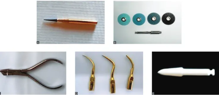
Adhesive remnant removal methods were (Fig 2): » TCB group: High speed tungsten carbide drill with 30 blades (#9214 FF/JET) during 30 seconds.
» SL group: Low speed large, middle, fine and su-per fine Sof-Lex discs (3M ESPE) for 40 seconds. » PL group: Adhesive removing plier (#193/ICE) in 10 seconds.
A
C
B
Figure 2 - Adhesive remnant removal methods: A) High speed tungsten carbide drill (TCB); B) Sof-Lex discs (SL); C) Adhesive removing plier (PL);
D) Ultrasound (US); and E) Fiberglass burs (FB).
» US group: Ultrasound on large, middle and fine tips (#02, 01 and 10-P/Gnatus) during 90 seconds. » FB group: Fiberglass burs in low speed with wa-ter for 20 seconds (#02/TDV).
Ater remnant adhesive removal, new assessments of enamel surface roughness (Ra) as well as new photo-graphs (ESI) were obtained. The presence of adhesive remnant was visually inspected under dental relector light (Dabi Atlante, Ribeirão Preto, SP, Brazil). Polish-ing was carried out with pumice paste (SS White, Rio de Janeiro, RJ, Brazil) and rubber cup (Microdont, São Paulo,/SP, Brazil) during 10 seconds. Ater polishing, new assessments of enamel surface roughness (Ra) as well as new photographs (ESI) were obtained.
Qualitative and quantitative analyses were performed before bracket bonding (initial Ra and ESI), ater debond-ing (adhesive removal Ra and ESI), and ater polishdebond-ing (Ra and ESI polishing). Data were statistically assessed. Roughness (Ra) values were submitted to analysis of variance (ANOVA F-test) and Tukey’s test, whereas ESI (scores) values were submitted to Kruskal-Wallis and Bonferroni test. Signiicance level was set at 5%.
RESULTS
Analysis of variance showed that enamel’s Ra was significantly influenced by the remnant adhesive re-moval method used (P < 0.001).
Initial Ra and ESI were significantly similar in all groups (Tables 1 and 2). Regular topography pre-vailed (Score 1) (Fig 3).
US Ra and ESI were greater than the other meth-ods (TCB, SL, PL and FB) with no difference among methods (P < 0.05). Initial Ra was greater than that showed after adhesive removal and after polishing in TCB, SL and FB groups (P < 0.05). Final PL Ra was significantly greater after polishing in comparison to initial Ra. After adhesive removal, Ra was signifi-cant greater in comparison to initial Ra (P < 0.05) for the US group. After adhesive removal, TCB and US methods caused more damages to dental enamel; thus, acceptable surfaces prevailed (Score 2).
Polishing was not significant in repairing Ra caused by removal methods (P < 0.05). However, in SL, FB and TCB groups, polishing reduced Ra when compared to initial Ra (P < 0.05), in addition
A
C
B
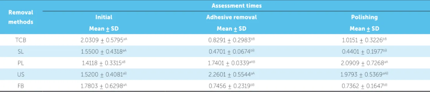
Table 1 - Roughness means and standard deviation for different adhesive removal methods at different assessment times.
Table 2 - Kruskal-Wallis analysis.
Removal
methods
Assessment times
Initial Adhesive removal Polishing
Mean ± SD Mean ± SD Mean ± SD
TCB 2.0309 ± 0.5795aA 0.8291 ± 0.2983bB 1.0151 ± 0.3226bB
SL 1.5500 ± 0.4318aA 0.4701 ± 0.0674bB 0.4401 ± 0.1977bB
PL 1.4118 ± 0.3315aB 1.7401 ± 0.0339aAB 2.0909 ± 0.7268aA
US 1.5200 ± 0.4081aB 2.2601 ± 0.5544aA 1.9793 ± 0.5369aAB
FB 1.7803 ± 0.6298aA 0.7456 ± 0.2319bB 0.7362 ± 0.1647bB
Different lower case letters in columns and capital letters in line are meaningfully different by Tukey’s test (P < 0.05). TCB: High speed tungsten carbide drill; SL: Sof-Lex discs; PL: Adhesive removing plier; US: Ultrasound; and FB: Fiberglass burs.
TCB: High speed tungsten carbide drill; SL: Sof-Lex discs; PL: Adhesive removing plier; US: Ultrasound; and FB: Fiberglass burs.
Groups Median 25% 75% Mean Statistics
TCB + initial 1 1 1 1.1 d
TCB + removal 2 2 3 2.3 abc
TCB + polishing 3 2 3 2.5 ab
SL + initial 1 1 1 1.2 d
SL + removal 1.5 1 2 1.5 bcd
SL + polishing 1 1 2 1.3 cd
PL + initial 1 1 1 0.9 d
PL + removal 1 1 2 1.6 bcd
PL + polishing 1 1 2 1.3 cd
US + initial 1 1 1 1.1 d
US + removal 3 3 4 3.2 a
US + polishing 3 2 3 2.8 a
FB + initial 1 1 1 1.1 d
FB + removal 2 1 2 1.6 bcd
FB + polishing 1 1 1 1 d
Figure 3 - Distribution of ESI frequency. Score 0 = Perfect, 1 = regular; 2 = acceptable; 3 = defective; 4 = unacceptable. TCB: High speed tungsten carbide drill; SL: Sof-Lex discs; PL: Adhesive removing plier; US: Ultrasound; and FB: Fiberglass burs.
Score 0 100
90 80 70 60 50 40 30
fr
equency (%)
20 10 0
Score 1 Score 2 Score Score 4
TC B +
initial
TC B +
removal
TC B +
polishingSL + initial
SL + removal
SL + polishing
PL + polishing
US + polishing
FB + polishing PL +
initial
US + initial FB + initial
PL + removal
US + removal
to recovering SL, FB and PL initial quality (ESI), al-though for TCB and US the polish effect was useless in restoring initial enamel features. Regular surfaces prevailed (Score 1).
DISCUSSION
Direct bracket bonding on enamel surface
contrib-uted to simplify bonding and debonding protocols.1,2
However, ater inishing orthodontic treatment, the
aim is to restore initial topographic quality,8,18 since
ir-reversible iatrogenic lesions might be caused (5.4%)6,13
due to a number of diferent factors, as reported in the
literature.11,12 Adhesive remnant and damages caused
to the enamel structure3,19 are unavoidable regardless of
the type of bracket and the removal technique. Adhesive remnant removal with rotating instru-ments causes enamel erosion at both high (19.2 μm)
and low (11.3 μm) speed.18 Nevertheless, the latter
causes more damage to pulp vitality6,8 due to heat
produced by the lack of air/water refrigeration. On the contrary, when manual instruments are used, prudence regards to force application is
recommend-ed in order to avoid enamel loss.11
Ideal removal material hardness has to be great-er than the adhesive remnant and lowgreat-er than the enamel structure.
The results yielded by the present research were based on roughness and enamel topographic quality parameters and allowed comparison between dif-ferent methods and potential individual variables; thereby proving unfeasible to determine the best or worst removal method.
Adhesive remnant removal was unfavorable in US groups due to presenting a significant increase in Ra. Conversely, AL groups presented a non-significant increase in Ra when removal methods were used.
US was considered harmful due to presenting defective and unacceptable surfaces with large and deep scratches. These findings are similar to those
by Hosein, Sheirriff and Ireland10 as well as Ireland,
Hosein and Sheirriff,14 and are due to difficulty in
re-moving adhesive remnants in consequence of the ac-tive insert tip not covering the entire work area. This requires a higher number of applications and more treatment time for complete reduction. Hardness of the tool used is also responsible for the aforemen-tioned results, as it is higher than that of the adhesive
remnant and the enamel, thereby causing enamel prism to break with vibrations.
Although PL produced large, deep vertical stretch-es and did not completely remove adhstretch-esive remnants, the conditions initially observed (regular topography)
remained, thereby corroborating Pignatta et al.7 This
technique had good performance due to being less in-vasive and providing more comfort to the patient as a result of relatively absence of vibrations when
com-pared to TCB4 and US activation. In addition, it
pro-duced little enamel roughness and demanded mini-mal time for reducing adhesive remnant, as stated by
Hosein, Sheirrif and Ireland,10 Albuquerque et al8 and
Tavares,3 and in disagreement with Miksic, Slaj and
Mestrovic4 and Rouleau Jr, Marshall Jr and Cooley9
who describe this method as the worst choice.
Those visible injuries were caused due to enamel surface convexity while the plier was being support-ed by the occlusal surface so as to allow the flat
ac-tive tip to remove residual adhesive by compression.8
This procedure also caused enamel thickness wear (μm) possibly detected by the rugosimeter, a manual tool that when subjected to excessive tension can lead to remarkable enamel delamination in
compari-son to other methods.11
The most favorable methods were TCB, SL and FB, as they had Ra reduction (P < 0.05) in compari-son to initial Ra. The least Ra was found in the SL
method, which is in accordance with Eliades et al.20
In comparison to SL and FB, the TCB method showed higher surface variance for marks as well as deep and large scratches (acceptable and defective sur-faces). These damages were assigned to higher TCB hardness in comparison to adhesive remnant and
enamel, which caused subjacent enamel loss10,14,17 after
residual adhesive removal when TCB was operated at high speed. This method, however, causes regular thickness wear (μm), which renders its identifica-tion unfeasible by the rugosimeter. Nevertheless, the
TCB method had minimal level of Ra,16 and proved
to be an effective method8,17,18 that causes less damage
with faster performance.1,6 Conversely, Karan,
Kir-celli and Tasdelen19 as well as Tavares3 assert that this
method, when compared to fiberglass burs as well as other methods, presents increased Ra.
of marks) was observed in SL and FB. However, SL preserved the initial features of experimental unities in 50% of cases (regular surface). These are considered desirable efects when compared to some
research-es5,6,15,18 that attribute decrease in surface variations to
ine and ultra-ine discs which reduce scratches caused by greater granulation discs (G-M). Nevertheless, it is a complex method for a practical procedure, as its four discs require extensive performance time once it passes
through bonding2,15 areas in sequence.
In accordance with Karan, Kircelli and Tasdelen,19
FB is recommended for adhesive remnant removal due to requiring less treatment time as a result of its ability of clearly differentiating adhesive from enamel, quickly wearing it without causing any le-sions when in contact with the surface. This is due to the glass fibers which are broken into fragments dur-ing abrasive movement and expel the adhesive part by part through grinding. After the procedure, the glass fiber segment is available again, and is improved within the same period.
On the other hand, Rastelli12 asserts that no
dam-age (scratches, cracks or wear) were found on enamel
surface. Moreover, Karan, Kircelli and Tasdelen19
re-port that removal by means of this method is delayed more than with TCB.
Pumice paste polishing is beneficial, fast and pleasant for the patient, as it straightens the majority of rough areas, giving special shine and decreasing
abrasive marks.5,6,7,9,18
For SL, FB and TCB, polishing reduced Ra sig-nificantly when compared to initial Ra. In addition, it restored SL and FB initial quality (regular surface). It is considered particularly advantageous for elimi-nating surface changes without injuring pulp tissues and causing minimal enamel loss in conformance
with Zarrinnia, Eid, and Kehoe18 as well as
Camp-bell.6 In the TCB group, deep, large scratches
re-mained due to pumice abrasiveness, thereby making restoration of early features (regular topography) un-feasible and featuring defective surfaces.
PL group resulted in significant increase of Ra in comparison to natural tooth. Although early enamel quality was restored (regular topography) and large
scratches were eliminated, as in agreement with Ryf
et al,11 deep scratches remained, similarly to Rouleau
Jr, Marshall Jr and Cooley9 and Pignatta et al.7 Enamel
layer remained with irregular thickness (μm), which was highlighted by the rugosimeter as Ra increase.
US group had Ra significantly increased after polishing when compared to initial Ra. Moreover, although it decreased defective and unacceptable surfaces, it did not restore enamel initial features (regular surfaces).
According to some researchers,11 no important
variances in Ra changes were found after polishing when comparing the interaction among TCB, SL and FB groups and between PL and US groups.
When comparing different removal methods, we found that polishing does not significantly increases or reduces Ra. For this reason, the polishing
proce-dure is optional, as stated by Zachrisson and Arthun.17
This fact can be explained by the speciic system of the polishing tool with its rough and porous fragments producing low abrasive power. Similarly, diferences in
TCB blades also afect the dental structure.11,17
Thus, orthodontists should attempt to choose a suitable protocol based on scientific evidence for ad-hesive remnant removal and initial tooth features res-toration so as to avoid undesirable results, reach pro-fessional and patient’s goals and ensure satisfactory,
conservative, successful treatment outcomes.2,5,13
CONCLUSIONS
Based on the results of this study it is reasonable to conclude that
1. All adhesive remnant removal methods changed enamel topography and roughness.
2. The US method is unsuitable to remove com-posite resin.
3. The methods of choice, in decreasing order, are: SL, FB, TCB and PL.
4. Pumice paste polishing was insignificant in restoring enamel initial conditions. Therefore, it is optional.
1. Mahdavie NN. The Efect of various debonding burs on the enamel surfaces of teeth after debonding metal brackets [tese]. Chicago: University of Illinois; 2012. 2. Santos Jr JH. Avaliação do esmalte dentário antes e após colagem e
descolagem de bráquetes ortodônticos [tese]. São Paulo (SP): Universidade de São Paulo; 2009.
3. Tavares SW. Análise in vitro de diferentes métodos da remoção da resina residual no esmalte dentário [tese]. Piracicaba (SP): Universidade Estadual de Campinas; 2006.
4. Miksic M, Slaj M, Mestrovic S. Qualitative analysis of the enamel surface after removal of remnant composite. Acta Stomatol Croat. 2003;37(3):247-51. 5. Macieski K, Rocha R, Locks A, Ribeiro GU. Efects evaluation of remaining resin
removal (three modes) on enamel surface after bracket debonding. Dental Press J Orthod. 2011;16(5):146-54.
6. Campbell PM. Enamel surfaces after orthodontic bracket debonding. Angle Orthod. 1995;65(2):103-10.
7. Pignatta LMB, Duarte Jr S, Santos ECA. Evaluation of enamel surface after bracket debonding and polishing. Dental Press J Orthod. 2012;17(4):77-84. 8. Albuquerque GS, Vedovello Filho M, Lucato AS, Boeck EM, Degan V, Kuramae M.
Evaluation of enamel roughness after ceramic bracket debonding and clean-up with diferent methods. Braz J Oral Sci. 2010;9(2):81-4.
9. Rouleau Jr BD, Marshall Jr GW, Cooley RO. Enamel surface evaluations after clinical treatment and removal of orthodontic brackets. Am J Orthod. 1982;81(5):423-6.
10. Hosein I, Sheirrif M, Ireland AJ. Enamel loss during bonding, debonding, and cleanup with use of a self-etching primer. Am J Orthod Dentofacial Orthop. 2004;126(6):717-24.
11. Ryf S, Flury S, Palaniappan S, Lussi A, Meerbeek BV, Zimmerli B. Enamel loss and adhesive remnants following bracket removal and various clean-up procedures in vitro. Eur J of Orthod. 2012;34(1):25-32.
REFERENCES
12. Rastelli MCS. Alterações morfológicas e da microdureza do esmalte dentário humano após utilização de pontas Fiberglass [tese]. Florianópolis (SP): Universidade Federal de Santa Catarina; 2008.
13. Zanarini M, Gracco A, Lattuca M, Marchionni S, Gatto MR, Bonetti A. Bracket base remnants after orthodontic debonding. Angle Orthod. 2013;83(5):885-91. 14. Ireland AJ, Hosein I, Sheirrif M. Enamel loss at bond-up, debonding and clean-up
following the use of a convencional light-cured composite and a resin-modiied glass polyalkenoate cement. Eur J Orthod. 2005;27(4):413-9.
15. Eminkahyagil N, Arman A, Cetinsahin A, Karabulut E. Efect of resin-removal methods on enamel and shear bond strength of rebonded brackets. Angle Orthod. 2006;76(2):314-21.
16. Ahari F, Akbari M, Akbari, Dabiri G. Enamel surface roughness after debonding of orthodontic brackets and various clean-up techniques. J Dent. 2013;10(1):82-93. 17. Zachrisson BU, Arthun J. Enamel surface appearance after various debonding
techniques. Am J Orthod. 1979;75(2):121-37.
18. Zarrinnia K, Eid NM, Kehoe MJ. The efect of diferent debonding techniques on the enamel surface: an in vitro qualitative study. Am J Orthod Dentofacial Orthop. 1995;108(3):284-93.
19. Karan S, Kircelli BH, Tasdelen B. Enamel surface roughness after debonding: comparison of two diferent burs. Angle Orthod. 2010;80(6):1081-8. 20. Eliades T, Gioka C, Eliades G, Makou M. Enamel surface roughness following
debonding using two resin grinding methods. Eur J Orthod. 2004;26(3):333-8. 21. Martins GC. Efeito do armazenamento em água na resistência de união de