ISSN 0100-879X
BIOMEDICAL SCIENCES
AND
CLINICAL INVESTIGATION
www.bjournal.com.br
www.bjournal.com.br
Volume 43 (11) 1010-1134 November 2010
Institutional Sponsors
The Brazilian Journal of Medical and Biological Research is partially financed by
Hotsite of proteomics metabolomics developped by:
Braz J Med Biol Res, November 2010, Volume 43(11) 1034-1041
Effect of hyperoxia on the intestinal IgA secretory component in
neonatal rats and on intestinal epithelial cells
in vitro
D.Y. Liu and J.J. Li
Effect of hyperoxia on the intestinal IgA
secretory component in neonatal rats
and on intestinal epithelial cells
in vitro
D.Y. Liu
1and J.J. Li
21Research Centre, 2Pediatry Department, China Medical University Affiliated Shengjing Hospital,
Shenyang City, Liaoning Province, China
Abstract
Oxygen therapy is essential for the treatment of some neonatal critical care conditions but its extrapulmonary effects have not been adequately investigated. We therefore studied the effects of various oxygen concentrations on intestinal epithelial cell function. In order to assess the effects of hyperoxia on the intestinal immunological barrier, we studied two physiological changes in neonatal rats exposed to hyperoxia: the change in intestinal IgA secretory component (SC, an important component of SIgA) and changes in intestinal epithelial cells. Immunohistochemistry and Western blot were used to detect changes in the intestinal tissue SC of neonatal rats. To detect intestinal epithelial cell growth, cells were counted, and 3-(4,5-dimethylthiazol-2-yl)-2,5-diphenyltetrazolium bromide (MTT) and Giemsa staining were used to assess cell survival. Immunohistochemistry was used to determine SC expression. The expression of intestinal SC in neonatal rats under hyperoxic conditions was notably increased compared with rats inhaling room air (P < 0.01). In vitro, 40% O2 was beneficial for cell growth. However, 60% O2 and 90%
O2 induced rapid cell death. Also, 40% O2 induced expression of SC by intestinal epithelial cells, whereas 60% O2 did not;
however, 90% O2 limited the ability of intestinal epithelial cells to express SC.In vivo and in vitro, moderate hyperoxia brought
about increases in intestinal SC. This would be expected to bring about an increase in intestinal SIgA. High levels of SC and SIgA would serve to benefit hyperoxia-exposed individuals by helping to maintain optimal conditions in the intestinal tract.
Key words: Intestinal epithelial cells; Cell growth; Secretory component; Hyperoxia; Secretory IgA
Introduction
Correspondence: D.Y. Liu, Research Center, China Medical University Affiliate Shengjing Hospital, No. 36, Sanhao Street, Heping District, Shenyang City, Liaoning Province, China. E-mail: [email protected]
Received June 9, 2010. Accepted September 28, 2010. Available online October 8, 2010. Published November 12, 2010.
Oxygen therapy (continuously inhaling a high concen-tration of oxygen, resulting in hyperoxia) is essential for the treatment of some neonatal critical care conditions, but its extrapulmonary effects have not been adequately investigated. We, therefore, studied the effects of various oxygen concentrations on intestinal epithelial cell functions. The lungs are the primary targets of hyperoxia. Pathophysi-ological manifestations of pulmonary oxygen toxicity have been extensively studied in neonatal rats (1), such as
hyperoxia-induced pulmonary injury, retinopathy (2,3) and
neurological disorders (4). There have been some reports about the effect of hyperoxia on intestinal epithelial cells
in vitro (5-7). In vivo, hyperoxia creates intestinal serosal and submucosal vasodilation, vascularization, and growth retardation in neonatal rats (8). Because the intestinal villi and mucosa continue to grow and differentiate after birth, the long-term effects of postnatal hyperoxia exposure may
be most relevant to the human infant (9).
Secretory IgA (SIgA) plays a critical role as an immuno-logical barrier in the intestine. It is the predominant intestinal
immunoglobulin, and acts as the first line of defense for
the intestinal mucosa. SIgA adheres mainly to bacteria or viruses on the intestinal epithelial surface. It guarantees both immune exclusion and neutralization of translocated bacteria, thus preserving the integrity of the intestinal
barrier by preventing bacterial-induced inflammation. The anti-inflammatory properties of SIgA are well-known
(10-12). SIgA secreted into the intestinal mucus is an important regulatory factor for maintaining the intestinal barrier (13).
Also, SIgA may weaken bacterial translation (14-16). The
Effect of hyperoxia on intestine epithelial cells 1035
SC is the extracellular component of the poly Ig recep-tor (pIgR), which is expressed on the intestinal epithelial surface. SC prevents proteolytic degradation of SIgA, and also increases the viscosity of the mucus enough to allow mucosal adhesion and mucosal immunological defense.
SC is a nonspecific scavenger of microorganisms and plays a key role in the protection of the intestinal mucous membrane, and also in limiting the inflammation process
there (18). SC is important for defense against bacteria (19-22) and parasites (23). Moreover, SC can play a criti-cal role in the immunologicriti-cally mediated neutralization of cholera toxin (24). We have found that ileal SIgA was
remarkably increased in neonatal rats under conditions of
hyperoxia (25). In the present study, neonatal rats inhaled continuously high concentrations of oxygen (hyperoxia) for 14 and 21 days. We then assayed for changes in the expression of intestinal SC induced by the hyperoxia. We used cell counting, along with 3-(4,5-dimethylthiazol-2-yl)-2,5-diphenyltetrazolium bromide (MTT) and Giemsa staining, to detect the growth, cell division, and survival of intestinal epithelial cells treated with hyperoxia, and im-munohistochemistry to detect SC expression in intestinal epithelial cells treated with hyperoxia. Our aim was to gain a better understanding of the effects of hyperoxia on the immunological barrier of the gut.
Material and Methods
Animal model
Wistar rats were obtained from the China Medical University (Shenyang, China). The study was approved by the animal Ethics Committee of China Medical University. Timed pregnant
Wistar rats were transported to our laboratory one week before
delivery and were individually housed in transparent cages.
Room temperature, humidity, and daily light-dark cycles were
automatically controlled. All pregnant rats delivered within a 12-h period and their pups were randomly divided into an air-inhaling group (exposed to room air, N = 20) and hyperoxia group (90-95% O2, N = 40). All pups were individually weighed and numbered on the back with a permanent ink marker and
then placed in environmentally controlled chambers. In the hyperoxia group, oxygen was continuously delivered into sealed environmental chambers to achieve a constant
concentra-tion of 90-95% oxygen, as confirmed by an oxygen monitor
(OM-25ME, MAXTEC, USA) daily. The air-inhaling group was exposed to similar environmental conditions, except for inhaling
room air. Oxygen and room air were filtered through natrolite to keep the CO2 concentration below 0.5% (as confirmed
using a DapexGas Monitor, USA). To equalize the effect of nursing on the pups’ development, at 24 h the mothers were cross-fostered to the opposite litter (the air-inhaling group to the hyperoxia group and vice versa). The dams were switched every 24 h throughout the protocol. Each pup was weighed
daily. Pups were sacrificed on the 14th and 21st day and their
intestinal tissues were dissected.
Immunohistochemistry for the detection of intestinal tissue SC
Paraffin-embedded sections of the intestinal tissues of rats were deparaffinized, rehydrated, and incubated with
rabbit anti-rat SC (Bethyl, USA). The slides were rinsed three times with PBS between incubations, and sections were counterstained with hematoxylin. The primary antibody was replaced with PBS as a negative control. The median absorbance value of SC was determined with the Image analysis software (Shanghai, China) after scanning.
Western blot analysis of SC protein of intestinal tissues
Proteins extracted from intestinal tissues were sepa-rated by sodium dodecyl sulfate (SDS) polyacrylamide gel
electrophoresis and transferred to polyvinylidene fluoride membranes. The membranes were blocked with Tris-buffer containing 50 ng/L skim milk and probed with rabbit anti-rat SC antibody (Bethyl) or β-actin (Sigma, USA) followed by a peroxidase-conjugated secondary antibody. They were then
incubated with an enhanced chemiluminescent substrate
and exposed to X-OMAT film (Perkin Elmer, USA). Images
were scanned and analyzed with the Tanon GIS-2020 software (Tanon, China).
Cell culture
The Caco-2 cell line was obtained from the Cell Biology Institute of Shanghai, China. Caco-2 cells were cultured on two 5-mm plastic Petri dishes at 37°C in air and 5% CO2 in 1640 medium (Gibco, Invitrogen Co., USA), supplemented
with 20% fetal calf serum (FCS), 100 µg/mL streptomycin
and 100 IU/mL penicillin, pH 7.4 (regulated with 3.7 g/L NaHCO3). We selected the Caco-2 cell line because the Caco-2 cell monolayer is a model of intestinal epithelial cells, with the same enzymes, transporters, and morphology
as intact human intestinal epithelial cells, with remarkable
morphological and biochemical similarity to the columnar
epithelium of the small intestine (26).
Growth curve constructed by counting cells
Prior to counting, cells were plated with fresh medium and cultured at 37°C in air and 5% CO2 for 24 h. On the 2nd day
after plating, all nonadherent cells were discarded and the remaining cells were equally divided into groups and gassed with 5% CO2:40% O2, 5% CO2:60% O2, or 5% CO2:90% O2.
Then, cells were counted on the 1st, 2nd, 3rd, 4th, 5th, and
6th days. Each sample was tested six times.
MTT
Prior to treatment, cells were plated onto 96-well microtiter
plates with fresh medium and cultured at 37°C in air and 5% CO2 for 24 h. On the 2nd day after plating, all nonadherent cells
were discarded and the remaining cells were equally divided into groups gassed with 5% CO2:40% O2, 5% CO2:60% O2,
treated with 20 μL MTT for 4 h at 37°C. The reactions were
stopped by adding DMSO. The absorbance of each well was determined at 450 nm. Each sample was tested six times.
Cell division experiment
Caco-2 cells were cultured on coverslips at 37°C in air and 5% CO2 for 24 h. On the 2nd day, cells on coverslips were
divided equally into groups gassed with 5% CO2:40% O2,
5% CO2:60% O2, or 5% CO2:90% O2. Then, on the 3rd and 6th days, cells were fixed on coverslips with methanol/glacial
acetic acid (3:1) for 30 min and counterstained with Giemsa for 10 min. Mitotic cells were counted. The mitotic index was calculated as mitotic cells/total cell number. Each sample was tested six times.
Immunohistochemistry for the detection of SC expression by Caco-2 cells
Cells on coverslips were fixed with 4% paraformaldehyde and incubated with 3% H2O2. Then, sections were treated with
10% goat serum for 30 min and incubated with goat anti-rat SC (Bethyl) overnight at 4°C. The sections were counterstained with hematoxylin. The primary antibody was replaced with PBS as a negative control.
Statistical analysis
Data are reported as means ± SD. Statistical differences between treatment groups were determined by the t-test.
Growth curves were calculated as the best fit for exponential
curves, and ANOVA with Bonferroni’s correction was used for coupled categories.
Results
Mortality rate and growth rate of neonatal rats exposed to hyperoxia
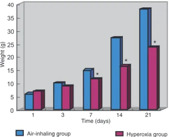
Weight loss was observed in neonatal rats after 7-10 days of hyperoxia exposure. The body weight of neonatal rats in the
hyperoxia group was lower than that of the air-inhaling group (Figure 1). The mortality of the neonatal rats in the hyperoxia
group was 45.9 ± 4.2% at day 14 and 62.5 ± 4.2% at day 21.
Weight gain and mortality of the air-inhaling group of neonatal rats were normal.
SC was enhanced in ileal tissue of neonatal rats exposed to hyperoxia
The expression of SC in the cytoplasm and on the
mem-branes of intestinal epithelial cells was remarkably enhanced
in the hyperoxia group compared to the air-inhaling group (Figure 2). In the hyperoxia group, the median absorbance
values for SC were 6.5 ± 1.2 at day 14 and 7.0 ± 1.5 at
day 21. In the air-inhaling group, the median absorbance
Figure 1. Weight of rats in the air-inhaling and hyperoxia groups. The body weight of the hyperoxia rats started to be lower than that of the air-inhaling rats on the 3rd day. From the 7th to the 21st day, the body weight of the hyperoxia rats was significantly lower com -pared to that of the air-inhaling group (*P < 0.01, t-test).
Effect of hyperoxia on intestine epithelial cells 1037
values for SC were 3.1 ± 1.1 at day 14 and 3.2 ± 1.3 at day 21. Also, the median absorbance value for SC in the hyperoxia group was notably increased compared with the air-inhaling group (P < 0.01; Figure 3).
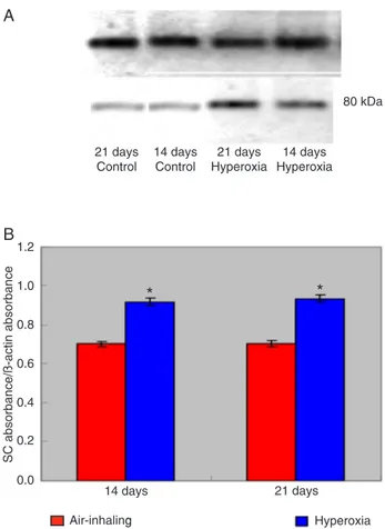
SC protein was increased in ileal tissue of neonatal rats exposed to hyperoxia
SC protein expression was increased in the hyperoxia group relative to the air-inhaling group. In the hyperoxia group, the relative expression rate of SC (absorbance value
of SC/absorbance value of β-actin) was 0.919 ± 0.060 at
day 14 and 0.934 ± 0.044 at day 21. In the air-inhaling group, the relative expression rate of SC (absorbance value
of SC/absorbance value of β-actin) was 0.702 ± 0.032 at
day 14 and 0.704 ± 0.022 at day 21. The relative expres-sion rate of SC was notably increased in the hyperoxia group compared to the air-inhaling group at days 14 and 21 (P < 0.01; Figure 4).
Effect of hyperoxia on Caco-2 cell growth
The growth of Caco-2 cells was significantly affected by 60 and 90% O2, as shown in Figure 5. Growth in 40% O2 was increased at day 2 and was exponentially
increased at days 4 and 5. When the gas was changed
to 60% O2, the growth rate decreased. Cells in 90% O2 had arrested growth followed by rapid death. Significant
differences in cell numbers were found between 21% O2 and both 60 and 90% O2 (P < 0.001). Significant differ -ences in cell numbers were also found between 40% O2 and both 60 and 90% O2 (P< 0.001). There was also a significant difference in cell numbers for 60 and 90% O2
(P < 0.001). These results suggest that cells can adapt to 40% O2, but 60 and 90% O2 cause severe cell damage and eventually cell death.
Effect of hyperoxia on Caco-2 cell survival
MTT was used to detect cell survival. At day 3, cells
that were cultured at 40 and 60% O2 were unchanged in population, but at 90% O2, cells underwent rapid death (P < 0.01). At day 6, cells that were cultured at 40% O2 increased in number (P > 0.01), but at 60 and 90% O2
their numbers were rapidly decreased compared to cells cultured at 21% O2 (P < 0.01; as shown in Figure 6). This showed that moderate oxygen induced intestinal epithelial cell growth.
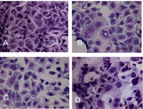
Effect of hyperoxia on Caco-2 cell division
At day 3, the mitotic indices of cells grown in 40, 60 and
90% O2 were significantly decreased compared to those grown in 21% O2. However, at day 6, the mitotic index of cells grown in 40% O2 was increased compared with
those grown in 21% O2. At 60% O2, the mitotic index was decreased, and some cell nuclei were larger than those seen under the 21% O2 conditions. At day 6, cell splitting was not observed at 90% O2 and the nuclei of living cells
Figure 3. Absorbance of the intestinal secretory component (SC) in the hyperoxia and air-inhaling groups. The mean absorbance value of SC was notably increased in the hyperoxia group com-pared to the air-inhaling group (*P < 0.01, t-test).
were larger than those seen under 21% O2 conditions (as
shown in Figure 7).
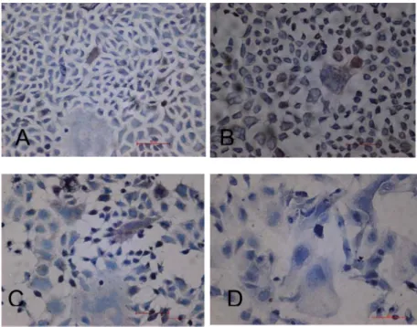
Immunochemical analysis of the effects of hyperoxia on SC-positive cells
SC is mainly localized in the cytoplasm and cell
mem-branes of Caco-2 cells, as shown in Figure 8. SC-positive cells constituted about 0.1-0.5% of the population of cells grown in 21% O2 and about 0.4-0.8% of the population
of cells grown in 40% O2. SC-positive cells constituted about 0.2-0.7% of the population of cells grown in 60%
O2. The expression of SC in Caco-2 cells was remarkably
Figure 5. Influence of O2 concentration on Caco-2 cell growth.
Each data point represents the mean cell number of 5-6 plastic Petri dishes from each of six preparations of mixed Caco-2 cells. Cells were maintained in 21, 40, 60, and 90% O2.
Figure 7. Effect of hyperoxia on Caco-2 cell division (40X). A, 21% O2: cells distributed
densely, some cells were splitting, and the mitotic index was 2.5%; B, 40% O2: compared
to 21% O2, cell splitting was increased and the mitotic index was 3.3%; C, 60% O2:
com-pared to 21% O2, cell splitting was decreased, the mitotic index was 1.3% and many larger
cell nuclei were observed; D, 90% O2: cell splitting was not observed and only larger cell
nuclei were seen.
Figure 6. Effect of hyperoxia on Caco-2 cell survival. On the 3rd day, cells rapidly died in the presence of 90% O2 (*P < 0.01, t
-test) and on the 6th day, at 60 and 90% O2 cell numbers were
rapid decreased compared to 21% O2 (+P < 0.01, t-test). At 40%
Effect of hyperoxia on intestine epithelial cells 1039
decreased as the percentage of oxygen was increased
above 60%, and SC-positive cells were not demonstrable
in cells grown in 90% O2.
Discussion
The intestinal tract faces physiological challenges in the human body. It must provide ready access to ingested nutrients and also protect against food-borne microbes. This
challenge is met by multiple specific and nonspecific host
defenses that establish and maintain a selective intestinal barrier. This immunological intestinal barrier is critical for defense against intestinal microbes and their toxins.
Oxygen therapy, leading to hyperoxia, is a necessary method for the treatment of some critical care conditions. The high concentration of oxygen inhaled, however, may be both a friend and a foe of the body. An adequate oxygen supply serves to maintain the physical integrity of the mu-cosa, thus decreasing invasion of the lumen by microbes. An adequate supply of oxygen is also important for effec-tive bactericidal function of resident neutrophils and other
phagocytic cells in the gut (7). However, hyperoxia also enhances the inflammatory response in adult mice infected with influenza A virus (27).It has been proposed that nor-mobaric hyperoxia might affect certain peripheral organs
(kidney, ileum) (8). The thickness of the ileal mucosa of
Figure 8. Effect of hyperoxia on the intestinal secretory component (SC) expression of Caco-2 cell (40X). A, The expression of SC in the cytoplasm and on the membranes of Caco-2 cell was pale brown. B, The expression of SC in the cytoplasm and on the membranes of Caco-2 cell was deep brown. C, The expression of SC in the cytoplasm and on the membranes of Caco-2 cell was brown. D, The expression of SC of Caco-2 cell was remarkably decreased. SC-positive cell was not seen in 90% O2.
neonatal rats under conditions leading to hyperoxia was
significantly greater (9). Hyperoxia may affect the barrier
function of the newborn rat’s intestine, rendering it sus-ceptible to bacterial insult (9). In this study, the expression of SC protein in neonatal rats under conditions leading to hyperoxia was increased compared to that of air-inhaling neonatal rats.The rise of SC may be due to the following
causes: first, hyperoxia renders the intestinal mucosa of
neonatal rats susceptible to bacterial insult.Bacterial inva-sion of the intestine stimulates intestinal epithelial cells to secretemore SC. Second, a direct effect of oxidative stress may induce changes in intestinal immunity cells, such as
greater secretion of cytokines. Some cytokines regulate
the expression of SC (28-30). Third, secondary systemic
inflammation may be another cause of the rise of SC. These
possibilities require further investigation. Increased SC
expression is beneficial to the transcytosis of pIgA and the
formation of SIgA. This is favorable to the defense against oxygen toxicity from hyperoxia and bacterial insults. Then, SC and SIgA both exert immunological functions in the exclusion and neutralization of bacteria.
In order to better understand the influence of hyperoxia
on intestinal epithelial cells,Caco-2 cells were treated in vitro
1. Warner BB, Stuart LA, Papes RA, Wispe JR. Functional and pathological effects of prolonged hyperoxia in neonatal mice.
Am J Physiol 1998; 275: L110-L117.
2. Smith LE. Pathogenesis of retinopathy of prematurity. Semin Neonatol 2003; 8: 469-473.
3. Madan A, Penn JS. Animal models of oxygen-induced retin-opathy. Front Biosci 2003; 8: d1030-d1043.
4. Kaindl AM, Sifringer M, Koppelstaetter A, Genz K, Loeber R, Boerner C, et al. Erythropoietin protects the developing brain from hyperoxia-induced cell death and proteome changes.
Ann Neurol 2008; 64: 523-534.
5. Baylor AE, Diebel LN, Liberati DM, Dulchavsky SA, Diglio CA, Brown WJ. The effects of varying oxygen conditions and immunoglobulin A on barrier defense to bacterial invasion.
Am Surg 2003; 69: 231-237.
6. Diebel LN, Liberati DM, Brown WJ, Dulchavsky SA, Painter TM, Diglio CA, et al. Secretory immunoglobulin A blocks hypoxia-augmented bacterial passage across Madin-Darby canine kidney cell monolayers. J Trauma 1997; 43: 759-763.
7. Diebel LN, Liberati DM, Dulchavsky SA, Diglio CA, Brown WJ. Synergistic effect of hyperoxia and immunoglobulin A on mucosal barrier defense. J Trauma 1999; 46: 374-378.
8. Torbati D, Tan GH, Smith S, Frazier KS, Gelvez J, Fakioglu H, et al. Multiple-organ effect of normobaric hyperoxia in neonatal rats. J Crit Care 2006; 21: 85-93.
9. Giannone PJ, Bauer JA, Schanbacher BL, Reber KM. Ef-fects of hyperoxia on postnatal intestinal development.
Biotech Histochem 2007; 82: 17-22.
10. Cunningham-Rundles C. Physiology of IgA and IgA defi -ciency. J Clin Immunol 2001; 21: 303-309.
11. Macpherson AJ, Hunziker L, McCoy K, Lamarre A. IgA responses in the intestinal mucosa against pathogenic and non-pathogenic microorganisms. Microbes Infect 2001; 3: 1021-1035.
12. Boullier S, Tanguy M, Kadaoui KA, Caubet C, Sansonetti P, Corthesy B, et al. Secretory IgA-mediated neutralization of Shigella flexneri prevents intestinal tissue destruction by down-regulating inflammatory circuits. J Immunol 2009; 183:
5879-5885.
13. Rey J, Garin N, Spertini F, Corthesy B. Targeting of secretory IgA to Peyer’s patch dendritic and T cells after transport by intestinal M cells. J Immunol 2004; 172: 3026-3033.
14. Bomsel M. Transcytosis of infectious human immunodefi -ciency virus across a tight human epithelial cell line barrier.
Nat Med 1997; 3: 42-47.
15. Mazanec MB, Nedrud JG, Kaetzel CS, Lamm ME. A three-tiered view of the role of IgA in mucosal defense. Immunol Today 1993; 14: 430-435.
16. Sawai T, Goldstone N, Drongowski RA, Coran AG, Harmon CM. Effect of secretory immunoglobulin A on bacterial translocation in an enterocyte-lymphocyte co-culture model.
Pediatr Surg Int 2001; 17: 275-279.
17. Norderhaug IN, Johansen FE, Schjerven H, Brandtzaeg P. Regulation of the formation and external transport of secretory immunoglobulins. Crit Rev Immunol 1999; 19: 481-508.
18. Phalipon A, Corthesy B. Novel functions of the polymeric Ig receptor: well beyond transport of immunoglobulins. Trends Immunol 2003; 24: 55-58.
19. Dallas SD, Rolfe RD. Binding of Clostridium difficile toxin A to human milk secretory component. J Med Microbiol 1998; 47: 879-888.
20. de Araujo AN, Giugliano LG. Lactoferrin and free secretory component of human milk inhibit the adhesion of entero -pathogenic Escherichia coli to HeLa cells. BMC Microbiol
2001; 1: 25.
21. Giugliano LG, Ribeiro ST, Vainstein MH, Ulhoa CJ. Free secretory component and lactoferrin of human milk inhibit the adhesion of enterotoxigenic Escherichia coli. J Med Microbiol 1995; 42: 3-9.
22. Hammerschmidt S, Talay SR, Brandtzaeg P, Chhatwal GS. SpsA, a novel pneumococcal surface protein with specific binding to secretory immunoglobulin A and secretory com-ponent. Mol Microbiol 1997; 25: 1113-1124.
23. Davids BJ, Palm JE, Housley MP, Smith JR, Andersen YS, Martin MG, et al. Polymeric immunoglobulin receptor in intestinal immune defense against the lumen-dwelling
pro-References
division rate reached 3.3%. At 60% O2, the cells continued
to divide, although at a reduced rate. However, at 90% O2, there was both growth arrest and cell death. This
demon-strates that severe hyperoxia may check cell growth and kill
intestinal epithelial cells, which in turn would facilitate the invasion of the gut by bacteria. In vitro, the optimal concen-tration of inhaled oxygen (about 0.4-0.8% for SC-positive cells at 40% O2 and about 0.2-0.7% for SC-positive cells at 60% O2) induced intestinal mucus to secrete SC, but severe hyperoxia (90% O2) limited the secretion of SC. However,
in vivo, the expression of intestinal SC protein in neonatal rats induced by hyperoxia was increased. The reason for this may be that, in vitro, 90% O2 acts directly on intestinal
epithelial cells, but in vivo, 95% O2 acts indirectly on the
intestine. We speculated that sublethal O2 exposure was
associated with induction of antioxidant defense systems
and the change in the intestinal environment. The change in intestinal environment may lead to intestinal bacterial disorders (9), which in turn may induce the intestinal epi-thelial cells to secrete SC.
In vivo and in vitroincreases in intestinal SC in response to exposure to moderate hyperoxia would be expected to
cause an increase in intestinal SIgA. High levels of SC and SIgA are beneficial to maintaining the optimal state of the
intestinal tract.
Acknowledgments
Effect of hyperoxia on intestine epithelial cells 1041
tozoan parasite Giardia. J Immunol 2006; 177: 6281-6290.
24. Uren TK, Wijburg OL, Simmons C, Johansen FE, Brandt -zaeg P, Strugnell RA. Vaccine-induced protection against gastrointestinal bacterial infections in the absence of secre-tory antibodies. Eur J Immunol 2005; 35: 180-188. 25. Liu DY, Chen XF, Li JJ. The change of intestinal mucous
secretory IgA in neonatal rats by hyperoxia. J China Med Univ 2009; 38: 45-46,49.
26. Yokomizo A, Moriwaki M. Transepithelial permeability of myricitrin and its degradation by simulated digestion in human intestinal Caco-2 cell monolayer. Biosci Biotechnol Biochem 2005; 69: 1774-1776.
27. O’Reilly MA, Marr SH, Yee M, Grath-Morrow SA, Lawrence BP. Neonatal hyperoxia enhances the inflammatory re
-sponse in adult mice infected with influenza A virus. Am J Respir Crit Care Med 2008; 177: 1103-1110.
28. Liu DY, Wang XL, Liu P. Tumor necrosis factor-alpha upregu-lates the expression of immunoglobulin secretory compo-nent. J Investig Allergol Clin Immunol 2007; 17: 101-106.
29. Denning GM. IL-4 and IFN-gamma synergistically increase total polymeric IgA receptor levels in human intestinal epithe-lial cells. Role of protein tyrosine kinases. J Immunol 1996;
156: 4807-4814.