Does plasma ANP participate in
natriuresis induced by
αα
αα
α
-MSH?
1Departamento de Fisiologia, Centro de Ciências Biológicas, Universidade Federal do Pará, 66076-900 Belém, PA, Brasil
2Departamento de Fisiologia, Faculdade de Medicina de Ribeirão Preto, Universidade de São Paulo, 14049-900 Ribeirão Preto, SP, Brasil
3Centre de Recherche Hôtel-Dieu de Montreal, Pavilion Marie-de-la-Ferre, Montréal, Quèbec H2W 1T8, Canada
4Neuropeptide Division, Department of Physiology,
The University of Texas Southwestern Medical Center, Dallas, Texas 75235, USA D.L.W. Picanço-Diniz1,
G. Ribeiro-Oliveira2, A.L.V. Favaretto2, J. Gutkowska3, S.M. McCann4 and J. Antunes-Rodrigues2
Abstract
α-Melanocyte-stimulating hormone (α-MSH; 0.6 and 3 nmol) micro-injected into the anteroventral region of the third ventricle (AV3V) induced a significant increase in diuresis without modifying natriure-sis or kaliurenatriure-sis. Intraperitoneal (ip) injection of α-MSH (3 and 9.6 nmol) induced a significant increase in urinary sodium, potassium and water excretion. Intraperitoneal (3 and 4.8 nmol) or iv (3 and 9.6 nmol) administration of α-MSH did not induce any significant changes in plasma atrial natriuretic peptide (ANP), suggesting that the natriure-sis, kaliuresis and diuresis induced by the systemic action of α-MSH can be dissociated from the increase in plasma ANP. These prelimi-nary results suggest that α-MSH may be involved in a γ -MSH-independent mechanism of regulation of hydromineral metabolism.
Correspondence
J. Antunes-Rodrigues Departamento de Fisiologia Faculdade de Medicina de Ribeirão Preto, USP Av. Bandeirantes, 3900 14049-900 Ribeirão Preto, SP Brasil
Presented at the International Symposium Neuroendocrine Control of Body Fluid Homeostasis, Ribeirão Preto, SP, Brasil, August 17-20, 1996. Research supported by FAPESP (Nos. 91/0567-0 and 94/3805-7) and CNPq (Nos. 50167/91-7 and 521593/94-8).
Received November 29, 1996 Accepted January 6, 1997
Key words •α-MSH
•AV3V
•ANP
•Natriuresis
•Kaliuresis
•Diuresis
Introduction
α-Melanocyte-stimulating hormone (α -MSH) is a peptide derived from proopiomel-anocortin (POMC), a precursor protein that contains the amino acid sequences of other peptides acting on salt and water balance in the organism, including ß- and γ-MSH (1).
The participation of α-MSH in the regu-lation of mineral metabolism has been dis-cussed by many investigators (2). Water dep-rivation for 12 h and oral (3) or intravenous (iv) injection of hypertonic NaCl solutions produced a marked depletion of pituitary MSH, suggesting a compensatory secretion in response to salt concentration in the or-ganism. Intraperitoneal (ip) injections of α -MSH exhibited a natriuretic and kaliuretic
effect in intact (4) hypophysectomized or adrenalectomized (5) water-loaded rats, in-dicating an independent action of pituitary and adrenal glands and suggesting its direct intervention at the renal level. An antidi-uretic effect was observed after ip injection of α-MSH into water-loaded rats, but the authors suggested that this effect resulted from lysine-vasopressin contamination in the preparation (6).
The participation of atrial natriuretic pep-tide (ANP) directly at the renal level and dependence on renal nerve activity are two possible mechanisms suggested for the natri-uresis induced by γ-MSH. However, only few studies on the mechanisms involved in
In the present study we examined the possibility of a modulatory action of α-MSH on hydromineral metabolism by a γ -MSH-independent mechanism of action.
Material and Methods
Adult male Wistar rats (230-260 g body weight) from our animal facilities, housed in individual cages under controlled light (14-h light, 10-h dark) and temperature conditions (23-25oC) with free access to food and wa-ter, were used.
For the experiments the animals were divided into three experimental groups: i) rats with chronically indwelling AV3V can-nulae were subjected 5 days later to α-MSH (0.6 and 3.0 nmol) microinjection to study its effect on sodium, potassium and water excretion. ii) Intact rats were injected ip with
α-MSH (3.0 and 9.6 nmol) to study its effect on sodium, potassium and water excretion. iii) Intact or jugular-cannulated rats were injected ip (3.0 and 4.8 nmol) or iv (3.0 and 9.6 nmol) with α-MSH, respectively, to study its effect on plasma atrial natriuretic peptide levels. The rats were sacrificed prior to (0 min) and 5 or 15 min after α-MSH injection.
Experimental procedures
Under anesthesia with tribromoethanol (2.5 mg/100 gbody weight; T4840-2; Aldrich, Milwaukee, WI), a stainless steel cannula (0.7 mm OD, 13 mm length) was stereotaxi-cally implanted into the AV3V according to a technique previously described (7), using the Paxinos and Watson stereotaxic coordi-nates (8). The animals were then handled daily and trained for gavage to reduce any stress associated with these experimental pro-cedures.
Five days after surgery, the rats were fasted for 14 h before the experiment with free access to water. On the next morning, each rat received a load of tepid tap water (5% body weight) by gavage, and was placed
in an individual metabolic cage without food or water. Urine passed through the funnel at the bottom of the cage into a graduated cen-trifuge tube. Urinary volume (reported as µl min-1 100 g body weight-1) was collected immediately before and 20, 60, and 120 min after drug microinjection into the AV3V (experiment I) or ip (experiment II) injection.
α-MSH (7251; Peninsula Laboratories, Belmont, CA) was injected into the AV3V (1 µl) through a stainless steel dental can-nula (0.31 mm OD, 14 mm length) con-nected to a 10-µl Hamilton microsyringe by a polyethylene tube (PE 10). Intraperitoneal
α-MSH injections (0.2 ml; experiment II) were performed through a 1-ml syringe into the abdominal region. Saline vehicle (0.15 M NaCl) was used as control for all experi-ments. Intravenous α-MSH injections (0.2 ml; experiment III) were performed through a silastic cannula implanted (1 day before) into the jugular vein connected to a polyeth-ylene tube (PE 50), as previously described (9). Routine histological procedures were done to localize the sites where the cannulae were implanted. After the experiments, all animals were killed under ether anesthesia and the brains were removed and fixed in 10% formol. Twenty-µm thick frontal paraf-fin sections were stained by the Nissl tech-nique.
Measurements
Each urine sample was analyzed for so-dium and potassium concentrations (µEq min-1 100 gbody weight-1) using a Micronal flame photometer (model B262).
plasma was stored at -70oC. Immunoreactive ANP was extracted from 1 ml of plasma by heat-activated Vycor glass (No. 7930, Mesh 140; Corning, New York), and the lyophilized residue was stored at -70oC. Each sample was resuspended in 400 µl ANP buffer (50 mM potassium phosphate, pH 7.4, contain-ing 0.15 M NaCl, 0.1% bovine serum albu-min (Sigma No. A-7888), 0.1% Triton X-100, and 0.02% NaN3), and aliquots of 50 and 100 µl were taken in duplicate for radio-immunoassay (10).
Statistical analysis
The statistical analysis of the data was performed by GBSTAT computer program. Data are reported as means ± SEM. Analysis of variance (ANOVA) was used for statisti-cal evaluation. A Pvalue less than 0.05 was considered to be significant.
Results
Effect of central (AV3V) or systemic (ip) injection of ααααα-MSH on natriuresis, kaliuresis and diuresis (experiments I and II)
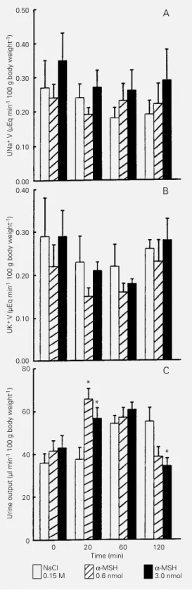
Figure 1A and 1B shows that microinjec-tions of α-MSH into the AV3V did not in-duce changes in sodium or potassium excre-tion. On the other hand, a significant in-crease in urine outflow occurred 20 min after microinjection of 0.6 nmol α-MSH (175%) and 3 nmol (151%) into the AV3V (Figure 1C).
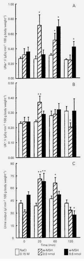
α-MSH at the 3-nmol dose induced a significant increase in sodium excretion 20 min (184%) and 60 min (84%) after ip ad-ministration. α-MSH at the 9.6-nmol dose induced a 176% and 75% increase in sodium excretion at 60 and 120 min, respectively, under the same experimental conditions (Fig-ure 2A). A two-phase effect on potassium excretion was observed after ip administra-tion of α-MSH at the 3-nmol dose. This dose induced an increase at 20 min (61%) and no
UNa
+.
V (µEq min
-1 100 g body weigh
t
-1) 0.50
0.40
0.30
0.20
0.10
0.00 0.40
0.30
0.20
0.10
0.00 80
60
40
20
0
123 123 123 123 NaCl
0.15 M
α-MSH 0.6 nmol
α-MSH 3.0 nmol
A
B
C
0 20 60 120
Time (min)
UK
+.V
(µEq min
-1 100 g body weight -1)
Urine output (µl min
-1 100 g body weigh
t
-1)
* *
*
Figure 1 - Effects of microinjec-tion of α-MSH into the AV3V on sodium (A), potassium (B) and urine output (C) in water-loaded rats. Urine samples were col-lected before and 20, 60, and 120 min after microinjection of the peptide into the AV3V. α− MSH (0.6 and 3.0 nmol) was de-livered in 1.0 µl saline. Data are reported as means ± SEM for 8 to 14 animals per group. UNa+.V or UK+.V = Sodium or potassium excretion expressed as the prod-uct of concentration of the ion and urine flow per min (V). *P<0.05 compared to control group (ANOVA).
UNa
+.V (µEq min -1 100 g body weight -1)
1.00
0.80
0.60
0.40
0.20
0.00 0.50
0.40
0.30
0.20
0.00
90
75
60
45
30
123 123 123 NaCl
0.15 M
α-MSH 3.0 nmol
α-MSH 9.6 nmol
A
B
C
0 20 60 120
Time (min)
UK
+.V (µEq min -1 100 g body weight -1)
Urine output (µl min
-1 100 g body weight -1)
0.10
15
0
*
* *
*
* **
**** *
*
phase effect on urine output was observed after ip injection of α-MSH at the 9.6-nmol dose. A significant increase was observed at 20 min (78%), no effect at 60 min and a significant decrease (52%) at 120 min (Fig-ure 2C).
Effect of systemic (iv or ip) injection of ααααα -MSH on plasma ANP levels (experiment III)
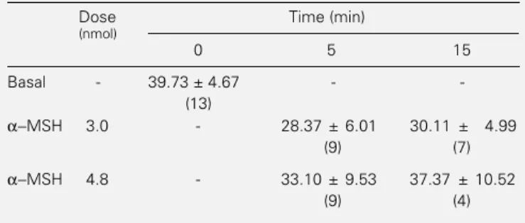
Table 1 shows that intraperitoneal injec-tions of α-MSH (3 and 4.8 nmol) did not affect ANP levels throughout the experimental peri-ods (5 and 15 min). Similar results were ob-tained with intravenous injections of 3 and 9.6 nmol of α-MSH (data not shown).
Discussion
Although it has been known for many years that systemic administration of α-MSH induces an increase in sodium and potas-sium urinary excretion (4-6), the mechanism of these effects remains unknown. The puta-tive participation of the central nervous sys-tem in these effects was investigated in the present study. Our results showed that α -MSH administration into the AV3V at the doses of 0.6 and 3 nmol induced an increase in urine outflow but no effect on natriuresis or kaliuresis (Figure 1A-C). These data sug-gest a positive and independent α-MSH regu-lation of urine outflow at the AV3V level, since a systemic effect of central administra-tion would be improbable because, under these experimental conditions, no changes in natriuresis or kaliuresis were observed. Probably an inhibitory influence on vaso-pressin secretion via inhibition of cholin-ergic and angiotensincholin-ergic neurons in the AV3V may explain the data observed (11).
Our results also show that plasma ANP is not involved in the increase of water and electrolyte excretion induced by systemic α -MSH administration because ip injection of
α-MSH (3 and 4.8 nmol doses) did not in-duce changes in plasma ANP levels during Figure 2 - Effects of ip
adminis-tration of α-MSH on sodium (A), potassium (B) and urine output (C) in water-loaded rats. Urine samples were collected before and 20, 60, and 120 min after injection. α-MSH (3.0 and 9.6 nmol) was delivered in 200 µl saline. Data are reported as means ± SEM for 5 to 7 animals per group. *P<0.05, **P<0.01 compared to control (ANOVA).
two-the experimental period analyzed (Table 1). The natriuretic effect of α-MSH has also been described in hypophysectomized or adrenalectomized rats, leading the authors to suggest that the kidneys were the most prob-able site of action of α-MSH (5). However, direct effects of α-MSH on renal function and ANP secretion have not been well ex-plored. In contrast, several lines of evidence indicate that γ-MSH-induced natriuresis may be mediated by ANP or occur by a direct action of the peptide at the renal level, being dependent on renal nerve activity (2). The uncorrelated increase in α-MSH-induced natriuresis with plasma ANP hereby reported suggests that this peptide may have mecha-nisms of action different from those of γ -MSH.
The diuretic effect observed after ip ad-ministration of α-MSH (3 nmol) in the pres-ent study is contrary to that observed by other investigators who suggested that anti-diuresis evoked by large doses of α-MSH may be caused by contamination with lysine-vasopressin, since the preparation supplied was reported to contain 0.1 mU of pressor activity per µg (6). The diuretic effect of α -MSH may result from a direct action at the renal level by inhibition of tubular sodium reabsorption (2) or by a central inhibition of vasopressin secretion.
In conclusion, our data indicate that α -MSH may induce diuresis by a central ac-tion, and natriuresis, kaliuresis and diuresis by a systemic action. These effects on so-dium and potassium excretion seem not to be mediated by changes in plasma ANP. These preliminary results suggest that α-MSH may be involved in a γ-MSH-independent mech-anism of regulation of hydromineral me-tabolism.
Acknowledgments
We thank Marina Holanda and Maria Valci Aparecida dos Santos Silva for techni-cal assistance.
Table 1 - Plasma ANP concentration (pg/ml) before and 5 and 15 min after ip injection of α-MSH.
Animals were decapitated and trunk blood was collected into cooled tubes with proteolytic enzyme inhibitors. Immunoreactive ANP was extracted from 1.0 ml of plasma with Vycor and assayed by RIA. α-MSH (3.0 and 4.8 nmol) was delivered in 200 µl saline. The number of animals is given in parentheses.
Dose Time (min)
0 5 15
Basal - 39.73 ± 4.67 -
-(13)
α−MSH 3.0 - 28.37 ± 6.01 30.11 ± 4.99
(9) (7)
α−MSH 4.8 - 33.10 ± 9.53 37.37 ± 10.52
(9) (4)
References
1. Eipper BA & Mains RE (1980). Structure and biosynthesis of pro-adrenocorticotro-pin/endorphin and related peptides. En-docrine Reviews, 1: 1-27.
2. Valentin JP, Wiedemann E & Humphreys MH (1993). Natriuretic properties of mel-anocyte-stimulating hormones. Journal of Cardiovascular Pharmacology, 22: S114-S118.
3. Kastin AJ (1967). MSH and vasopressin activities in pituitary of rats treated with hypertonic saline. Federation Proceed-ings, 26: 255 (Abstract).
4. Orias R & McCann SM (1970). Natriuretic effect of α-MSH in the water-loaded rat. Proceedings of the Society for Experimen-tal Biology and Medicine, 133: 469-474.
5. Orias R & McCann SM (1972). Natriuretic effect of alpha melanocyte stimulating hormone (α-MSH) in hypophysectomized or adrenalectomized rats. Proceedings of the Society for Experimental Biology and Medicine, 139: 872-876.
6. Orias R & McCann SM (1972). Natriuresis induced by alpha and beta melanocyte stimulating hormone (MSH) in rats. Endo-crinology, 90: 700-706.
7. Antunes-Rodrigues J & McCann SM (1970). Water, sodium chloride, and food intake induced by injections of cholinergic and adrenergic drugs into the third ven-tricle of the rat brain. Proceedings of the Society for Experimental Biology and Medicine, 133: 1464-1470.
8. Paxinos G & Watson C (1986). The Rat Brain in Stereotaxic Coordinates. 2nd edn. Academic Press, Orlando.
9. Harms PG & Ojeda SR (1974). A rapid and simple procedure for chronic cannulation of the rat jugular vein. Journal of Applied Physiology, 36: 391-392.
10. Gutkowska J (1987). Radioimmunoassay for atrial natriuretic factor. Nucleotide Medical Biology, 14: 323-331.
11. Hoffman WE, Phillips MI & Schmid PG (1977). Antidiuretic hormone release and the pressor response to central angio-tensin II and cholinergic stimulation. Neu-ropharmacology, 16: 463-472.