Cholinergic-opioidergic interaction
in the central amygdala induces
antinociception in the guinea pig
1Departamento de Morfologia, Estomatologia e Fisiologia,
Faculdade de Odontologia de Ribeirão Preto, Universidade de São Paulo, Ribeirão Preto, SP, Brasil
2Departamento de Fisiologia, Faculdade de Medicina de Ribeirão Preto,
Universidade de São Paulo, Ribeirão Preto, SP, Brasil C.R.A. Leite-Panissi1,
M.R. Brentegani2 and
L. Menescal-de-Oliveira2
Abstract
Several studies have demonstrated the involvement of the central nucleus of the amygdala (CEA) in the modulation of defensive behavior and in antinociceptive regulation. In a previous study, we demonstrated the existence of a cholinergic-opioidergic interaction in the CEA, modulating the defensive response of tonic immobility in guinea pigs. In the present study, we investigated a similar interaction in the CEA, but now involved in the regulation of the nociceptive response. Microinjection of carbachol (2.7 nmol) and morphine (2.2 nmol) into the CEA promoted antinociception up to 45 min after microinjection in guinea pigs as determined by a decrease in the vocalization index in the vocalization test. This test consists of the application of a peripheral noxious stimulus (electric shock into the subcutaneous region of the thigh) that provokes the emission of a vocalization response by the animal. Furthermore, the present results demonstrated that the antinociceptive effect of carbachol (2.7 nmol; N = 10) was blocked by previous administration of atropine (0.7 nmol; N = 7) or naloxone (1.3 nmol; N = 7) into the same site. In addition, the decrease in the vocalization index induced by the microinjection of morphine (2.2 nmol; N = 9) into the CEA was prevented by pretreat-ment with naloxone (1.3 nmol; N = 11). All sites of injection were confirmed by histology. These results indicate the involvement of the cholinergic and opioidergic systems of the CEA in the modulation of antinociception in guinea pigs. In addition, the present study suggests that cholinergic transmission may activate the release of endorphins/ enkephalins from interneurons of the CEA, resulting in antinocicep-tion.
Correspondence
L. Menescal-de-Oliveira Departamento de Fisiologia FMRP, USP
14049-900 Ribeirão Preto, SP Brasil
Fax: +55-16-633-0017 E-mail: [email protected] Research supported by CNPq, CAPES, FAEPA and FAPESP (No. 97/14286-9).
Received October 10, 2003 Accepted July 5, 2004
Key words
•Antinociception •Vocalization test •Cholinergic-opioidergic
interaction •Amygdala •Naloxone •Atropine
Introduction
The central nucleus of the amygdala (CEA) is involved in diverse emotional and cognitive functions related to responses of fear and ori-entation (1,2), defensive behavior (3,4), and
(trigemio)pontoamygdaloid pathway with relay in the parabrachial area (9). This path-way is probably involved in the affective-emotional (fear, aggression), behavioral (vo-calization, fight, freezing) and autonomic (cardiovascular and respiratory responses) reactions to noxious events (9).
Fox and Sorenson (7) have shown that lesion of the CEA promoted a reduction in antinociception induced by meeting a preda-tor and also in antinociception promoted by classical conditioning evaluated by the tail-flick test. These results suggest the involve-ment of the CEA in the regulation of antino-ciception induced by aversive events.
Some reports have demonstrated the ex-istence of peptides and amino acids in the amygdala, that participate in neurotransmis-sion and also of various neurotransmitters such as acetylcholine and enkephalin. In-deed, enzymes related to acetylcholine pro-cessing (acetylcholinesterase and choline acetyltransferase) are present in the lateral part of the CEA, indicating that acetylcho-line synthesis occurs in the amygdala (10). In addition, some studies have demonstrated the existence of diverse neurons containing enkephalin in the amygdaloid complex (11). In this respect, it has been shown that the administration of morphine into the amyg-dala promotes an increase of the nociceptive threshold in different algesimetric tests (12, 13). Pavlovic et al. (14) observed that micro-injection of morphine or ß-endorphin into the amygdala produced a significant increase of latency in the tail-flick test as well as in the jump test in rats. Furthermore, the in-crease in tail-flick latency and jump thresh-old was prevented by pretreatment with opioid antagonists (naltrexone, ß-funaltrex-amine and naltrindole isothiocyanate) in-jected into the periaqueductal gray matter (PAG). Taken together, these results suggest that the existence of opioidergic synapses in the PAG is essential for the antinociceptive effect on opioidergic stimulation of the amyg-dala in rats.
It has been demonstrated that microin-jection of the cholinergic agonist, carbachol, into the CEA, but not into the basolateral or lateral nuclei of the amygdala, produced an increase in tail-flick latency (3). In addition, Ahn et al. (8) showed that carbachol admin-istered into the CEA promoted antinocicep-tion in rats after the applicaantinocicep-tion of a noxious electric stimulus to the dental pulp and this effect was blocked by pretreatment with at-ropine at the same site. Furthermore, in a previous study (15) we demonstrated that the cholinergic system of the CEA partici-pates in the modulation of defensive behav-ior and also in antinociception through a functional and anatomical connection with the ventrolateral PAG, because pretreatment with lidocaine injected into the ventrolateral PAG blocked the decrease in the duration of the defensive behavior of tonic immobility and the reduction of the vocalization index after carbachol microinjection into the CEA. The existence of a circuit in the CEA that modulates the defensive response of tonic immobility has been described (16). We showed that a cholinergic-opioidergic inter-action in the CEA is involved in the modula-tion of this defensive behavior. Thus, cholin-ergic and opioidcholin-ergic stimulation of the CEA exerts an inhibitory action on the duration of tonic immobility episodes in guinea pigs. Furthermore, the effect of the administration of carbachol and morphine into the CEA was blocked by pretreatment with naloxone mi-croinjected into the same site.
Material and Methods
Adult male guinea pigs (Cavia porcellus) weighing 400-500 g were obtained from the animal care facility of the Faculty of Medi-cine of Ribeirão Preto (FMRP). The animals were kept in Plexiglas wall cages (56 x 37 x 39 cm, 5 animals per cage) in a room main-tained at 24 ± 1ºC, on a 12-h light cycle, with free access to water and food. Experiments were carried out according to the ethical recommendations of the Committee for Re-search and Ethical Issues of the International Association for the Study of Pain (17).
Antinociception was determined by the vocalization test. Guinea pigs are not com-monly used in experiments destined to the study of the neural mechanisms of analgesia. Algesimetric tests currently used in rats and mice are not applicable to guinea pigs for anatomical reasons (tail-flick) or because they have not been standardized (writhing). In our laboratory, we have been using for some years tests that have proved to be ap-propriate for guinea pigs (15,18,19). The vocalization test consists of the application of a peripheral noxious stimulus (electric shock) that provokes the emission of a vo-calization response by the animal, which is interpreted as a manifestation of pain.
For peripheral noxious stimulation, a pair of non-insulated electrodes was implanted into the subcutaneous region of the thigh. The animal was then placed in an acrylic box lined with nylon foam where some move-ment was possible. After 20 min of animal habituation to the experimental situation, the electrode was connected to an electronic stimulator that released pulses (square waves, 100-Hz frequency, 0.5-ms duration) of vary-ing intensity (0.5 to 4.0 V) sufficient to induce vocalization. Once the threshold value was established, voltage was maintained at a constant level throughout the experiment. The electric shock (3-s duration) induced brief motor and vocalization responses that did not persist in the intervals between stimuli.
The peripheral noxious stimulus was then applied 5, 15, 30, 45, 60, and 75 min after the different treatments. Vocalization was re-corded with an Aiwa DM-64 microphone connected to the pre-amplifier of a poly-graph. In the polygraphic recording of vocal-ization, the peak amplitude is proportional to the intensity of animal vocalization. The mean peak of each response is a reliable index of the magnitude of vocalization. The peak amplitude of the graphic vocalization re-cordings was measured in millimeters and the mean of each response was used for quantitative evaluation. As a control, a base-line test was performed to determine the smallest noxious stimulus necessary to pro-duce a vocalization response by the animal. Three consecutive noxious stimuli were ap-plied and the mean vocalization amplitude was calculated for control periods (without saline or drug administration).
For guide cannula implantation, the ani-mals were anesthetized with sodium pento-barbital (Nembutal, 40 mg/kg, ip) and placed in a stereotaxic apparatus (David-Kopf In-struments, Tujunga, CA, USA) with the mouthpiece 21.4 mm below the interauricular line, and one guide cannula prepared from a hypodermic needle (measuring 14 mm in length and 0.6 mm OD) was implanted into the left hemisphere toward the CEA. Ac-cording to the atlas of Rössner for guinea pigs (20), the stereotaxic coordinates for placement of the guide cannula implanted toward the CEA were 3.4 mm caudal to the bregma, 6.1 mm lateral to the midline, and 7.5 mm below the cortical surface. The guide cannula was lowered to a depth of 1 mm above the target regions and fixed to the skull with self-polymerizing resin and an additional anchoring screw.
2 animals (N = 10) were microinjected with carbachol (2.7 nmol), and group 3 animals (N = 7) received atropine (0.7 nmol), fol-lowed 10 min later by carbachol (2.7 nmol) into the CEA. Group 4 animals (N = 7) were microinjected with naloxone (1.3 nmol), fol-lowed 10 min later by microinjection of carbachol (2.7 nmol) into the CEA. Group 5 animals (N = 9) received morphine sulfate (2.2 nmol), and group 6 animals (N = 11) were microinjected with naloxone (1.3 nmol), followed 10 min later by morphine sulfate (2.2 nmol) into the CEA. Finally, to evaluate the separate effect of atropine and naloxone, two experimental groups were microinjected with atropine (N = 5) or naloxone (N = 5), followed 10 min later by saline into the CEA.
The microinjections were performed with a Hamilton microsyringe (10 µl) connected to a PE-10 polyethylene catheter, which in turn was coupled to a Mizzy needle segment (0.3 mm OD; 1 mm longer than the guide cannula). In all experimental groups, a vol-ume of 0.2 µl was microinjected over a period of 1 min, and the Mizzy needle was left in place for an additional 40 s to prevent reflux.
Carbachol, atropine, naloxone, and mor-phine sulfate were obtained from Sigma, St. Louis, MO, USA. The drugs were dis-solved in saline (0.9%, w/v, NaCl). The doses employed were based on previous studies (4,16).
After the end of the experiments, the animals were anesthetized with 50 mg/kg thionembutal and perfused intracardially with saline followed by 10% formalin. The brains were removed and fixed in 10% formalin. The material was then submitted to routine histological processing and sections were observed under a microscope to determine the locations of the stimulated sites accord-ing to the Rössner atlas (20). Only animals whose microinjection reached the target structure were used for data analysis.
The results of the amplitude of
tion (mm) were transformed into a vocaliza-tion index (VI) using the following formula: VI = mean vocalization - control value/con-trol value. Data are reported as mean VI ± SEM and were analyzed by repeated meas-ures multivariate analysis of variance (MANOVA), with time (repeated factor) and treatment (independent factor) as variables, followed by one-way ANOVA and by the Duncan test. The level of significance was set at P < 0.05.
Results
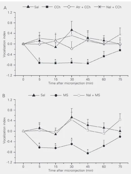
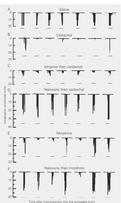
Microinjection of carbachol and mor-phine sulfate into the CEA promoted antino-ciception in guinea pigs, as determined by the decrease of VI in the vocalization test (Figures 1A,B and 2). Furthermore, the pres-ent results demonstrated that the antinoci-ceptive effect of carbachol (Figure 2B) was blocked by the previous administration of atropine (a muscarinic receptor antagonist) or naloxone (an opioidergic antagonist) into the same site (Figures 1A and 2C,D). In addition, the decrease in VI induced by mi-croinjection of morphine sulfate (Figure 2E) into the CEA was prevented by pretreatment with naloxone (Figures 1B and 2F). Saline administration into the CEA did not alter the VI after administration of a noxious periph-eral stimulus to the animal (Figures 1A,B and 2A), and this group was used as control in all other experiments. Furthermore, treat-ment with atropine or naloxone alone fol-lowed by saline did not promote significant alterations in the VI at any of the experimen-tal time points.
Individual MANOVA applied to the dif-ferent experimental groups revealed that car-bachol and morphine sulfate administration into the CEA promoted a significant differ-ence in the VI along the experiment (F6,54 =
6.12, P < 0.01 for carbachol and F6,48 = 5.48,
min after microinjection (P < 0.05, Duncan test), and this effect was observed up to 45 min after microinjection (Figure 1A,B). For the groups receiving saline, atropine + car-bachol, naloxone + carcar-bachol, and naloxone + morphine sulfate, statistical analysis did not show differences in VI with time after the microinjections (F6,48 = 0.92, P > 0.05;
F6,36 = 0.52, P > 0.05; F6,54 = 0.80, P > 0.05,
and F6,60 = 1.44, P > 0.05, respectively,
MANOVA).
For analysis of the experimental groups as a whole regarding VI variation after the different treatments, two-way ANOVA was performed, which showed a significant dif-ference among treatments with time (F36,336
= 1.75, P < 0.05, MANOVA; Figure 1A,B). We then applied one-way ANOVA followed by the Duncan test to each experimental time interval. For the 5-min interval, there was a significant difference (F6,56 = 3.7, P< 0.05,
ANOVA) between the carbachol and mor-phine sulfate treatments when compared to the other groups (P < 0.05, Duncan test); however, these groups did not differ from one another. Furthermore, saline, atropine + carbachol, naloxone + carbachol, and nalox-one + morphine sulfate treatments did not differ from one another. This difference was also observed at the 15-, 30- and 45-min time intervals (F6,56 = 5.6, P < 0.05; F6,56 = 4.8,
P < 0.05, and F6,56 = 4.7, P < 0.05, ANOVA,
respectively). The post hoc Duncan test showed a difference (P < 0.05) between the carbachol and morphine sulfate treatments and all other treatments at the same time intervals as mentioned above (Figure 1A,B); however, there was no difference among the saline, atropine + carbachol, naloxone + carbachol, and naloxone + morphine sulfate groups. No difference between the experi-mental groups was observed at 60 or 75 min (F6,56 = 1.5, P > 0.05; F6,56 = 1.6, P >
0.05, ANOVA, respectively; Figure 1A and B).
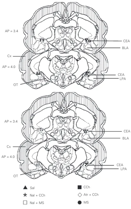
Figure 3 shows the sites of drug microin-jection in the CEA.
Figure 1. Cholinergic-opioidergic interaction in the central amygdala induces antinocicep-tion in the guinea pig. Data are reported as means ± SEM of the vocalizaantinocicep-tion index (VI) under control conditions (time zero, before microinjection) and after the administration of different drugs into the central nucleus of the amygdala (CEA) obtained for different experimental groups of conscious guinea pigs submitted to a peripheral noxious stimulus. The number of animals in each group was 7 to 11. A, VI after microinjection of saline (Sal, 0.9%, 0.2 µl; triangles); after carbachol microinjection (CCh, 2.7 nmol/0.2 µl; squares); after atropine (Atr, 0.7 nmol/0.2 µl) microinjection followed by CCh into the CEA (lozenges); after naloxone (Nal, 1.3 nmol/0.2 µl) microinjection followed by CCh into the CEA (sextile). B, VI after microinjection of Sal (0.9%, 0.2 µl; triangles); after morphine sulfate (MS; 2.2 nmol/ 0.2 µl; circles) and after Nal followed by MS (squares). *P < 0.05 compared to respective control (time zero) and to the Sal, Atr + CCh, Nal + CCh, and Nal + MS groups at each time of the experiment (Duncan test).
Vocalization index
1.2
0.8
0.4
0.0
-0.4
-0.8
-1.2
1.2
0.8
0.4
0.0
-0.4
-0.8
-1.2
Vocalization index
0 5 15 30 45 60 75
Time after microinjection (min)
Sal CCh Atr + CCh Nal + CCh
* * * *
* *
*
*
Sal MS Nal + MS
0 5 15 30 45 60 75
Time after microinjection (min)
Discussion
Various studies have demonstrated that chemical or electric stimulation of different
A
brain sites is able to promote antinocicep-tion, with the PAG, dorsal raphe nucleus and magnus raphe nucleus having been exten-sively explored (21). On the other hand, some reports have shown the role of the amygdala in antinociception as well as its involvement in emotional modulation (2) and defensive responses (4). Probably, the amygdaloid circuitry that contributes to anti-nociception involves a connection with the neural substrate that is activated in defensive antinociception following fear behaviors (3,12). On this basis, it is possible that the same neural substrate will be responsible for the modulation of defensive behavior and antinociception. In this respect, several stud-ies have reported the involvement of some brain structures including the parabrachial area (22,23), PAG (24,25), lateral hypothala-mus (26,27), and amygdala (4,6,14) in the modulation of defensive behavior and anti-nociception.
The involvement of the amygdaloid com-plex in nociceptive regulation has been dem-onstrated in different investigations (3,6,12, 13), but guinea pigs and the vocalization test are not commonly used in experiments des-tined to the study of the neural mechanisms of analgesia. In this respect, Oliveira and Prado (3) demonstrated that microinjection of carbachol into different amygdaloid nu-clei (basolateral, lateral posterior and cen-tral) induced an increase of the withdrawal reflex in the tail-flick test in rats. Further-more, the same treatment promoted behav-ioral alterations such as motor hyperactivity and masticatory movements. The authors suggested that the antinociception and be-havioral alterations induced by the intra-amygdaloid administration of carbachol de-pend on the activation of muscarinic recep-tors, because pretreatment with atropine pre-vented the behavioral and antinociceptive effect of carbachol. In addition, ip adminis-tration of diazepam blocked only the behav-ioral responses induced by carbachol admin-istered into the CEA, indicating that the Figure 2. Schematic drawing of the vocalization amplitude (mm) after administration of
different drugs into the central nucleus of the amygdala (CEA) of guinea pigs. A, Animal microinjected with saline; B, animal treated with carbachol (CCh) into the CEA; C, animal treated with atropine (Atr) followed by CCh; D, animal microinjected with naloxone (Nal) followed by CCh; E, animal microinjected with morphine sulfate (MS); F, animal treated with Nal followed by MS into the CEA. The horizontal bar indicates the application of the noxious stimuli (3 s) at the different time intervals (min). The zero represents the vocaliza-tion amplitude during the control period (without saline or drug administravocaliza-tion). The quanti-ties of the drugs employed are given in the legend to Figure 1.
Vocalization amplitude (mm)
D
0
A
0
B
0
C
0
E
0
F
0
10
20
30 10
20
30 10
20
30 10
20 10
20
30 10
20
40
Saline
Carbachol
Atropine then carbachol
Naloxone then carbachol
Morphine
Naloxone then morphine
antinociceptive and behavioral responses induced by carbachol stimulation depend on different circuitries in the amygdala (3). These findings support the present results obtained for guinea pigs, which showed that carba-chol stimulation of the CEA promoted anti-nociception evidenced by a decrease in the VI (Figure 1A), and also agree with the results of our previous study (16) in which carbachol promoted a decrease in the defen-sive response of tonic immobility. Taken together, these results suggest that the acti-vation of the cholinergic system of the cen-tral amygdala is involved in the modulation of defensive behavior and antinociception.
Also, the results reported by Oliveira and Prado (28) showing that electric stimulation of the CEA promoted antinociception in the tail-flick test in rats and that this effect was blocked by the systemic administration of atropine, naloxone and propranolol indicate the participation of the cholinergic, opio-idergic and adrenergic systems in the antino-ciception induced by CEA stimulation. In this context, in addition to the involvement of the cholinergic system of the CEA in nociceptive regulation, other neurotransmit-ters of this substrate are strongly involved in nociception. Manning (29) observed the lat-eralized loss of opioid antinociception (ip
administration of morphine) after unilateral inactivation of the CEA in the formalin test. These results suggest a topographic ipsilat-eral organization in the descendent control of nociception. These data agree with those of the present study in which antinocicep-tion induced by the administraantinocicep-tion of carba-chol or morphine sulfate into the CEA was observed in guinea pigs after application of a noxious electric stimulus to the thigh ipsilat-erally to the microinjection site of the drug. Moreover, Pavlovic et al. (14), studying the opioidergic system of the amygdala, showed that the administration of morphine or ß-endorphin into the CEA promoted antinoci-ception in the tail-flick test and in the jump test in rats. These results agree with our
AP = 4.0 AP = 3.4
AP = 3.4
Cx
AP = 4.0
OT
OT
CEA
BLA
CEA LPA
CEA
CEA LPA
Sal
BLA
Nal + CCh
Nal + MS
Atr + CCh CCh
MS Cx
findings showing that morphine sulfate ad-ministration into the CEA induced antinoci-ception in the vocalization test, as demon-strated by a decrease in VI (Figure 1B), and that this effect could be prevented by pre-administration of naloxone into the same site, suggesting that the effect of morphine sulfate occurs at the level of local opioider-gic receptors.
Our results again showed that the antino-ciception induced by carbachol or morphine sulfate administered into the CEA is pre-vented by pretreatment with naloxone at the same site, suggesting that a cholinergic-opio-idergic interaction in this substrate is respon-sible for the nociceptive modulation. In this respect, Guimarães et al. (24) reported that antinociception induced by carbachol ad-ministration into the dorsal PAG of rats evalu-ated by the vocalization test was blocked by treatment with atropine, mecamylamine or naloxone at the same site. These results dem-onstrated the interaction of the cholinergic and opioidergic systems in dorsal PAG modu-lation of antinociception. The same interac-tion was also observed in our previous report (19) but in the ventral PAG when carbachol or morphine sulfate stimulation promoted a decrease of VI in guinea pigs. Again, a simi-lar cholinergic-opioidergic interaction was demonstrated in the CEA, but involved in the control of the defensive behavior of tonic
immobility (16). On this basis, we propose that cholinergic transmission might activate the release of endorphins/enkephalins from interneurons of the CEA, resulting in a de-creased VI. Finally, in agreement with Har-ris (30), it is possible that the CEA partici-pates in descending antinociceptive mechan-isms through its anatomical-functional con-nection with the PAG. In this respect, Harris (30) suggested that defensive systems in the amygdala are able to activate PAG antinoci-ceptive mechanisms via direct, possibly en-kephalinergic, projections that inhibit the local GABAergic neurons in the PAG. In addition, the descending projections from the PAG to intraspinal antinociceptive mech-anisms are under the tonic inhibitory influ-ence of local GABAergic neurons.
The results of the present study showed that the activation of the cholinergic or opio-idergic system of the CEA promotes antino-ciception in guinea pigs as evidenced by a decrease of the vocalization index. In addi-tion, antinociception produced by cholin-ergic stimulation of the CEA depends on opioid synapses present at the same site.
Acknowledgments
We thank Mr. Rubens Fernando de Melo for histological processing of the specimens.
References
1. Kapp BS, Whalen PJ, Supple WF & Pascoue JP (1992). Amygdaloid contributions to conditioned arousal and mental and sensory infor-mation processing. In: Aggleton JP (Editor), The Amygdala: Neuro-biological Aspects of Emotions, Memory and Mental Dysfunction.
Wiley-Liss, New York.
2. Gallangher M & Chiba AA (1996). The amygdala and emotion.
Current Opinion in Neurobiology, 6: 221-227.
3. Oliveira MA & Prado WA (1994). Antinociception and behavioral manifestations induced by intracerebroventricular or intra-amygda-loid administration of cholinergic agonists in the rat. Pain, 57: 383-391.
4. Leite-Panissi CRA, Monassi CR & Menescal-de-Oliveira L (1999).
Role of amygdaloid nuclei in the modulation of tonic immobility in guinea pigs. Physiology and Behavior, 67: 717-724.
5. Danielson EH, Magnunson DJ & Gray TS (1989). The central amyg-daloid nucleus innervation of the dorsal vagal complex in rat: a
Phaseolus vulgaris, leukoagglutinin lectin anterograde tracing study.
Brain Research Bulletin, 22: 705-715.
6. Oliveira MA & Prado WA (2001). Role of PAG in the antinociception evoked from the medial or central amygdala in rats. Brain Research Bulletin, 54: 55-63.
7. Fox RJ & Sorenson CA (1994). Bilateral lesions of the amygdala attenuate analgesia induced by diverse environmental challenges.
8. Ahn DK, Kim YS & Park JS (1999). Central-amygdaloid carbachol suppressed nociceptive jaw opening reflex in freely moving rats.
Progress in Neuro-Psychopharmacology and Biological Psychiatry, 23: 685-695.
9. Bernard JF & Besson JM (1990). The spino(trigemio)pontoamyg-daloid pathway: electrophysiological evidence for an involvement in pain processes. Journal of Neurophysiology, 63: 473-490. 10. Chow TW & Cummings JL (2000). The amygdala and Alzheimer’s
disease. In: Aggleton JP (Editor), The Amygdala: A Functional Analy-sis. Oxford University Press, New York.
11. Cassel MD, Gray TS & Kiss JZ (1986). Neuronal architecture in the rat central nucleus of the amygdala: a cytological, hodological and immunocytochemical study. Journal of Comparative Neurology, 246: 278-299.
12. Manning BH & Mayer DJ (1995). The central nucleus of the amyg-dala contributes to the production of morphine antinociception in the formalin test. Pain, 63: 141-152.
13. Helmstetter FJ, Bellgowan PSF & Poore LH (1995). Microinfusion of
mu but not delta or kappa opioid agonist into the basolateral amyg-dala results in inhibition of the tail-flick reflex in pentobarbital-anes-thetized rats. Journal of Pharmacology and Experimental Therapeu-tics, 275: 381-388.
14. Pavlovic ZW, Cooper ML & Bodnar RJ (1996). Opioid antagonists in the periaqueductal gray inhibit morphine and ß-endorphin analgesia elicited from the amygdala of rats. Brain Research, 741: 13-26. 15. Leite-Panissi CRA, Coimbra NC & Menescal-de-Oliveira L (2003).
The cholinergic stimulation of the central amygdala modifying the tonic immobility response and antinociception in guinea pigs de-pends on the ventrolateral periaqueductal gray. Brain Research Bulletin, 60: 167-178.
16. Leite-Panissi CRA & Menescal-de-Oliveira L (2002). Central nucleus of the amygdala and tonic immobility in guinea pigs. Brain Research Bulletin, 58: 13-19.
17. Zimmermann M (1983). Ethical guidelines for investigations of ex-perimental pain in conscious animals. Pain, 16: 109-110.
18. Menescal-de-Oliveira L & Lico MC (1977). Pain modulation in the adrenergically stimulated area postrema in the guinea pig. Physiolo-gy and Behavior, 19: 359-364.
19. Menescal-de-Oliveira L, Brentegani MR, Leite-Panissi CRA & Monassi CR (1999). Cholinergic-opioidergic interaction in the ven-trolateral periaqueductal gray matter inducing antinociception in guinea pigs. 9th World Congress on Pain, August 22-29, 1999, Vienna, Austria, 148 (Abstract 58).
20. Rössner W (1965). Stereotaktischer Hirnatlas vom Meersch-weinchen. Palla Verlag, Munich, Germany.
21. Besson JM & Chaouch A (1987). Peripheral and spinal mechanisms of nociception. Physiological Reviews, 67: 167-186.
22. Katayama Y, Watkins LR, Becker DP & Hayes RL (1984). Non-opiate analgesia induced by carbachol microinjection into the pontine para-brachial region of the cat. Brain Research, 296: 269-283.
23. Menescal-de-Oliveira L & Hoffmann A (1993). The parabrachial re-gion: a possible substrate shared by the systems that modulate pain and tonic immobility. Behavioural Brain Research, 56: 127-132. 24. Guimarães APC, Guimarães FS & Prado WA (2000). Modulation of carbachol-induced antinociception from the rat periaqueductal gray.
Brain Research Bulletin, 51: 471-478.
25. Monassi CR, Leite-Panissi CRA & Menescal-de-Oliveira L (1999). Ventrolateral periaqueductal gray matter and the control of tonic immobility. Brain Research Bulletin, 50: 201-208.
26. Dafny N, Dong WQ, Prieto-Gomez C, Reyes-Vasquez C, Stanford J & Qiao JT (1996). Lateral hypothalamus: site involved in pain modu-lation. Neuroscience, 70: 449-460.
27. Oliveira L, Hoffmann A & Menescal-de-Oliveira L (1997). The lateral hypothalamus in the modulation of tonic immobility in guinea pigs.
NeuroReport, 8: 3489-3493.
28. Oliveira MA & Prado WA (1998). Antinociception induced by stimu-lating amygdaloid nuclei in rats: Changes produced by systemically administered antagonists. Brazilian Journal of Medical and Biologi-cal Research, 31: 681-690.
29. Manning BH (1998). A lateralized deficit in morphine antinociception after unilateral inactivation of the central amygdala. Journal of Neu-roscience, 18: 9453-9470.