ISSN 0100-879X
BIOMEDICAL SCIENCES
AND
CLINICAL INVESTIGATION
www.bjournal.com.br
www.bjournal.com.br
Volume 43 (4) 268-380 April 2011
Braz J Med Biol Res, April 2011, Volume 44(4) 327-331
doi: 10.1590/S0100-879X2011007500021
Effect of neurotrophic factor, MDP, on rats' nerve regeneration
A.A. Fornazari, M.R. de Rezende, R. Mattar Jr., R.I. Taira, G.B. dos Santos and R.G. Paulos
Faculdade de Medicina de Ribeirão Preto Campus
Ribeirão Preto
Institutional Sponsors
The Brazilian Journal of Medical and Biological Research is partially financed by
analiticaweb.com.br S C I E N T I F I C Hotsite of proteomics metabolomics
Effect of neurotrophic factor, MDP, on rats’
nerve regeneration
A.A. Fornazari, M.R. de Rezende, R. Mattar Jr., R.I. Taira,
G.B. dos Santos and R.G. Paulos
Laboratório de Microcirurgia, Instituto de Ortopedia e Traumatologia, Hospital das Clínicas, Universidade de São Paulo, São Paulo, SP, Brasil
Abstract
Our objective was to determine the immune-modulating effects of the neurotrophic factor N-acetylmuramyl-L-alanyl-D-isoglu-tamine (MDP) on median nerve regeneration in rats. We used male Wistar rats (120-140 days of age, weighing 250-332 g) and compared the results of three different techniques of nerve repair: 1) epineural neurorrhaphy using sutures alone (group
S - 10 rats), 2) epineural neurorrhaphy using sutures plus fibrin tissue adhesive (FTA; group SF - 20 rats), and 3) sutures plus
FTA, with MDP added to the FTA (group SFM - 20 rats). Functional assessments using the grasp test were performed weekly
for 12 weeks to identify recovery of flexor muscle function in the fingers secondary to median nerve regeneration. Histological analysis was also utilized. The total number and diameter of myelinated fibers were determined in each proximal and distal nerve segment. Two indices, reported as percentage, were calculated from these parameters, namely, the regeneration index and the diameter change index. By the 8th week, superiority of group SFM over group S became apparent in the grasping test (P = 0.005). By the 12th week, rats that had received MDP were superior in the grasping test compared to both group S (P < 0.001) and group SF (P = 0.001). Moreover, group SF was better in the grasping test than group S (P = 0.014). However, no
significant differences between groups were identified by histological analysis. In the present study, rats that had received MDP obtained better function, in the absence of any significant histological differences.
Key words: Grasping test; Neurotrophic factor; N-acetylmuramyl-L-alanyl-D-isoglutamine (MDP)
Introduction
Correspondence: A.A. Fornazari, Laboratório de Microcirurgia, Instituto de Ortopedia e Traumatologia, HC, USP, Rua Ovídio Pires
de Campos, 333, 05403-010 São Paulo, SP, Brasil. Fax: +55-11-3067-7000. E-mail: [email protected]
Received May 15, 2010. Accepted February 3, 2011. Available online February 25, 2011. Published April 11, 2011. When peripheral nerve is damaged it undergoes the
de-generation process known as Wallerian dede-generation. After
nerve injury, a local inflammatory reaction occurs that involves
macrophages. These cells, in turn, are responsible for many of the fundamental events that occur during the regenera-tive process. Macrophages play a crucial role in processes of Wallerian degeneration and subsequent regeneration of peripheral nerves (1). The use of neurotrophic factors has been of special interest to researchers investigating the best ways to stimulate nerve regeneration. N-acetylmuramyl-L-alanyl-D-isoglutamine (MDP) is an immuno-adjuvant that promotes increased macrophage activity (2). When injected into the crushed sciatic nerve, MDP activates macrophages and promotes recovery of walking locomotion in rats (3). Some results indicate that locally applied MDP stimulates peripheral nerve regeneration in rats (4). Based on the
feasibility of using fibrin glue mixed with the neurotrophic
factor, we decided to study the immunomodulatory effect of
MDP on the process of peripheral nerve regeneration in a
rat experimental model.
Material and Methods
Animals
Healthy male Wistar rats between 120 and 140 days of age and weighing 250-332 g were used, all without
mo-tor deficits. Rats were maintained in cages in groups of 5.
The cages were maintained in an acclimatized atmosphere under hygienic conditions, with food and water provided ad libitum. The study was approved by the Ethics Committee of Departamento de Ortopedia e Traumatologia, Hospital das Clínicas, Universidade de São Paulo (USP), Brazil, and was conducted according to the ethical guidelines for animal
experimentation established by the Brazilian School of Animal Experimentation and by the Institute of Laboratory Animal
328 A.A. Fornazari et al.
Groups
Rats were anesthetized intraperitoneally (ip) with 30 to 40 mg/kg pentobarbital. An incision was made in the prepped
and sterilized axillary area of the right upper extremity, and the median nerve was identified and dissected. The nerve was transected with micro-scissors 5 mm proximal to the
medial epicondyle of the humerus and the lesion was im-mediately repaired always by the same surgeon.
Three groups of rats were used: group S (N = 10) was subjected to end-to-end epineural neurorrhaphy without ten-sion, using four simple 10-0 mononylon sutures. Group SF (N = 20) was subjected to epineural neurorrhaphy without
tension, using two simple sutures plus 0.10 mL fibrin tissue adhesive (FTA; Tissucol®, Immuno AG, Austria). Group SFM
(N = 20) was subjected to the same procedure as group SF,
except that 0.1 mL neurotrophic factor MDP (Sigma Chemi -cal Co., USA) was added to the FTA. MDP was maintained at 15°C and diluted in 1 mL saline to a 2-mM concentration.
After adding an equal volume of FTA, the final concentration
of MDP was 1 mM. Surgery was performed under a light
microscope at 12X magnification. After nerve repair, the
surgical wound was closed with 3.0 mononylon sutures.
Rats were excluded from analysis for the following reasons:
perioperative or post-operative death of the animal, tissue
loss in the injured area, autophagy or other significant mu -tilation in an animal, deep infection at the site of the lesion, and postoperative loss of more than 10% of preoperative
weight. For the three groups, 50 rats were used. Six rats were excluded from the SFM group (N = 20). Two weeks postoperatively, 3 rats exhibited autophagy in the area of the surgical wound and significant motor alteration. During
the 5th postoperative week, one rat died of an idiopathic
cause. Two rats failed to exhibit any measurable flexor
strength during the grasping test over the entire 12-week
postoperative period and, once euthanized and examined,
both dehiscence of the sutures and tissue loss in the area of the median nerve lesion were noted.
Functional analysis
All rats were submitted to a grasping test for functional evaluation, as classically described (5). For the test, each rat was elevated by the tail and stimulated to grab a metal
hook that comprised the apex of a metal pyramidal frame.
The total frame weighed 700 g and rested on a conventional
digital scale. The left upper extremity of the rat had been previously immobilized with adhesive tape. To examine finger flexion, we observed the rat grasping the hook while being held. At the same time, a second observer filmed
the procedure and recorded the weight registered on the
scale at the exact moment that the rat released the hook.
The difference between the weight of the metal frame (700 g) and the weight registered by the second observer was
used to estimate the force generated by the muscle flexors of the fingers. When there was flexion of the palm or wrist, the data were excluded.
Rats were submitted to the grasp test both preoperatively and postoperatively. After repair of the nerve injury, each rat underwent the grasping test three times every 2 weeks through the 12th week, with the average of each three
grouped tests used to represent the mean flexor strength
at each time. These values were compared between the three groups to determine the rate and degree of functional recovery. Rats were weighed before each grasp test. At the
end of the 12th postoperative week, rats were sacrificed by
ip injection of a lethal dose of pentobarbital.
Histological analysis
Ten rats were chosen at random from each group. The
nerve segments proximal and distal to the repair were dis -sected. A 2-cm segment of the nerve including the repair region was removed. Two 4-mm long segments were
ob-tained, 4 mm proximal and 4 mm distal to the repair site, and fixed in a solution of 2% osmium tetroxide. Transverse sectioning was performed and 1-μm sections were obtained
and stained with 1% toluidine blue. For histological analysis, each section was photographed using a digital camera
(Coolpix; Nikon, Japan) coupled to a light microscope, the objective of which had a magnification of 40 times. One random field of each proximal and distal segment was photographed. Each picture captured an area of 36,000
µm2. The pictures from the proximal and distal segment of each nerve were analyzed using the Sigma Scan Pro 5.0 software (SPSS Inc., USA). We obtained the total number
and diameter of myelinated fibers. Two indices, expressed
as a percentage, were calculated from these parameters,
namely, the regeneration index and the diameter change index. The regeneration index was calculated by dividing the total number of regenerated axons in the distal segment by the total number of regenerated axons in the proximal segment, resulting in the percentage of axons that crossed the repair site. The diameter change index was calculated by dividing the mean diameter of regenerated axons in
the distal segment by the mean diameter of regenerated
axons in the proximal segment (6). These indexes were
compared to the results obtained in the functional
evalua-tions to determine the extent to which functional recovery reflected the histological findings (6).
Statistical analysis
Data are reported as means ± standard deviation. For the functional test, data were analyzed by two-way repeated measures analysis of variance (ANOVA) followed by the Tukey post hoc test. For histological analysis,
one-way ANOVA was used with the level of significance set at
P < 0.05.
Results
Functional analysis
weeks, and therefore it was not necessary to exclude any
animal due to weight loss.
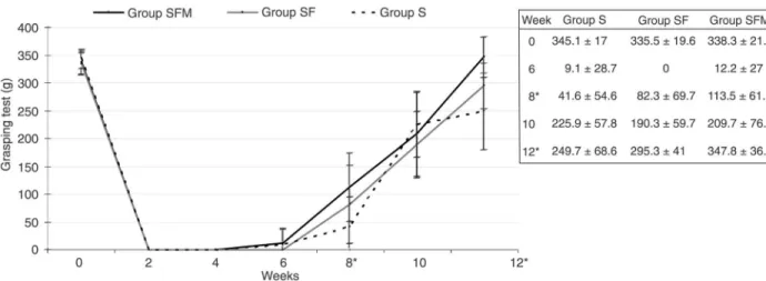
Figure 1 illustrates the median nerve profiles of the three study groups obtained at the time of the first evaluation (t0) and at the end of the 6th, 8th, 10th, and 12th weeks
after surgery. After surgery, all groups ultimately displayed evidence of increasing median nerve function over time.
Statistically significant differences were apparent, however,
at week 8 (P = 0.019) and week 12 (P < 0.001). Post hoc analysis of week 8 data revealed greater grasping strength in group SFM than in group S (P = 0.05). At week 12, the grasping strength of group SFM was greater than that of
both group S (P < 0.001) and group SF (P = 0.001) and, at the same time, the strength of group SF was greater than that of group S (P = 0.014). At week 12, groups SF and S failed to achieve their respective average values estimated before surgery (t0). The SFM group showed total functional recovery.
Histological analysis for axonal regeneration
Table 1 presents descriptive statistics for the three study groups. No difference was observed between the parameters evaluated. The requirement for similar variances between groups was met, justifying the use of ANOVA.
Figure 1. Median nerve profiles during the grasping test in the three study groups. Data are reported as means ±standard deviation.
Group S = epineural neurorrhaphy; group SF = epineural neurorrhaphy plus tissue fibrin adhesive; group SFM = epineural neuror
-rhaphy plus tissue fibrin adhesive and neurotrophic factor. *At week 8, grasping strength was greater in group SFM than in group S (P = 0.05). *At week 12, grasping strength was greater in group SFM than in both group S (P < 0.001) and group SF (P = 0.001). Data
were analyzed by two-way repeated measures ANOVA followed by the post hoc Tukey test.
Table 1. Results of histomorphometric evaluation after statistical comparison between the groups.
Group parameter Group S Group SF Group SFM
PA 520.20 ± 89.21 489.30 ± 59.91 511.00 ± 227.60 DA 849.00 ± 191.20 724.70 ± 107.10 757.50 ± 156.90
RI (%) 164 ± 27 150 ± 27 172 ± 71
PD (μm) 5.89 ± 1.24 5.78 ± 1.24 6.56 ± 1.45
DD (μm) 3.57 ± 0.56 3.77 ± 0.37 3.96 ± 0.74
DCI (%) 62 ± 11 67 ± 10 67 ± 40
330 A.A. Fornazari et al.
Discussion
Group SFM was functionally superior to the other two groups at weeks 8 and 12 and showed total functional recovery over 12 weeks, although histological analysis
failed to reveal any significant difference between the three
groups. Fukuyama et al. (3) compared intrafascicular injec-tion of 50 µL Ringer soluinjec-tion (Lactec® injection, Ohtsuka,
Japan) with or without MDP at 2 mg/mL into the crushed sciatic nerve of rats. The injection was done with a syringe
attached with an ultrafine needle (29G) slightly distal to the
crushed site. In the MDP group, they reported an increase
in the axonal elongation measured by the pinch test 5 days
after the crush operation (P < 0.05) and increased functional
recovery measured by the sciatic nerve functional index
in walking track analysis, 3 weeks after the operation and injection (P < 0.01) (3).
In our study, histological analysis failed to confirm any
immunoadjuvant activity of MDP. The discrepancy between the results of the functional and histological analysis
might be explained by the grasping test being able to as
-sess viable nerve fibers among those still regenerating. Moreover, regenerating axons might undergo misdirected
growth leading to aberrant muscle reinnervation (7,8) and
the presence of polyneuronally innervated muscle fibers may reduce the efficiency of peripheral nerve regeneration
(9). The group with the worst results was the suture alone group (S), especially compared to the MDP-treated group (SFM) at weeks 8 and 12. As some investigators have speculated, neurorrhaphy could hinder nerve regeneration
in several ways: by escalating the inflammatory response at
the repair point, by causing deviations in the path of those
axons otherwise aiming towards the distal segment of the nerve (10), by temporarily reducing blood flow through the
microcirculation (11), and via the formation of mechanical obstructions such as scar tissue.
The use of neurotrophic substances has been of special interest to those investigating ways by which to optimize nerve regeneration. MDP is a minimum structural unit re-sponsible for the immunoadjuvant activities of bacterial cell wall peptidoglycans in a variety of species (4). It is known that MDP can be considered a functional lipopolysaccha-ride (LPS) analogue (12). MDP is an immuno-adjuvant that promotes increased activity among microglial phago-cytes, Schwann cells and macrophages (2). Wallerian degeneration involves cellular and molecular activities
coordinated by a complex network of cytokines that also
contribute to macrophage recruitment and activation and, consequently, to macrophage-dependent functions like myelin removal by phagocytosis (13). A decreased response of these cells in injured nerves has been associated with
decreased axonal regeneration (14). However, an interven
-tion can accelerate the regenerative process by introducing
inflammatory cells or an enhanced inflammatory response
(14). The macrophages rapidly remove myelin debris that contain neurite growth inhibitors like myelin-associated glycoprotein. They release cytokines such as interleukin
-1 (IL-1) and macrophage inflammatory protein-1 (MIP-1),
which probably stimulate Schwann cells and other types
in the nerves (3) and growth factors that facilitate axonal
regeneration (14). One of the activation mechanisms of macrophages occurs through tumoral necrosis factor-alpha
(TNF-α), which is one of the first cytokines synthesized by
Schwann cells during the Wallerian degeneration process
(15). TNF-α directly stimulates the macrophages (16)
and induces the production of granulocyte-macrophage
colony-stimulating factor (GM-CSF) by fibroblasts, promot -ing macrophage activation. MDP is capable of induc-ing
the formation of several cytokines like TNF-α or stimu -lates cells to produce them (17), indirectly contributing to macrophage activation. Spinal cord cells were induced to proliferate temporarily by the peripheral administration of certain stimulators of the innate immune system, including MDP (18). Toll-like receptor (TLR) and NOD2 ligands are key receptors that allow mammals to detect the presence
of infection by recognizing specific microorganism-derived
biomolecules, the so-called pathogen-association molecular patterns (PAMPs). MDP is an NOD2 ligand. Recognition of PAMPs through TLR and NOD2 receptors appears to
link the inflammatory and regenerative process. However,
other studies have shown that myelin fraction-stimulated macrophages release neurotrophic factors that increase the survival and neurite regeneration of adult dorsal root ganglion neurons in culture (19). Adding MDP or LPS to the culture medium did not enhance neurotrophic secre-tion by macrophages. These observasecre-tions suggest that
myelin may be a specific activator of macrophages in nerve
regeneration.
In summary, using a transected median nerve rat model, we demonstrated enhanced functional recovery twice along the postoperative period of 12 weeks in rats treated with
the combination of suturing, fibrin tissue adhesive and the
neurotrophic factor MDP compared with rats treated either with neurorrhaphy alone or with sutures plus FTA without MDP. Histological analysis, however, did not identify any
difference by which to explain the enhanced functional
recovery.
Acknowledgments
References
1. Bruck W. The role of macrophages in Wallerian degenera-tion. Brain Pathol 1997; 7: 741-752.
2. Parant MA, Parant FJ, Le Contel C, Lefrancier P, Chedid L. MDP derivatives and resistance to bacterial infections in mice. Adv Exp Med Biol 1992; 319: 175-184.
3. Fukuyama R, Takeda H, Fushiki S, Yamamoto T. Muramyl dipeptide injected into crushed sciatic nerve, activates mac-rophages and promotes recovery of walking locomotion in rats. Restor Neurol Neurosci 1998; 13: 213-219.
4. Inoe AP, Pereira FC, Stopiglia AJ, Da-Silva CF. Pharma-cological immunomodulation enhances peripheral nerve regeneration. Pesq Vet Bras 2007; 27: 363-369 (http://www. scielo.br/pdf/pvb/v27n9/v27n9a02.pdf).
5. Bertelli JA, Mira JC. The grasping test: a simple behavioral method for objective quantitative assessment of peripheral nerve regeneration in the rat. J Neurosci Methods 1995; 58: 151-155.
6. Martins RS, Siqueira MG, Da Silva CF, Plese JP. Overall
assessment of regeneration in peripheral nerve lesion repair
using fibrin glue, suture, or a combination of the 2 techniques
in a rat model. Which is the ideal choice? Surg Neurol 2005;
64 (Suppl 1): S1-S6.
7. Dellon AL, Mackinnon SE. Sciatic nerve regeneration in the rat. Validity of walking track assessment in the presence of chronic contractures. Microsurgery 1989; 10: 220-225. 8. Bodine-Fowler SC, Meyer RS, Moskovitz A, Abrams R, Botte
MJ. Inaccurate projection of rat soleus motoneurons: a com-parison of nerve repair techniques. Muscle Nerve 1997; 20: 29-37.
9. Benfrech E, Alnot JY, Henin D. [An experimental study of
nerve sutures and grafts of the sciatic nerve in the rat using
fibrin glue]. Ann Chir Main 1989; 8: 296-299.
10. Smahel J, Meyer VE, Bachem U. Glueing of peripheral
nerves with fibrin: experimental studies. J Reconstr Micro-surg 1987; 3: 211-220.
11. Moy OJ, Peimer CA, Koniuch MP, Howard C, Zielezny Katikaneni PR. Fibrin seal adhesive versus nonabsorbable microsuture in peripheral nerve repair. J Hand Surg Am
1988; 13: 273-278.
12. Hoffmann JA, Kafatos FC, Janeway CA, Ezekowitz RA. Phylogenetic perspectives in innate immunity. Science 1999; 284: 1313-1318.
13. Ramon y Cajal S. Degeneration and regeneration of the nervous system. London: Oxford University Press; 1928. 14. Kiefer R, Kieseier BC, Stoll G, Hartung HP. The role of
mac-rophages in immune-mediated damage to the peripheral nervous system. Prog Neurobiol 2001; 64: 109-127. 15. Shamash S, Reichert F, Rotshenker S. The cytokine network
of Wallerian degeneration: tumor necrosis factor-alpha, interleukin-1alpha, and interleukin-1beta. J Neurosci 2002;
22: 3052-3060.
16. Moonis M, Ahmad I, Bachhawat BK. Macrophages in host
defence - an overview. Indian J Biochem Biophys 1992; 29: 115-122.
17. Wolfert MA, Murray TF, Boons GJ, Moore JN. The origin of
the synergistic effect of muramyl dipeptide with endotoxin
and peptidoglycan. J Biol Chem 2002; 277: 39179-39186. 18. Su Y, Zhang Z, Trautmann K, Xu S, Schluesener HJ. TLR
and NOD2 ligands induce cell proliferation in the rat intact spinal cord. J Neuropathol Exp Neurol 2005; 64: 991-997. 19. Hikawa N, Takenaka T. Myelin-stimulated macrophages