J of Evolution of Med and Dent Sci/ eISSN- 2278-4802, pISSN- 2278-4748/ Vol. 4/ Issue 38/ May 11, 2015 Page 6572
CUT THROAT INJURIES AND IT MANAGEMENT: A CLINICAL STUDY OF 48
PATIENTS
Md. Naveed Ahmed1, M. Sreedhar Rao2, S. Muneeruddin Ahmed3, M. Mahendra Kumar4
HOW TO CITE THIS ARTICLE:
Md. Naveed Ahmed, M. Sreedhar Rao, S. Muneeruddin Ahmed, M. Mahendra Kumar. Cut Throat Injuries and it Management: A Clinical Study of 48 Patients . Journal of Evolution of Medical and Dental Sciences 2015; Vol. 4, Issue 38, May 11; Page: 6572-6581, DOI: 10.14260/jemds/2015/953
ABSTRACT: INTRODUCTION: Cut throat injuries are one of the emergency conditions managed by E.N.T. specialists. They are clinically interesting and pose a challenge to the surgeon in the management. Six out of the ten cut throat injuries treated at casualty department are homicidal in nature. Such injuries can involve any part of the upper airway from the supra hyoid region to upper trachea. Commonest site of involvement in our series was between thyroid cartilage and cricoid cartilage. Initial management was done by securing the airway with preliminary tracheostomy below the cut in the neck and introducing a cuffed tracheostomy tube of appropriate size. Thorough exploration of the wound to note other vascular or neural injuries and injuries to esophagus was carried out. Initial management consisted of Correction of hypovolaemia, ligation of the bleeding vessels and primary repair of the wound in layers. The cut edges of the pre laryngeal and pre tracheal muscles are identified and sutured to strengthen the gap between the cartilages. The mortality is low if the treatment protocols are followed meticulously. MATERIALS AND METHODS: 48 Patients presenting with cut throat injuries at the casualty are taken for the study. 28 cases are homicidal, 14 are homicidal and 3 are accidental and 3 are road traffic accidents. All the injuries are open variety. They are examined, resuscitated and treated by primary suture. The data is classified and analyzed, depending upon level of the cut injury in the airway, the length and depth of the cut injury. The important structures damaged involvement of air and food passages. Is observed and recorded. Complications during the management are analyzed and treated. OBSERVATION: The cause of injury is homicidal, suicidal and RTA in nature. All the cut throat injuries encountered in this study are open variety. Complications included mediastinal emphysema, hypovolemic shock, wound dehiscence and laryngo-tracheal stenosis. Survival rate is improved by prompt treatment DISCUSSION: As all the cut throat injuries presented airway compromise we managed the patients by securing the airway by tracheostomy below the level of injury to airway. Apart from providing airway, this would give rest to the site of injury & avoid aspiration. Hypovolaemia was corrected with volume expanders and blood transfusion. After initial management patients were shifted to the operation theatre. Most of the patients were managed under General Anesthesia. Tracheostomy provided the route for administration of General Anesthesia. Wound debridement was done as most of the cases presented more than 6 to 8 hrs from the time of injury. Exploration of the wound was done to identify vascular or neural damage and treated accordingly. RESULTS: In the present study mortality rate is 10%. Complications are managed by conservative methods. Two patients required laryngeal and tracheal stents to correct stenosis.
J of Evolution of Med and Dent Sci/ eISSN- 2278-4802, pISSN- 2278-4748/ Vol. 4/ Issue 38/ May 11, 2015 Page 6573
INTRODUCTION: The incidence of Cut throat injuries (CTI) is not so uncommon in Telangana. CTIs are potentially dangerous, If not treated in time may lead to death of the patient due to asphyxia and Hemorrhage. Cut throat injuries are a sub-classification of neck injuries and are usually open type. The depth of the cut varies depending upon the actual cause behind the cut; accidental or homicidal or suicidal. It also depends upon the nature of the weapon.(1) In our country, closed injuries are more common, usually resulting from traffic accidents. The management of neck injuries is controversial in terms of both diagnosis and treatment.(2) Few authors consider exploration of the wound necessary if it crosses platysma. The external wound may appear small, but the nature of vascular injury and injury to the important structures like recurrent laryngeal nerve, Larynx can be known only after exploration. Conservative management is possible only if the airway is uninvolved.(3-5) The commonly used sharp objects such as razor, knives, or broken bottle pieces or glasses that may cause superficial or penetrating injuries.
This may result from accident, homicide, or a suicide attempt.(6) The common causes of CTIs in this part of the world are suicide attempts. Family problems, psychiatric illness, unemployment, and poverty may be the triggering factors in suicide attempts.(5) Exposed Hypopharyx and or larynx, hemorrhage, shock, and asphyxia from aspirated blood are the common causes of death following a CTI. It is known that appropriate measures can save lives in the majority of cases.(7,8) The value of tracheostomy in the management of CTI has been highlighted in the literature. All patients who have attempted suicide should undergo a psychiatric evaluation. The objective of the present study is to analyze the clinical presentation of cut throat injuries, to propose a management protocol in the treatment. As there are a very few scientific papers published in the management of cut throat injuries in this part of the State, this paper is an attempt to highlight the issue. The present study includes 44 patients of CTIs to review the causes, nature of injury to important structures in the neck.
MATERIALS AND METHODS: The present study is conducted between February 2013 and March 2015 at Government ENT Hospital, Koti attached to Osmania Medical College, Hyderabad. It includes 48 patients. After recording the demographic data, the actual cause of the injury whether homicidal or accidental or suicidal is elicited form the attendants of the patient. The nature of weapon used or involved is enquired from the attendants. All the patients presented with injury to the front of neck region involving the airway. No injury of the neck on the posterior side is encountered. Time elapsed between the actual injuries and reporting to the present hospital is noted. The level of consciousness, inebriation and grading of the hypovolaemia shock is done. Clinical photographs are taken. During exploration of the wound the level of injury or cut involving the Hypo pharynx, Larynx and trachea are recorded. They are classified as injuries above Hyoid, across Hyoid, Thyro-hyoid membrane, across thyroid cartilage, Crico thyroid membrane, Across Cricoid cartilage, Crico tracheal membrane and Upper trachea. Hematological investigations are done record the Hemoglobin, PCV, RBC count and MCHC. All the patients are subjected to X-ray neck, Chest to rule out associated Pneumothorax or mediastinitis. Post-operative complications are recorded. Time taken for weaning and decann-ulations is recorded. Late complications like laryngeal, tracheal stenosis are noted.
J of Evolution of Med and Dent Sci/ eISSN- 2278-4802, pISSN- 2278-4748/ Vol. 4/ Issue 38/ May 11, 2015 Page 6574 asphyxia. Patient is shifted to the OR. Under endotracheal intubation where possible and otherwise by performing an Emergency tracheostomy below the cut injury General Anesthesia is given. Wound is explored.
Bleeding points are ligated. Wound debridement is done. In patients where the injury involved the hyoid membrane, primary repair of the wound was done by suturing the thyro-hyoid membrane with Perichondrium of the thyroid cartilage with 3-0 Prolene. Fasciae & muscle were sutured with 3-0 catgut in layers. Skin closed with non-absorbable silk after keeping the red rubber drain. In patients with involvement of thyroid cartilage primary repair was done by approximating the cut ends with 3-0 Prolene. Fasciae & pre laryngeal muscles were sutured in layers with 3-0 catgut. Skin closed with silk after keeping the red rubber drain. In patients with cricothyroid membrane injury, cut ends were sutured and the damaged cartilage structures were approximated with 3-0 Prolene. Fasciae & pre laryngeal muscles were repaired in layers with 3-0 catgut. Skin closed with silk after keeping the drain. In patients with cricoid cartilage injury, approximation of the cut ends done by suturing the Perichondrium with 3-0 Prolene. Fasciae & pre tracheal muscles were repaired with catgut in layers. Skin closed with silk after keeping a drain.
Post operatively patients were strictly advised not to extend the neck, as it may result in
wound dehiscence. Feeding was taken care through the Ryle’s tube. Broad spectrum antibiotics were
given to take of the associated bacterial infection. Regular dressings were done. Suture removal done after seven days. Wound debridement wound suturing in layers is the mainstay o the treatment. All the cases were followed with endoscopic examination of the airway. Vocal cord mobility is observed through video laryngoscopy in all the patients. Recurrent laryngeal nerve injury is seen in 7 patients. Endoscopy also helped to look for Stenosis of the airway. In our series all patients recovered well with no voice abnormalities except the 2 patients who had RLN injury. Decannulation of Tracheostomy was uneventful in all the patients. There is no involvement of the major vessels of carotid sheath. The entire course of events like presenting symptoms, surgical emphysema, X-rays, hematological tests, treatment adopted, complications presented are recorded. The observations are entered in to SPSS v15. 0 database, to carry out their descriptive statistical analysis.
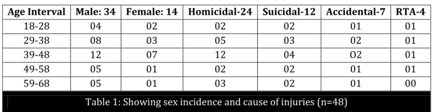
OBSERVATIONS: Among the 48 patients included in the present study, majority are males 34(70.83%) and 14(29.16%) are females. The age of the patients ranged between 18 and 68 years, with a mean age of 45.52, median 46 and mode is 52. The age group of 39 to 48 years is found to be involved in 19(39.58%) of the CTIs. The cause of injury was a homicidal in 24(50%), suicidal in 12 patients (25%), accidental in (14.58%) and traffic accident in (8.33%) (Table 1).
Age Interval Male: 34 Female: 14 Homicidal-24 Suicidal-12 Accidental-7 RTA-4
18-28 04 02 02 02 01 01
29-38 08 03 05 03 02 01
39-48 12 07 12 04 O2 01
49-58 05 01 02 02 01 01
59-68 05 01 03 02 01 00
Table 1: Showing sex incidence and cause of injuries (n=48)
J of Evolution of Med and Dent Sci/ eISSN- 2278-4802, pISSN- 2278-4748/ Vol. 4/ Issue 38/ May 11, 2015 Page 6575
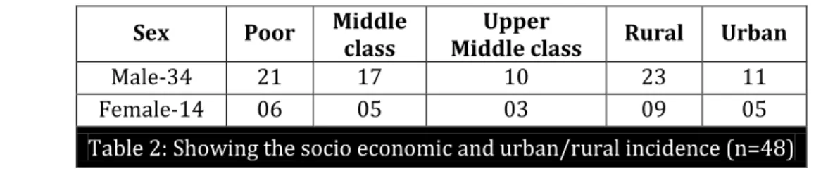
Sex Poor Middle class
Upper
Middle class Rural Urban
Male-34 21 17 10 23 11
Female-14 06 05 03 09 05
Table 2: Showing the socio economic and urban/rural incidence (n=48)
The most common clinical presentation in the study is bleeding; 34(70.83%) from the neck wound gushing mixed with saliva and mucus. Noisy breathing with blood stained frothy secretions from the cut wound is the next common symptom; 29(60.41%). Aphonia is observed in 25 patients (52.08%) if the injury is involving larynx. On examination, the most frequent sign is subcutaneous emphysema around the wound, sometimes extending the entire neck, chest and upper abdomen; 32 (66.66%). Glasgow coma scale is used to calculate the level of consciousness, 42(87.50%) of patients showed a scale above 7. In four patients as they are in inebriated condition it could not be calculated. In two patients GCS is above 7. Radiological examination of Neck and Chest is done in all the patients after the resuscitation. Hemoglobin is found below 7Gms percent in 30(62.5%) patients.
The PCV values below21 are seen in 29(60.41%) of the patients. The MCHC values below 20 are seen in 31(64.58%) of the patients. Surgical emphysema around the wound extending to the neck above & chest and upper abdomen below is seen in 41(85.41%) of the patients. Vocal cord paralysis is seen in 11(22.91%) of the patients. Emergency tracheostomy is done in 40(83.33%) of the patients (Table 3). On exploration of the wounds it is found that, in 4(8.33%) patients the cut is above the Hyoid bone, in 6(12.5%) patients across the Hyoid bone, and in 12(25%) patients above the Thyroid cartilage.
The injury cutting across the thyroid cartilage (Fig. 1 & 2) is seen in 10(2.83%) patients, followed by injury to the crico thyroid membrane seen in 5 patients. In 4 patients the injury is seen across the cricoid cartilage. 3 patients showed across crico tracheal membrane (Table 4). 4 patients showed involvement of trachea. 8 patients with (16.66%) suspected laryngo-tracheal trauma underwent CT imaging before treatment, most often a neck CT. In 4 (8.33%) of the patients tracheostomy was not done as there is no respiratory obstruction and primary wound closure is done. In 5(10.41%) of the patients a laryngeal or tracheal stent has to be used for managing the
stenosis, as the stenosis segment is more than Cms and causing Meyerhof’s grade III stenosis.
J of Evolution of Med and Dent Sci/ eISSN- 2278-4802, pISSN- 2278-4748/ Vol. 4/ Issue 38/ May 11, 2015 Page 6576
Observation Number Percentage
Bleeding 34 70.83
Asphyxia 29 60.41
Aphonia 25 52.08
Hypovolemic shock 27 56.25 Level of consciousness > 7 42 87.50 Hemoglobin levels < 7 Gms 30 62.50
PCV < 20 21 43.75
MCHC <20 24 50
Surgical Emphysema 41 85.41 Mediastinal Emphysema 09 18.75 Vocal cord palsy 11 22.91 Emergency Tracheostomy 43 89.58
Table 3: Showing incidence of symptoms and signs (n=48)
Sex Supra
Hyoid Hyoid
Thyro-hyoid Thyroid
Crico-thyroid Cricoid
Crico-tracheal Tracheal Total
Male 03 04 10 07 03 02 02 03 34
Female 01 02 02 03 02 02 01 01 14
Table 4: Showing sex incidence and level of injuries (n=48)
Nature of Laryngeal Frame Work Injury Male Female Percentage %
Cut across Thyroid cartilage 10 04 29.16 Avulsion of arytenoids 07 03 20.83 Lacerated vocal cords 01 02 6.25 Fracture of Cricoid cartilage 06 01 14.53
Crico tracheal avulsion 04 -- 8.33
Table 5: Showing nature of laryngeal framework injuries
Post-operative complications Male-34 Female-14
Unilateral RLN palsy 04 02
Stenosis 04 01
Wound dehiscence 17 10
Hoarse voice 13 08
Bilateral RLN palsy 03 02
J of Evolution of Med and Dent Sci/ eISSN- 2278-4802, pISSN- 2278-4748/ Vol. 4/ Issue 38/ May 11, 2015 Page 6577 The post-operative complications noted are unilateral RLN palsy in 6 (12.5%), Bilateral RLN palsy in 5 (10.41%) and Stenosis in 5 patients. Wound dehiscence is noted in 27 (56.25%) of then patients. Hoarseness of voice is seen in 21 (43.75%) of the patients (Table 6).
DISCUSSION: Cut throat Injuries are not discussed elaborately in the text books of Otolaryngology. They are only found as a passing note. CTIs are common in the present day of life of civilization especially in India as the crime rate increasing day by day. They can lead to a piquant situation and if not handled properly culminating to immediate death or produce a permanent airway stenosis.(9) The forensic implications are well emphasized along with the principles of management of CTIs in the medical literature from West Africa. In the present study 48 patients are brought to the Casualty department of Regional ENT Hospital in Hyderabad.(10) Onatoi et al found male preponderance in CTIs in their study and concluded that it may be due to their risk taking behavior, active participation and frequent involvement in interpersonal violence.(11)
Fig. 1: Showing A- the Cut Throat Injury across the Thyroid Cartilage
Fig. 1: Showing B- Cut showing above Thyroid cartilage
Fig 2: Cut injury exposing the Larynx
J of Evolution of Med and Dent Sci/ eISSN- 2278-4802, pISSN- 2278-4748/ Vol. 4/ Issue 38/ May 11, 2015 Page 6578
In a study by Aich et al majority of the patient’s belonged to young age between to
years. In contrast our study shows the commonest age group involved is 19(39.58%). Homicide 24 (50%) is seen more commonly, as the motive behind the CTIs in the present study than, attempted suicide or accidental cause (Table 1).(9) Mohanty et al. in their analysis of 588 suicide victims found that financial burden (37%) and marital disharmony (35%) were the principal reasons for suicide attempts. In our study, the causes of suicide are family troubles, sibling rivalry and psychiatric illness. The causes of Homicidal injuries are varied like land disputes, riots, factionist disputes and bank robbery.(12) Modi and Pandy observed that in India, suicidal wounds of the throat are rare.(13,14) This is in contrast with western studies where suicidal CTIs are common.(10) From their study Onotai and Ibekwe concluded that CTIs can be better managed by a multidisciplinary team and if the patients are shifted earlier to the Hospital.(15) When suicidal cut throat injuries occur, a multidisciplinary approach is required in the effective management of victims.
This requires the close collaboration of the Otorhinolaryngologist, the anesthesiologist and the psychiatrist. The same authors found incidence of suicidal and homicidal cases equally in their study. All the patients had their pharynx and larynx opened due to CTIs.(16) Herzog et al mention in their study, dividing the Injuries of the neck into three anatomic zones for the purpose of ease of assessment; 1. Zone I injuries occur at the thoracic inlet. This zone extends from the level of the cricoid cartilage to the clavicles. 2. Zone II injuries are those occurring in the region between the cricoid cartilage and the angle of the mandible. Injuries in this zone are the easiest to expose and evaluate. 3. Zone III injuries occur between the angle of the mandible and the base of the skull. Unlike zone II, zones I and III are protected by bony structures making zone II more vulnerable to injuries.(17)
Peralta et al described some of the problems encountered during general anesthesia in treating CTIs are anatomical distortion of the neck and tracheal displacement making intubation difficult; intravenous induction agents producing hypotension, which is undesirable for a bleeding patient; and danger of vomiting and aspiration. Inhalation induction is also difficult, due to partial breathing 'through the neck' and uncertain control of airway.(18,19) Manilal and Okayo, in their study found majority of injuries in this study were in Zone II and most of them had laryngeal injury which is in keeping with other studies. Sometimes it is difficult to predict the turn of events in some patients, as initially they do not present signs of vascular injury and have patent airway. Later on these patients, as the sepsis sets in may go in to carotid blow out as seen in one patient in this study.(20)
Roberto et al concluded that orotracheal intubation is possible in a high number of patients, without significant laryngeal lesions because the latter case may aggravate the consequences. But in our study in spite of airway injuries orotracheal intubation could be undertaken by manipulation at the neck. In their series one fourth of the patients required performance of an emergency tracheotomy to maintain airway patency. In the present study except in four patients in all the patients tracheostomy is done. All the patients should be subjected for thorough examination to rule out vascular, esophageal injuries while exploring the wound. Also care should be taken to exclude cervical spine injuries.
J of Evolution of Med and Dent Sci/ eISSN- 2278-4802, pISSN- 2278-4748/ Vol. 4/ Issue 38/ May 11, 2015 Page 6579 Flexible Nasopharyngoscope is useful to identify laryngeal injuries and movement of vocal
cords. Schaefer’s classification as amended by Fuhrman is useful in grading the severity of CTIs.
Grade I are treated with antibiotics following suturing the wound in layers. Grade II requires tracheostomy and conservative treatment. In Grade III and IV require exploration and repair. Avulsion of arytenoids cartilage is seen in 20.83% of the patients I this study, which is treated by direct suturing to the cricoid cartilage with 3-0 Prolene. Thyroid cartilage cut across its width is sutured with a 2-0 wire. Lacerated vocal cords are left alone and postoperatively monitored for granulation tissue formation. Cricoid cartilage fractures and crico tracheal avulsions are sutured with 3-0 Prolene. All the explorations are done through anterior approach to the sternoceidomastoid (Table 5).
In the present study no esophageal cut is seen. These injuries usually occur due to penetrating neck injuries. They will lead to rapid development of Mediastinitis. Patients will present with complaints of hemetemesis and drooling blood stained saliva.(21) A certain degree of hoarseness of voice remains in these patients at the time of discharge. They will have a certain degree of difficulty in protecting the airway from aspiration. In our study the incidence is in 21 patients (43.75%). There is no complaint of aspiration at the time of discharge in the present study.(22) Pharyngo -cutaneous fistulae are encountered with CTIs. Avoidance is by meticulous suturing the tissue layers re-enforcing each other, avoiding overlapping of suture lines and choosing correct suture material. They can be
prevented by Ryle’s tube feeding and avoiding oral feeds, in the post-operative period. If the fistulae persist the can be managed by Cutaneous flaps. If Ryles tube is to be avoided a feeding jejunostomy is advised by Darlong et al.
CONCLUSION: Nowadays cut throat injuries are increasingly being managed by ENT surgeons. The management knowhow is essential to a successful outcome. Those patients presenting with injury involving the pharynx, Larynx or upper trachea need a preliminary tracheostomy. This will enable the surgeon to undertake primary closure of the wound and in addition the actual site of wound gets rest
necessary for early healing. The patient’s alimentary needs are better managed with Ryle’s tube
feeding. Post-operative endoscopy identifies the nerve injuries and Stenosis problems. Decannulation is never a problem if initial primary suturing is accurate in approximating the cut ends of the airway. Most of the patients in this series went home with a reasonably good voice. It is important to record protocols used and correct operative findings for medical legal purpose and future reviews.
REFERENCES:
1. Miller RH, Duplechain JK. Heridas cervicales penetrantes. Otolaryngol Clin N Am. 1991. 13-27. 2. Temas actuales en traumatismos de cabeza y cuello. 2. Tisherman SA, Bokhari F, Collier B,
Cumming J, Ebert J, Holevar M, et al. Clinical practice guideline: penetrating zone II neck trauma. J Trauma. 2008. 64. 1392- 5.
3. Penden M Macgee K, Sharma G, The injury chart book; A graphical overview of the global burden of injuries: Geneva; World Health Organization. 2002.
4. Ladapo AA. Open injuries of the anterior neck. Ghana Med J 1979. 18. 182-186. 5. Duncan JAT. A case of severely cut throat. Br J Anaesth 1975. 47. 1327-1329.
6. Beigh Z, Ahmad R. Management of cut-throat injuries. Egypt J Otolaryngo 2014. 30. 268-71. 7. Ezeanolue BC. Management of the upper airway in severe cut throat injuries. Afr J Med Med Sci
J of Evolution of Med and Dent Sci/ eISSN- 2278-4802, pISSN- 2278-4748/ Vol. 4/ Issue 38/ May 11, 2015 Page 6580 8. Eshiet A, Antiaue SG, Onoym IV, Edentekhe TA. Surgical airway problems and their
management. The University of Calaban Teaching Hospital experience. Niger Postg Med J 1979; 4: 15-18.
9. Mohanty S, Sahu G, Mohanty MK, Patnaik M. Suicide in India: a four year retrospective study. J Forensic Leg Med 2007. 14. 185-189.
10.Onotai LO, Ibekwe U. The pattern of cut throat injuries in the University of Port-Harcourt Teaching Hospital, Portharcourt. Niger J Med. 2010. 19. 264-266.
11.M Aich, ABM Khorshed Alam, DC Talukder, MA Rouf Sarder, AY Fakir, M Hossain. Cut throat injury: review of 67 cases. Bangladesh J Otorhinolaryngol 2011. 17. 5-13.
12.Modi JP, AS Pandy. MODI's medical jurisprudence and toxicology. 20th ed. Bombay, India: Butterworths publications. 1977. 256-275.
13.Gordon O, Shapiro HA, Berson SD. Forensic medicine - a guide to principles. 3rd ed. Edinburgh, London: London Churchill Livingstone. 1988. 300-319.
14.Simpson CK. Simpsons forensic medicine. Severa Bureau, Layla Vanderberh editor. Bernard knight. 10th ed. London: Eward Arnold, Hodder and Stoughton Ltd. 1991. 101-102.
15.Bhattacharjee N, Arefin SM, Mazumder SM, Khan MK. Cut throat injury: a retrospective study of 26 cases. Bangladesh Med Res Coun bulletin. 1997 Dec. 23. 3. 87-90.
16.Herzog M, Hoppe F, Baier G, Dieler R: Injuries of the head and neck in suicidal intention. Laryngorhinootologie 2005. 84. 3. 176-81.
17.Peralta R, Hurford WE. Airway trauma. Int Anesthesiol Clin 2000. 38. 111-27.
18.Manilal A, Khorshed ABM, Talukder DC, Sarder RMA, Fakir AT, Hossain M: Cut throat injury: review of 67 cases. Bangladesh J Otorhinolaryngol 2011, 17. 5-13.
19.Okoye BC, Oteri AJ: Cut throat injuries in Port Harcourt. Sahel Med J 2001, 4. 207-209.
20.Roberto García-Zornoza, ∗ Carmelo Morales-Angulo, Rocío González-Aguado, Leticia Acle Cervera, Eloy Cortizo Vázquez, Sergio Obeso Agüera. Neck Injuries Servicio de Otorrino-laringología. Acta Otorrinolaringol Esp. 2012. 63. 1. 47-54.
21.Glinjongol C, Pakdirat B. Management of tracheobrochial injuries: A 10-year experience at Ratchaburi hospital. J Med Assoc Thai 2005; 88: 32-40.
22.Morales Angulo C, Rodríguez Iglesias J, Mazón Gutiérrez A, Rubio Suárez A, Rama J. Diagnóstico y tratamiento de las perforaciones de esófago cervical en adultos. Acta Otorrinolaringol Esp. 1999; 50: 142-6.
J of Evolution of Med and Dent Sci/ eISSN- 2278-4802, pISSN- 2278-4748/ Vol. 4/ Issue 38/ May 11, 2015 Page 6581
AUTHORS:
1. Md. Naveed Ahmed 2. M. Sreedhar Rao 3. S. Muneeruddin Ahmed 4. M. Mahendra Kumar
PARTICULARS OF CONTRIBUTORS:
1. Professor, Department of ENT, Gandhi Medical College, Hyderabad.
2. Assistant Professor, Department of ENT, Kurnool Medical College, Kurnool. 3. Formerly Professor & HOD, Department
of ENT, Osmania Medical College, Hyderabad, Telangana.
FINANCIAL OR OTHER
COMPETING INTERESTS: None
4. Assistant Professor, Department of ENT, Kurnool Medical College, Kurnool, Andhra Pradesh.
NAME ADDRESS EMAIL ID OF THE CORRESPONDING AUTHOR:
Dr. S. Muneeruddin Ahmed, # 44/118, Prakash Nagar, Kurnool-518004.
E-mail: [email protected]