ABSTRACT
Objective: Lung cancer is a global public health problem and is associated with high mortality. Lung cancer could be largely avoided by reducing the prevalence of smoking. The objective of this study was to analyze the effects of social, behavioral, and clinical factors on the survival time of patients with non-small cell lung cancer treated at Cancer Hospital I of the José Alencar Gomes da Silva National Cancer Institute, located in the city of Rio de Janeiro, Brazil, between 2000 and 2003. Methods: This was a retrospective hospital cohort study involving 1,194 patients. The 60-month disease-speciic survival probabilities were calculated with the Kaplan-Meier method for three stage groups. The importance of the studied factors was assessed with a hierarchical theoretical model after adjustment by Cox multiple regression. Results: The estimated 60-month speciic-disease lethality rate was 86.0%. The 60-month speciic-disease-speciic survival probability ranged from 25.0% (stages I/II) to 2.5% (stage IV). The performance status, the intention to treat, and the initial treatment modality were the major prognostic factors identiied in the study population. Conclusions: In this cohort of patients, the disease-speciic survival probabilities were extremely low. We identiied no factors that could be modiied after the diagnosis in order to improve survival. Primary prevention, such as reducing the prevalence of smoking, is still the best method to reduce the number of people who will suffer the consequences of lung cancer.
Keywords: Lung neoplasms/epidemiology; Carcinoma, non-small-cell lung; Survival analysis.
Factors associated with disease-speciic
survival of patients with non-small
cell lung cancer
Mirian Carvalho de Souza1, Oswaldo Gonçalves Cruz2, Ana Glória Godoi Vasconcelos3
Correspondence to:
Mirian Carvalho de Souza. Rua Marquês de Pombal, 125, 7º andar, Centro, CEP 20230-240, Rio de Janeiro, RJ, Brasil. Tel.: 55 21 3207-5667. E-mail: [email protected]
Financial support: None.
INTRODUCTION
Lung cancer is the most common type of cancer worldwide; it is estimated that, in 2012, there were 1.8 million new cases.(1) In Brazil in 2015, 27,000 new cases
were estimated.(2)
Although lung cancer has various histological types,
the most widely used classiication system is that which
divides tumors into small cell carcinomas (15%) and non-small cell carcinomas (85%).(3)
The 60-month survival probability of patients with non-small cell lung carcinoma is lower than 15% in Europe.(4)
A study conducted in the United States obtained estimates ranging from 66% (stage Ia) to 4% (stage IV). (5) A study
involving patients from a university hospital in the city of Rio de Janeiro, Brazil, found that the 60-month survival probability was 6%, with it being 14% for the early stages and 5% for the advanced stages.(6)
Among the prognostic factors studied for lung cancer patients(7) are stage, performance status,(8) weight loss,
gender, age, smoking, smoking history, quality of life, marital status, depression, and genetic mutations.(6,9-11)
Epidemiological studies have indicated that the effects of socioeconomic factors on health outcomes are indirect, occurring through behavioral and clinical factors. In this context, it is important to establish the hierarchy
of these factors in determining the occurrence of lung cancer and in the survival probability of patients with this type of cancer.(12,13)
The objective of the present article was to analyze the importance of social, behavioral, and clinical factors on the survival time of patients with non-small cell lung cancer treated at Hospital do Câncer I do Instituto Nacional de CâncerJosé Alencar Gomes da Silva (HCI/INCA, Cancer Hospital I of the José Alencar Gomes da Silva National Cancer Institute), located in the city of Rio de Janeiro, Brazil, between 2000 and 2003.
METHODS
This was a retrospective observational hospital cohort study in which the object of interest was the time from diagnosis to death from lung cancer or metastasis.
The target population consisted of patients diagnosed with primary non-small cell lung carcinoma, between
2000 and 2003, who were registered in the Registro
Hospitalar de Câncer (RHC, Hospital Cancer Registry) of HCI/INCA, which is a tertiary referral hospital for the treatment of cancer in the state of Rio de Janeiro, Brazil.
Eligible patients were deined as those from the state
of Rio de Janeiro, where HCI/INCA is located, in whom
diagnosis was conirmed by either anatomic pathological 1. Divisão de Pesquisa Populacional,
Coordenação de Pesquisa e Educação, Instituto Nacional de Câncer José Alencar Gomes da Silva – INCA – Rio de Janeiro (RJ) Brasil. 2. Programa de Computação Cientíica,
Fundação Oswaldo Cruz – Fiocruz – Rio de Janeiro (RJ) Brasil. 3. Departamento de Métodos
Quantitativos em Saúde, Escola Nacional de Saúde Pública Sérgio Arouca – ENSP – Fundação Oswaldo Cruz – Fiocruz – Rio de Janeiro (RJ) Brasil.
Submitted: 30 March 2015. Accepted: 27 January 2016.
or cytological examination of the tumor and who had not been previously treated. The list of patients who met the eligibility criteria was extracted from the RHC of HCI/INCA, with the primary source of cancer registry information being medical records. At HCI/INCA, medical records were not electronic. To update data on patient survival, we searched the Rio de Janeiro State Mortality Database, and, for patients for whom the information was missing, we conducted an active search according to the RHC routine.(14) In addition, medical
records were abstracted for information about smoking history and performance status, which is measured with scales that are used to evaluate how the disease progresses and affects the daily living abilities of the patient, in order to determine appropriate treatment and prognosis.(8) Patients who had ever smoked were
considered smokers.
Of the 1,502 cases of non-small cell lung cancer registered in the RHC between 2000 and 2003, 1,394 lived in the state of Rio de Janeiro. Of those, 200 were excluded because it was impossible to determine disease stage by reviewing the medical records.
To reduce the inluence of anticipation bias,(15) all analyses were stratiied by clinical stage group, as determined by the tumor-node-metastasis classiication
system(16): stages I/II (early stage); stage III; and
stage IV. Stages I and II were gathered into one group in order to provide greater stability to the results of the models.
The factors identiied in the review of the literature
were organized into a hierarchical theoretical model (Figure 1).(1-6) Distal factors included sociodemographic
characteristics and family history of cancer; intermediate factors included behavioral characteristics as well as access to and effectiveness of the health care system, all
of which are generally inluenced by sociodemographic
characteristics; and proximal factors included patient clinical characteristics, disease characteristics, and
treatment characteristics, all of which can be inluenced by the previous level factors. Of the 28 factors identiied,
10 were not analyzed because they were unavailable or because they were available in very few medical records. In the categorization of the studied factors, priority was given not only to coherence in the object of study but also to data stability as a function of the sample, especially in relation to stages I/II.
Age at diagnosis was included in all multiple regression models because it is directly related to death (from a biological standpoint), because it characterizes the
birth cohort, and because it inluences other factors
(smoking, occupation, etc.)
The 60-month disease-speciic survival probability
in lung cancer was estimated with the Kaplan-Meier method, on the basis of the following criteria: i)
initial event: diagnosis of lung cancer; ii) inal event:
death from lung cancer or metastasis; iii) survival
time: time from initial to inal event or time to loss to
follow-up; and iv) censored cases: cases that were lost to follow-up over the 60-month period; cases in
Distal Factors
Intermediate Factors
Proximal
Factors Outcome
Behavioral Factors
Access to the Health Care System • Family history of cancerb
Sociodemographic Characteristics
• Age
• Level of education • Race
• Gender • Marital status • Incomea
Family History
• Diet rich in vegetablesa • Alcoholismb
• Occupationb • Smoking
• Exposure to environmental pollutiona
• Diagnosis occurring prior to admission to the tertiary hospital • Distance from the hospital to the patient's home
• Time from
• first visit to diagnosis • first visit to first treatment • diagnosis to first treatment Effectiveness of the Health Care System
Clinical characteristics
• Patient performance status (ER) • Reported morbidities (asbestosisa, COPDb, silicosisa,
and tuberculosisb) • Mutation in EGFRa
Characteristics of the Disease
• Stage • Laterality • Detailed location • Histological type
Treatment
• Treatment employed • Initial treatment intent • Disease status at the end of first treatmentb
• Number of treatment linesa
Stage will be used as a stratification variable in order to reduce anticipation bias. aVariables unavailable for analysis.
bVariables unavailable in a high proportion of cases.
Death from Lung Cancer
which death from lung cancer or metastasis was not
conirmed; and cases in which patients survived the
60-month follow-up.
Differences between the estimated probability curves were determined via log-rank test. Variables that had a value of p < 0.20 in the log-rank test were included in the Cox models.(17) The adjusted effects of factors
on survival time were calculated, for each stage, by using the Cox model, on the basis of the hierarchical theoretical model proposed in Figure 1.
In the preparation of the models, variables were entered in blocks. First, distal factors were entered; those with a value of p < 0.10 were maintained in model 1. Subsequently, intermediate factors were added to model 1, and the same selection criterion was applied (model 2). The same procedure was repeated
for proximal factors, and the inal model was obtained.
In the modeling process, the previous level factors that
lost signiicance upon inclusion of more proximal-level factors were maintained. The goodness-of-it of the
models was determined by calculating the likelihood ratio, the probability of agreement, and the overall
goodness-of-it.(17)
The research project that generated the present article is registered with the research ethics committees of the INCA and the Sérgio Arouca National School of Public Health (Protocol nos. CAAE-012.0.007.031-11 and CAAE-0163.0.031.007-11).
RESULTS
A comparison of cases included and excluded as per
the eligibility criteria indicated no statistically signiicant
differences (chi-square test) in the distribution by gender, level of education, smoking, histological type, or treatment.
The mean age of the 1,194 patients included in the study was 62 years, and it decreased with the severity of tumor stage (stages I/II, 65 years; stage III, 62 years; and stage IV, 60 years). Most subjects were
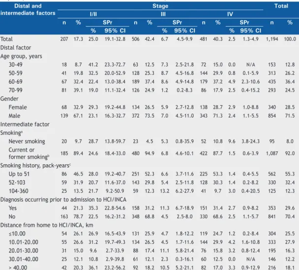
Table 1. Distribution and 60-month disease-speciic survival probability of the study cohort of patients with non-small cell lung cancer by clinical stage, as well as by distal factor and by intermediate factor of the proposed model. Cancer Hospital I, José Alencar Gomes da Silva National Cancer Institute, 2000-2003.
Distal and intermediate factors
Stage Total
I/II III IV
n % SPr n % SPr n % SPr n %
% 95% CI % 95% CI % 95% CI
Total 207 17.3 25.0 19.1-32.8 506 42.4 6.7 4.5-9.9 481 40.3 2.5 1.3-4.9 1,194 100.0
Distal factor Age group, years
30-49 18 8.7 41.2 23.3-72.7 63 12.5 7.3 2.5-21.8 72 15.0 0.0 N/A 153 12.8
50-59 41 19.8 32.5 20.0-52.9 128 25.3 8.7 4.5-16.8 144 29.9 0.8 0.1-5.9 313 26.2
60-69 67 32.4 22.4 13.0-38.4 189 37.4 8.6 4.9-14.8 179 37.2 4.9 2.3-10.6 435 36.4
70-99 81 39.1 19.0 11.1-32.4 126 24.9 1.2 0.2-8.3 86 17.9 2.5 0.4-15.2 293 24.5
Gender
Female 68 32.9 29.3 19.2-44.8 134 26.5 5.9 2.7-12.8 138 28.7 2.9 1.0-8.8 340 28.5
Male 139 67.1 23.1 16.3-32.7 372 73.5 7.0 4.5-11.0 343 71.3 2.4 1.1-5.5 854 71.5
Intermediate factor Smokinga
Never smoking 20 9.7 28.7 13.8-59.7 23 4.5 5.3 0.8-35.9 52 10.8 9.6 3.8-24.3 95 8.0
Current or
former smokingb 185 89.4 24.6 18.4-33.0 480 94.9 6.8 4.6-10.1 422 87.7 1.5 0.6-3.9 1,087 92.0
Smoking history, pack-yearsc
Up to 51 86 46.5 28.0 19.2-40.7 251 52.3 6.6 3.7-11.6 225 53.3 1.4 0.4-5.5 562 55.3
52-103 59 31.9 20.7 11.6-37.0 143 29.8 5.4 2.5-11.8 128 30.3 1.4 0.2-8.2 330 32.4
104-360 25 13.5 21.7 9.2-50.9 59 12.3 13.2 6.2-27.9 41 9.7 3.0 0.4-20.5 125 12.3
Diagnosis occurring prior to admission to HCI/INCA
Yes 44 21.3 35.3 22.8-54.6 158 31.2 11.3 6.7-18.9 151 31.4 2.7 0.9-8.2 353 29.6
No 163 78.7 22.5 16.2-31.2 348 68.8 4.5 2.5-8.0 330 68.6 2.5 1.1-5.7 841 70.4
Distance from home to HCI/INCA, km
≤10.00 54 26.1 26.9 16.5-43.9 131 25.9 4.7 1.8-12.2 119 24.7 1.2 0.2-8.4 304 25.5
10.01-20.00 55 26.6 31.2 19.7-49.3 134 26.5 4.5 1.7-11.6 144 29.9 4.2 1.6-10.8 333 27.9
20.01-30.00 31 15.0 9.6 2.7-33.9 88 17.4 11.1 5.8-21.4 76 15.8 3.2 0.8-12.4 195 16.3
30.01-40.00 25 12.1 10.8 2.9-39.8 61 12.1 2.3 0.3-16.1 60 12.5 0.0 N/A 146 12.2
male and smokers; in stage III, the smoker/nonsmoker ratio reached its maximum value (20.9:1.0). The mean smoking history was 60 pack-years, and less than one
third of the patients had a conirmed diagnosis prior
to admission to HCI/INCA (Table 1).
At diagnosis, more than half of the patients in stages I/II and III presented with restrictions for performing
vigorous physical activities. The most common irst
treatment was radiotherapy, and, in stages I/II,
surgery was the irst treatment in only one fourth of
the cases. Adenocarcinoma predominated in stages I/II and stage IV, followed by squamous carcinoma,
chiely in stage III (Table 2).
By the end of the 60-month follow-up, 1,027 patients (86.0%) had died from lung cancer, 66 (5.5%) had died from other causes, 70 (5.9%) had survived, and 31 (2.6%) had been lost. The estimated 12-month
and 60-month disease-speciic survival probabilities
were 32.7% (95% CI: 30.0-35.5%) and 7.9% (95% CI: 6.3-9.7%), respectively. The median survival time was estimated to be 17.7 months for stages I/II, 8.0 months for stage III, and 5.5 months for stage IV.
Patients with stage IV disease who were nonsmokers had a better prognosis than did those who were smokers/former smokers. Being admitted to HCI/
INCA with a conirmed diagnosis doubled the survival
probability of stage III patients (Table 1). Survival decreased with increasing limitation as assessed by the performance status scale, regardless of stage. Among the patients for whom information on tumor location was available, stage I/II and stage III patients had a better prognosis (Table 2).
In the modeling process, the cases with missing
values for the variables included in the inal models
were excluded to allow comparability between the models of the different levels. We excluded 20 stage
Table 2. Distribution and 60-month disease-speciic survival probability of the study cohort of patients with non-small cell lung cancer by clinical stage and by proximal factor of the proposed model. Cancer Hospital I, José Alencar Gomes da Silva National Cancer Institute, 2000-2003.
Proximal factor Stage Total
I/II III IV
n % SPr n % SPr n % SPr n %
% 95% CI % 95% CI % 95% CI
Performance statusa
Fully active 34 16.4 53.5 38.2-74.9 39 7.7 10.7 3.7-30.7 48 10.0 6.3 1.8-22.2 121 11.6
Limited in
vigorous activities 114 55.1 24.7 17.3-35.2 308 60.9 8.0 5.2-12.2 233 48.4 2.5 1.1-5.9 655 62.8 Able of self-care
but unable to work
39 18.8 0.0 N/A 93 18.4 1.3 0.2-9.0 105 21.8 1.3 0.2-8.8 237 22.7
Bedridden at least 50% of the day
0 0.0 N/A N/A 8 1.6 0.0 N/A 22 4.6 0.0 N/A 30 2.9
Tumor lateralityb
Unilateral 192 92.8 26.2 20.0-34.3 467 92.3 6.5 4.4-9.6 423 87.9 2.9 1.5-5.5 1,082 98.5
Bilateral 1 0.5 0.0 N/A 4 0.8 25.0 4.6-100.0 11 2.3 0.0 N/A 16 1.5
Availability of detailed information on tumor location
Yes 165 79.7 28.2 21.4-37.2 305 60.3 7.9 5.1-12.4 236 49.1 3.0 1.2-7.2 706 59.1
No 42 20.3 10.7 3.7-30.7 201 39.7 4.6 2.1-9.9 245 50.9 2.3 0.9-5.9 488 40.9
Initial treatment intentc
Curative 132 78.6 33.8 25.9-44.3 231 55.8 9.7 6.2-15.3 82 22.9 4.8 1.4-16.5 445 47.3
Palliative 22 13.1 0.0 N/A 149 36.0 2.7 0.9-8.1 275 76.8 2.4 1.0-5.7 446 47.4
Neoadjuvant 14 8.3 33.8 13.5-84.5 34 8.2 13.6 5.5-33.8 1 0.3 0.0 N/A 49 5.2
Initial treatment modality
Surgery 52 25.1 63.5 50.3-80.3 9 1.8 28.6 8.9-92.2 9 1.9 42.9 18.2-100.0 70 5.9
Radiotherapy 89 43.0 6.7 2.2-20.2 247 48.8 5.7 3.1-10.5 233 48.4 1.1 0.3-4.5 569 47.7
Chemotherapy 38 18.4 30.0 17.2-52.5 181 35.8 9.1 5.3-15.5 158 32.8 2.6 0.9-7.8 377 31.6
No treatment 28 13.5 0.0 N/A 69 13.6 0.0 N/A 81 16.8 0.0 N/A 178 14.9
Histological type
Adenocarcinoma 87 42.0 30.6 21.3-43.9 198 39.1 6.8 3.7-12.6 237 49.3 2.9 1.3-6.9 522 43.7
Squamous
carcinoma 81 39.1 17.3 9.9-30.2 207 40.9 6.7 3.7-12.2 130 27.0 1.8 0.3-10.3 418 35.0 Other
I/II cases, 108 stage III cases, and 75 stage IV cases. Excluded and analyzed cases were compared, and no
statistically signiicant differences were observed for
the variables gender, age, level of education, race, marital status, smoking, histological type, treatment, vital status, or follow-up period.
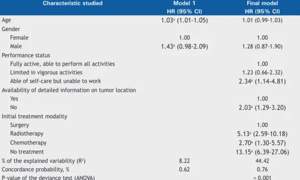
In stages I/II, age, gender, performance status, detailed tumor location, histological type, initial treatment intent, and initial treatment modality proved to be important prognostic factors in the crude analysis of the Cox models and were used in the hierarchical modeling.
None of the intermediate factors were included in the multiple regression model of stages I/II (p > 0.20 in the log-rank test). When the proximal factors were added to model 1, the risk estimates decreased. The risk of death, adjusted for the other variables of the
inal model, was 2.34 times higher among patients who
were unable to perform work activities than among those who were active, was twice as high among those for whom information on tumor location was unavailable as among those for whom this information was available, and was thirteen times higher among untreated patients than among operated patients. The risk of death associated with the use of radiotherapy or chemotherapy was high when compared with that related to surgery (Table 3).
In stage III, the prognostic factors used in the hierarchical modeling were age, diagnosis occurring prior to admission, distance from home to HCI/INCA, performance status, tumor laterality, tumor location, initial treatment intent, and initial treatment modality.
In the adjusted model for stage III, there were no differences in the estimates when the intermediate factors were added to model 1; however, when the proximal factors were included, the age-related risk estimate decreased. The effect of diagnosis occurring prior to admission increased when the proximal factors
were included. The inal model showed an excess risk of
death of 70.0%, adjusted for the other factors, among the patients who had not been given a diagnosis prior to admission (Table 4).
Performance status maintained a strong association
with outcome in the inal model and showed an
increasing gradient of risk of death/worsening of the patient’s physical state. Patients who underwent palliative treatments had a risk of death, adjusted for
the other factors in the inal model, 2.48 times that
of those who underwent curative treatments. Among the untreated patients, this estimate was even higher (Table 4).
In stage IV, the study characteristics gender, smoking, smoking history, diagnosis occurring prior to admission, distance from home to HCI/INCA, performance status, tumor laterality, histological type, initial treatment intent, and initial treatment modality were used in the hierarchical modeling. The age-related risk estimate
decreased and gained statistical signiicance when the proximal factors were included in the inal model.
The effect associated with smoking decreased and
lost statistical signiicance when the proximal factors
were entered. This behavior probably occurs because the effect of smoking is mediated by proximal factors. Adjusted for the other factors, the risk of death by
Table 3. Results of the hierarchical Cox model of patients with stage I/II non-small cell lung cancer. Cancer Hospital I, José Alencar Gomes da Silva National Cancer Institute, 2000-2003.
Characteristic studied Model 1 Final model
HR (95% CI) HR (95% CI)
Age 1.03a (1.01-1.05) 1.01 (0.99-1.03)
Gender
Female 1.00 1.00
Male 1.43b (0.98-2.09) 1.28 (0.87-1.90)
Performance status
Fully active, able to perform all activities 1.00
Limited in vigorous activities 1.23 (0.66-2.32)
Able of self-care but unable to work 2.34a (1.14-4.81)
Availability of detailed information on tumor location
Yes 1.00
No 2.03a (1.29-3.20)
Initial treatment modality
Surgery 1.00
Radiotherapy 5.13a (2.59-10.18)
Chemotherapy 2.70a (1.30-5.57)
No treatment 13.15a (6.39-27.06)
% of the explained variability (R2) 8.22 44.42
Concordance probability, % 0.62 0.76
P-value of the deviance test (ANOVA) < 0.001
the end of the 60-month follow-up was found to be 50.0% higher among those who had not been given a diagnosis prior to admission. Those who lived within a radius of 20-30 km of HCI/INCA had an adjusted risk of death that was 28.0% lower than that for those who lived closer to HCI/INCA (Table 5).
Performance status maintained a strong association with outcome after adjustment and showed an increasing gradient of risk of death/worsening of the patient’s physical state. The risks associated with nonsurgical treatments, as compared with surgical treatments, are extremely high in that stage. In addition, the risk of death by the end of the 60-month follow-up is estimated to be eight times higher among untreated patients than among operated patients (Table 5).
The inclusion of the proximal factors in the more
distal-level models signiicantly increased the likeli
-hood ratio of the inal models, regardless of stage.
In addition, the probability of agreement of these
models can be classiied as very good in stages I/II
and stage III and as coherent in stage IV. The all-level
adjusted models were signiicantly different from the
null model (p < 0.001) in the three stages analyzed (Tables 2, 3, and 4).
DISCUSSION
Among the factors evaluated, performance status, initial treatment intent, and initial treatment modality
stood out for inluencing the survival time of patients
with non-small cell lung cancer treated at HCI/INCA between 2000 and 2003, in all stage groups. The magnitudes and directions of the estimated effects related to these factors in the present study are consistent with those reported in other studies and will be addressed below.(6,18,19)
Survival studies of lung cancer patients commonly involve clinical trial patients. A review of the literature revealed two survival studies of patients with non-small cell lung cancer, both of which were conducted in the city of Rio de Janeiro, Brazil: one in a public hospital, in which 60-month survival probabilities were estimated(6); and one in a private clinic,(19) in
which 24-month survival probabilities were estimated.
The predominance of advanced stage disease (stage IV, 40.3%), males (71.5%), and smokers (92.0%) observed in the present study is consistent with the characteristics of other study populations.(1,6,18-20)
Results that are consistent and in agreement with
indings of previous studies were observed for the Table 4. Results of the hierarchical Cox model of patients with stage III non-small cell lung cancer. Cancer Hospital I, José Alencar Gomes da Silva National Cancer Institute, 2000-2003.
Characteristic studied Model 1 Model 2 Final model
HR (95% CI) HR (95% CI) HR (95% CI)
Age 1.02a (1.01-1.03) 1.02a (1.01-1.03) 1.01 (1.00-1.02)
Diagnosis occurring prior to admission to HCI/INCA
Yes 1.00 1.00
No 1.63a (1.30-2.06) 1.70a (1.34-2.15)
Distance from HCI/INCA to home, km
≤ 10.00 1.00 1.00
10.01-20.00 0.86 (0.64-1.16) 0.88 (0.65-1.19)
20.01-30.00 0.75b (0.54-1.04) 0.71a (0.43-0.85)
30.01-40.00 1.10 (0.77-1.58) 0.95 (0.65-1.37)
> 40.00 0.71a (0.52-0.99) 0.64a (0.46-0.90)
Performance status
Fully active, able to perform all activities 1.00
Limited in vigorous activities 1.33 (0.89-2.00)
Able of self-care but unable to work 2.70a (1.73-4.21)
Bedridden at least 50% of awake hours 4.56a (1.93-10.75)
Tumor laterality
Unilateral 1.00
Bilateral 0.32b (0.10-1.02)
Initial treatment intent
Curative 1.00
Palliative 2.48a (1.93-3.21)
Neoadjuvant 0.97 (0.63-1.48)
No treatment 3.67a (2.56-5.25)
% of the explained variability (R2) 2.62 8.37 32.86
Concordance probability, % 0.56 0.60 0.71
P-value of the deviance test(ANOVA) < 0.001 < 0.001
distribution of the study population by smoking history (mean, 60 pack-years),(6) performance status
(approx-imately 90.0% were limited),(6,18,19) and histological
type (more than 40.0% had adenocarcinoma).(5,18,20)
The median estimated survival time was slightly higher than that found in the study conducted in a public hospital in Rio de Janeiro, for all stages.(6) It is
likely that, because HCI/INCA is an oncology referral center, it has a health care infrastructure that favors a better prognosis, in comparison with the other public hospital not specializing in oncology. In contrast, the low proportion of operated patients, especially stage I/II patients, indicates limited access to this treatment modality.
The 60-month survival probabilities below 25.0% found in most of the analysis categories in stages I/ II illustrate how devastating cancer is, regardless of what factors are evaluated, even when the disease is
diagnosed in early stages. This inding underscores
the importance of primary prevention with two major strategies: encouraging smoking cessation and
increasing young people’s awareness regarding the dangers of smoking in order to prevent them from acquiring this behavior.
In the evaluation of prognostic factors through the use of the Cox models, results that are consistent with those of other studies were observed for performance status,(18) that is, patients who are more limited have
a lower survival probability.
Regarding initial treatment intent and initial treatment
modality, the adjusted results of the inal models of the
present study are consistent with what is expected in oncology,(21,22) that is, survival probabilities are higher
among those initially treated with curative intent and among those treated with surgery, which, for lung cancer, is the treatment modality that is most likely to result in a cure.
In stage IV, a mediation effect of smoking was observed for the proximal factors. Considering that variables are entered in blocks, it is impossible to determine which factor is responsible for this effect.(13) Table 5. Results of the hierarchical Cox model of patients with stage IV non-small cell lung cancer. Cancer Hospital I, José Alencar Gomes da Silva National Cancer Institute, 2000-2003.
Characteristic studied Model 1 Model 2 Final model
HR (95% CI) HR (95% CI) HR (95% CI)
Age 0.99 (0.98-1.00) 0.99 (0.98-1.00) 0.98a (0.97-0.99)
Gender
Female 1.00 1.00 1.00
Male 1.21c (0.97-1.52) 1.13 (0.89-1.43) 1.11 (0.87-1.41)
Smoking
Never smoking 1.00 1.00
Current or former smokingb 1.46a (1.03-2.07) 1.23 (0.86-1.77)
Diagnosis occurring prior to admission to HCI/INCA
Yes 1.00 1.00
No 1.39a (1.12-1.73) 1.50a (1.19-1.88)
Distance from HCI/INCA to home, km
≤ 10.00 1.00 1.00
10.01 -20.00 0.79 (0.60-1.05) 0.83 (0.63-1.10)
20.01 -30.00 0.79 (0.57-1.08) 0.72a (0.52-0.99)
30.01-40.00 1.18 (0.82-1.69) 1.00 (0.69-1.45)
> 40.00 0.76c (0.55-1.04) 0.85 (0.62-1.18)
Performance status
Fully active, able to perform all activities 1.00
Limited in vigorous activities 1.46a (1.04-2.06)
Able of self-care but unable to work 2.58a (1.72-3.85)
Bedridden at least 50% of awake hours 3.87a (2.24-6.68)
Initial treatment modality
Surgery 1.00
Radiotherapy 4.70a (1.70-12.97)
Chemotherapy 3.28a (1.20-9.03)
No treatment 8.03a (2.85-22.66)
% of the explained variability (R2) 1.21 6.14 25.68
Concordance probability, % 0.53 0.58 0.69
P-value of the deviance test(ANOVA) < 0.001 < 0.001
The results associated with the distance from the patient’s home to HCI/INCA can be explained on the basis of how the health care system in the state of Rio de Janeiro was organized in terms of lung cancer treatment during the study period. A medical referral, together with test results indicating the presence of a malignant tumor, was required for admission to HCI/INCA. Patients who lived outside the city of Rio de Janeiro and were referred to HCI/INCA usually
beneited from a free shuttle service organized and
provided by each municipal government. In general, this service facilitated continuation of treatment and follow-up. Perhaps other ways of evaluating access to the hospital in relation to place of residence, taking into account the route traveled and the transport used as reported by patients, can provide survival analysis results that are more consistent.
In the present study, the incompleteness of the medical records limited the use of some factors in the model proposed in Figure 1 and case inclusion in the analyses. Since the clinical stage was not recorded, we lost 14.3% of the eligible cases, which affected the stability of the estimates for some analysis categories. An evaluation of cases included and excluded on the basis of missing information revealed a statistically
sig-niicant difference regarding treatment—approximately
50.0% of the patients excluded from the analysis had not been treated, whereas among the cases analyzed, the proportion was 14.9%. This loss could
inluence the results obtained, but it is impossible to
determine the magnitude of this effect because we do not know the stage of the patients who were not analyzed. Progressive improvement in the quality of data entry into medical records should be encouraged, since these documents are often used as a source for database building. Despite the limitations inherent to retrospective studies that use medical records, such studies are of great value in increasing knowledge about disease involvement in populations that are treated at health care clinics.
In summary, it can be noted that the estimated
60-month disease-speciic survival probabilities were very low, even in stages I/II. In addition, we identiied no factors that could be modiied after the diagnosis
in order to improve survival. Lung cancer is a silent disease whose symptoms are associated with other less lethal diseases, which can lead to a delay in diagnosis in relation to the natural history of the disease. The best method to reduce the number of people who will suffer the consequences of lung cancer is primary prevention, reducing smoking.
REFERENCES
1. Ferlay J, Soerjomataram I, Ervik M, Dikshit R, Eser S, Mathers C, Rebelo M, Parkin DM, Forman D, Bray, F. GLOBOCAN 2012 v1.0, Cancer incidence and mortality worldwide. IARC CancerBase [serial on the Internet]. 2013 [cited 2015 Jul 1];11. Lyon, France: International Agency for Research on Cancer; 2013. Available from: http://globocan.iarc.fr
2. Instituto Nacional de Câncer José Alencar Gomes da Silva. Estimativa 2014: Incidência de câncer no Brasil. Rio de Janeiro: INCA; 2014.
3. Travis WD. Pathology of lung cancer. Clin Chest Med. 2011;32(4):669-92. http://dx.doi.org/10.1016/j.ccm.2011.08.005
4. Verdecchia A, Francisci S, Brenner H, Gatta G, Micheli A, Mangone L, et al. Recent cancer survival in Europe: a 2000-02 period analysis of EUROCARE-4 data. Lancet Oncol. 2007;8(9):784-96. http:// dx.doi.org/10.1016/S1470-2045(07)70246-2
5. Yang P, Allen MS, Aubry MC, Wampler JA, Marks RS, Edell ES, et
al. Clinical features of 5,628 primary lung cancer patients: experience at Mayo Clinic from 1997 to 2003. Chest. 2005;128(1):452-62. http://dx.doi.org/10.1378/chest.128.1.452
6. Mora P. Análise de sobrevida em pacientes com câncer de pulmão [dissertation]. Rio de Janeiro: Universidade Federal do Rio de Janeiro; 2004.
7. León-Atance P, Moreno-Mata N, González-Aragoneses F, Ca-izares-Carretero MÁ, García-Jiménez MD, Genovés-Crespo M, et al. Multicenter analysis of survival and prognostic factors in pathologic stage I non-small-cell lung cancer according to the new 2009 TNM
classiication. Arch Bronconeumol. 2011;47(9):441-6. http://dx.doi.
org/10.1016/j.arbres.2011.04.004
8. Zubrod CG, Schneiderman M, Frei III E. Brindley C. Lennard Gold G, Shnider B, et al. Appraisal of methods for the study of chemotherapy of cancer in man: Comparative therapeutic trial of nitrogen mustard and triethylene thiophosphoramide. J Chron Dis. 1960;11(1):7-33. http://dx.doi.org/10.1016/0021-9681(60)90137-5
9. Brundage MD, Davies D, Mackillop WJ. Prognostic factors in non-small cell lung cancer: a decade of progress. Chest. 2002;122(3):1037-57. http://dx.doi.org/10.1378/chest.122.3.1037
10. Soria JC, Massard C, Le Chevalier T. Should
progression-free survival be the primary measure of eficacy for advanced
NSCLC therapy? Ann Oncol. 2010;21(12):2324-32. http://dx.doi. org/10.1093/annonc/mdq204
11. Jazieh AR, Hussain M, Howington JA, Spencer HJ, Husain M, Grismer JT, et al. Prognostic factors in patients with surgically resected stages I and II non-small cell lung cancer. Ann Thorac Surg. 2000;70(4):1168-71. http://dx.doi.org/10.1016/S0003-4975(00)01529-0
12. Victora CG, Huttly SR, Fuchs SC, Olinto MT. The role of conceptual frameworks in epidemiological analysis: a hierarchical approach. Int J Epidemiol. 1997;26(1):224-7. http://dx.doi.org/10.1093/ ije/26.1.224
13. Lima S, Carvalho ML, Vasconcelos AG. Proposal for a hierarchical framework applied to investigation of risk factors for neonatal mortality [Article in Portuguese]. Cad Saude Publica. 2008;24(8)1910-6.
14. Instituto Nacional de Câncer José Alencar Gomes da Silva. Registros Hospitalares de Câncer - planejamento e gestão. Vol 1. 2nd ed. Rio de Janeiro: INCA; 2010.
15. Szklo M, Nieto FJ. Epidemiology: beyond the basics. 2nd ed. Sudbury (MA): Jones & Bartlett Learning; 2007.
16. Sobin LH, Wittekind Ch, editors. TNM: classiicação de tumores
malignos. 6th ed. Rio de Janeiro: INCA; 2004.
17. Carvalho MS, Andreozzi VL, Codeço CT, Campos DP, Barbosa MT, Shikamura SE. Análise de sobrevivência: teoria e aplicaç̃es em saúde. 2nd ed. Rio de Janeiro: Fiocruz; 2011.
18. Kawaguchi T, Takada M, Kubo A, Matsumura A, Fukai S, Tamura A, et al. Performance status and smoking status are independent favorable prognostic factors for survival in non-small cell lung cancer: a comprehensive analysis of 26,957 patients with NSCLC. J Thorac Oncol. 2010;5(5):620-30. http://dx.doi.org/10.1097/ JTO.0b013e3181d2dcd9
19. Araujo L, Baldotto C, Zukin M, Vieira F, Victorino A, Rocha VR, et al. Survival and prognostic factors in patients with non-small cell lung cancer treated in private health care. Rev Bras Epidemiol. 2014;17(4):1001-1014. http://dx.doi.org/10.1590/1809-4503201400040017
with non-small cell lung cancer? Observations from the mayo clinic lung cancer cohort. Oncologist. 2007;12(12):1456-63. http://dx.doi. org/10.1634/theoncologist.12-12-1456
21. DeVita Jr VT, Lawrence TS, Rosenberg SA, DePinho RA, Weinberg RA, editors. Cancer : principles & practice of oncology. 9th ed.
Philadelphia (PA): Wolters Kluwer Health/Lippincott Williams & Wilkins; 2011.