Lúcia Marinilza Beccaria
I, Thays Marley Antonio Doimo
II, Nádia Aparecida Antonia Polletti
I,
Tais Pagliuco Barbosa
II, Daniele Cristiny da Silva
II, Alexandre Lins Werneck
II Fundação Faculdade de Medicina de São José do Rio Preto, Medical School. São José do Rio Preto, São Paulo, Brazil.
II Fundação Faculdade de Medicina de São José do Rio Preto, Base Hospital, Intensive Care Unit.
São José do Rio Preto, São Paulo, Brazil.
How to cite this article:
Beccaria LM, Doimo TMA, Polletti NAA, Barbosa TP, Silva DC, Werneck AL. Tracheal cuff pressure change before and after the performance of nursing care. Rev Bras Enferm [Internet]. 2017;70(6):1145-50.
DOI: http://dx.doi.org/10.1590/0034-7167-2016-0486
Submission: 12-06-2016 Approval: 07-19-2017
ABSTRACT
Objective: Verify the changes of endotracheal cuff pressure before and after oral hygiene, head-of-bed elevation at 0°, 30°, and 60°, change in body position, aspiration of the endotracheal tube, and in-bed bathing. Method: The study sample was composed of 88 patients. We performed 3,696 checks from July to September 2014. Results: Pressure values were analyzed in seven nursing care in the morning. Six of them were significantly altered before and after nursing procedure. In the afternoon, five of the health care provided were altered, and in the evening, only two. Most of pressure values were below recommended. Conclusion: There were differences before and after health care provided, showing changes in cuff pressure. In-bed bathing and head-of-bed elevation at 30° were the ones that most altered pressure values in the three working shifts. Therefore, it is necessary to measure cuff pressure at least twice per working shift, preferably after bathing.
Descriptors: Intubation, Intratracheal; Pressure; Intensive Care Units; Nursing; Patient.
RESUMO
Objetivo: Verificar As mudanças de pressão do balonete traqueal antes e após higiene oral, elevação da cabeceira do leito a 0 °, 30 ° e 60 °, mudança de decúbito, aspiração traqueal e banho no leito. Método: A população foi composta por 88 pacientes, totalizando 3696 verificações de julho a setembro de 2014. Resultados: Os valores de pressão foram analisados em sete procedimentos de cuidados de enfermagem realizados na parte da manhã. Seis apresentaram estavam alterações significativas antes e após a realização dos procedimentos de enfermagem. No período da tarde, cinco dos procedimentos de cuidados de enfermagem realizados apresentaram alterações, e à noite, apenas dois. A maioria dos valores de pressão estava abaixo dos valores recomendados. Conclusão: Houve diferenças antes e após a realização dos cuidados, demonstrando alteração da pressão do balonete. O banho no leito e a elevação da cabeceira do leito a 30 ° apresentou valores de pressão mais alterados nos três turnos de trabalho. Portanto, é necessário medir a pressão do balonete pelo menos duas vezes por turno de trabalho, de preferência após o banho.
Descritores: Intubação Endotraqueal; Pressão; Unidades de Terapia Intensiva; Enfermagem; Paciente.
RESUMEN
Objetivo: verificar los câmbios de presión del manguito traqueal antes y después de la higiene oral, elevación de la cabecera del lecho a 0º, 30º y 60º, cambio de decúbito, aspiración traqueal y baño en el lecho. Método: La población fue compuesta por 88 pacientes, totalizando 3696 verificaciones de julio a septiembre de 2014. Resultados: Los valores de presión fueron analisados em siete procedimentos de atención de enfermeira realizados em la parte de la mañana. Seis apresentaron alteraciones significativas antes y después de la realización de los procedimentos de enfermería. En el periodo de la tarde, cinco de los procedimentos de atención de enfermería realizados apresentaron alteraciones, y por la noche, sólo dos. Conclusión: Hubo diferencias antes y después de la
Tracheal cuff pressure change before and after
the performance of nursing care
INTRODUCTION
Tracheal intubation with a cuffed tube is considered definitive airway management in adults. The tube cuff is designed to pro-vide a seal against aspiration and to prevent leaks during posi-tive pressure ventilation(1-3). Hemodynamic instability, hypoxemia and acidosis in ICU patients indicate the need for maintaining a secure airway for these patients(3). In mechanically ventilated pa-tients, the endotracheal tube cuff should remain inflated in order to prevent gas leakage and aspiration of oropharyngeal contents into the lungs(4). Clinical complications range from a mild sore throat to tracheoesophageal fistula, tracheocarotid artery erosion, tracheal stenosis, and even tracheal rupture(1).
Poor physical conditions, emergent intubation, and medica-tion in ICU bring the high risk of intubamedica-tion complicamedica-tions as high as 54%(3). Sore throat, dysphagia, and/or dysphonia are reported in approximately 50% of cases. Cough can be more problem-atic, leading to hemodynamic alterations, arrhythmias, increase in intraocular and intracranial pressures, bronchospasm, and post-operative surgical complications. According to the literature, the incidence of cough at emergence ranges from 40 to 96%(5).
The development of increasingly more sophisticated ventila-tors allow for fine adjustment of sensitivity and include several trigger mechanisms, different inspiratory flow speeds, accelera-tion, mechanisms for ending inspiratory time, and monitoring options, which enable adjustment of the patient-ventilator syn-chrony and MV as a function of the patient’s disease. In this re-gard, the possibility of providing differential ventilatory support for restrictive and obstructive conditions stands out(6).
Different methods have been proposed to decrease the adverse effects associated with mucosal irritation caused by the endotra-cheal tube (ETT), including the administration of opioids, extuba-tion under deep anesthesia, use of fluticasone, and injecextuba-tion of intravenous lidocaine(5). On the other hand, high incidence of tra-cheal mucosal lesions is attributed to high cuff pressure compres-sion(3). Hyperinfation of the endotracheal cuff to pressures more than 25 cm H2O or greater is contributed to mucosal ischemia and subsequent destruction of the tracheal wall(7).
The pressure exerted on the trachea must be maintained within a therapeutic range (25 - 30 cmH2O or 18 - 22 mmHg) that is high enough to ensure delivery of mechanical ventilation and prevention of marked aspiration, but low enough to ensure perfusion to the tracheal capillaries without causing injury(2,8). The most common cause of non-malignant tracheoesophageal fistula remains cuff-related tracheal injury. Laryngeal stenosis is the most serious long-term complication of tracheal intubation in children due to scarring of the ulcerated mucosa(1).
Nowadays endotracheal tubes have high volume low pressure cuffs, generally made of polyvinylchloride or polyurethane. Cuff pressure is essential in endotracheal tube management. The tubes facilitate positive-pressure ventilation and reduce aspiration of
subglottic secretions(9). Several interventions have been used to reduce the cuff pressure and decrease the incidence of associated adverse consequences. Providing the stable cuff pressure by fill-ing the cuffs with anesthetic gas mixture control of cuff pressure was effective in minimizing the incidence and severity of tracheal mucosal damage(3). Tracheal mucosal blood flow is impaired when the tracheal cuff pressure is above 30 cm of water, which may lead to ischemia ulceration(1).
Cuff pressure can be measured with a simple and cheap anaeroid manometer that does not require maintenance. It is quick and easy to use and excessive pressures can be adjusted immediately. A cuff pressure between 20 and 30 cm of water has been proposed guideline published by the Royal College of Anaesthetists(10). Apart from maintaining correct cuff pres-sure, it is important to perform cuff pressure measurements at 6 - 12-hourly intervals and to use the correct method(2).
This study is relevant because there is no ideal pressure ume related to anatomical differences in patients. The ideal vol-ume is that one, which the cuff allows ventilation with positive pressure without air leakage. The extent, if any, that cuff shape influences cuff pressure after changes in body position is un-known. The object of this study was to verify whether there has been a change in cuff pressures before and after the nursing care.
OBJECTIVE
Verify the changes of endotracheal cuff pressure before and after oral hygiene, head-of-bed elevation at 0°, 30°, and 60°, change in body position, aspiration of the endotracheal tube, and in-bed bathing.
METHOD
Ethical aspects
The study meets the Resolution 466/2012, considering re-searches involving human beings, received approval of the Re-search Ethics Committee.
Design, study local, period
This is a prospective, retrospective, and cross-sectional study. The study was carried out in four intensive care units (ICU) of a teaching hospital in the Northwest region of São Paulo State. The participant facilities were the ICU prepaid health plan, the ICU from the Unified Health System (SUS), the emergency ICU, and the car-diology ICU. The study sample consisted of 88 patients comprising 3,696 checks and it was carried out from July to September 2014.
Study population/sampling
In order to determine the study sample, we used the intentional non-probabilistic sampling technique. The inclusion criteria were adult patients intubated via endotracheal or nasotracheal under
Lúcia Marinilza Beccaria Email: lucia@famerp.br
CORRESPONDING AUTHOR
mechanical ventilation using a tube with high-volume low-pressure cuff and a diameter ranging from 7.0 to 8.5 cm. Exclusion criteria were patients in whom the decu-bitus changes were contraindicated; inadequate integrity of the external cuff, favoring a false positive result, and patients with tracheostomy tube.
Study protocol
Cuff pressure was measured using a manometer or a cufflator (VBM Cuff Pressure Gauge) with an integrated pressure monitor, which indicates the optimal range of pressure; a pressure calibration pump with a release/ bleed valve; an analog cuff pressure gauge and inflator for tracheal tubes, ranging from 0 to 120 cm H2O with color ranges on the scale for optimum pressure read-ings, and an inflator bulb to adjust the proper pressure with a release valve.
The pressure checking was recorded on a check-list prepared by the authors containing variables such as gender, age, and lumen diameter. Nursing care in-cluded in-bed bathing, oral hygiene, suction tube, or cannula, patients’ head-of-bed elevation at 0°, 30°, and 60°, and change of positioning of patients in bed. We also verified whether this change occurred inde-pendently of the tracheal intubation time. Nurses who carried out the research and worked at the facilities per-formed records every 8 hours.
All selected patients were initially placed in the 35 degrees semi-Fowler position (the Semi-Fowler position is the position of a patient who is lying in bed in a supine position with the head-of-bed at ap-proximately 30 to 45 degrees). At first, it was verified and recorded the cuff internal pressure. Then, it was adjusted to 25 cmH20 before the beginning of nurs-ing procedures. This parameter was suggested by the Brazilian recommendations of mechanical ventila-tion 2013(9)
. Just after the procedure is accomplished, all the parameters were rechecked and corrected when necessary, which occurred consecutively in three times (day, evening, and night).
Results and statistical analyses
Data are expressed as percentage, mean ± standard deviation or median (range) as appropriate. Proportions were compared before and after intervention. The com-parison of average results was made by the Wilcoxon test (p≤0.05). Data were analyzed using paired non-parametric statistical methods. The statistical software Prism 6.0 was used for all tests.
RESULTS
The study sample was composed of 88 patients. We performed 3,696 checks from July to September 2014 at the four ICU facilities. Patients’ age ranged from 56 to 60 years and from 71 to 75 years, 10% e 11%, re-spectively, and 58% were male.
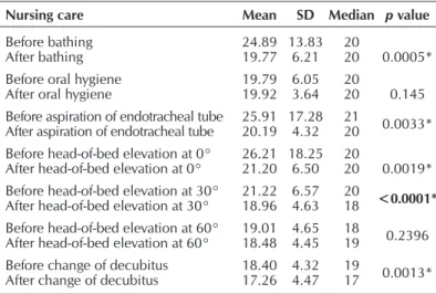
Table 2 – Cuff pressure: mean ± standard deviation and median referring to the night shift, São José do Rio Preto, São Paulo, Brazil, 2016
Nursing care Mean SD Median p value
Before bathing
After bathing 24.8919.77 13.836.21 2020 0.0005*
Before oral hygiene
After oral hygiene 19.7919.92 6.053.64 2020 0.145
Before aspiration of endotracheal tube
After aspiration of endotracheal tube 25.9120.19 17.284.32 2120 0.0033*
Before head-of-bed elevation at 0°
After head-of-bed elevation at 0° 26.2121.20 18.256.50 2020 0.0019*
Before head-of-bed elevation at 30°
After head-of-bed elevation at 30° 21.2218.96 6.574.63 2018 <0.0001*
Before head-of-bed elevation at 60°
After head-of-bed elevation at 60° 19.0118.48 4.654.45 1819 0.2396
Before change of decubitus
After change of decubitus 18.4017.26 4.324.47 1917 0.0013*
Note: * p values ≤ 0.05 significant generated by the Wilcoxon test; SD = Standard deviation.
Table 3 – Cuff pressure: mean ± standard deviation and median referring to the night shift, São José do Rio Preto, São Paulo, Brazil, 2015
Nursing care Mean SD Median p value
Before bathing
After bathing 30.6318.50 22.073.61 2318 <0.0001*
Before oral hygiene
After oral hygiene 18.6219.07 4.244.08 1820 0.2214
Before aspiration of endotracheal tube
After aspiration of endotracheal tube 19.2018.88 4.063.74 2019 0.0663
Before head-of-bed elevation at 0°
After head-of-bed elevation at 0° 18.8218.27 3.833.71 1918 0.1462
Before head-of-bed elevation at 30°
After head-of-bed elevation at 30° 18.4617.80 4.063.76 1818 0.0309*
Before head-of-bed elevation at 60°
After head-of-bed elevation at 60° 17.8217.55 3.813.42 1818 0.3404
Before change of decubitus
After change of decubitus 17.5917.02 3.383.37 1817 0.2189
Note: * p values ≤ 0.05 significant generated by the Wilcoxon test; SD = Standard deviation. Table 1 – Cuff pressure: mean ± standard deviation and median referring
to morning shift, São José do Rio Preto, São Paulo, Brazil, 2016
Nursing care Mean SD Median p value
Before bathing
After bathing 29.3219.37 19.064.16 2020 <0.0001*
Before oral hygiene
After oral hygiene 20.0520.42 7.644.19 2020 0.0482*
Before aspiration of endotracheal tube
After aspiration of endotracheal tube 20.3519.32 3.933.61 209 0.0075*
Before head-of-bed elevation at 0°
After head-of-bed elevation at 0° 20.5318.72 8.143.40 18.519 0.0489*
Before head-of-bed elevation at 30°
After head-of-bed elevation at 30° 19.0018.19 3.583.62 1918 0.0039*
Before head-of-bed elevation at 60°
After head-of-bed elevation at 60° 18.0219.29 3.684.33 1817 0.9906*
Before change of decubitus
After change of decubitus 18.3116.53 34.373.96 1716 0.0005*
Table 1 shows tracheal cuff pressure values in the morning. Table 2 shows tracheal cuff pressure values at the afternoon shift. Table 3 shows tracheal cuff pressures at the night shift.
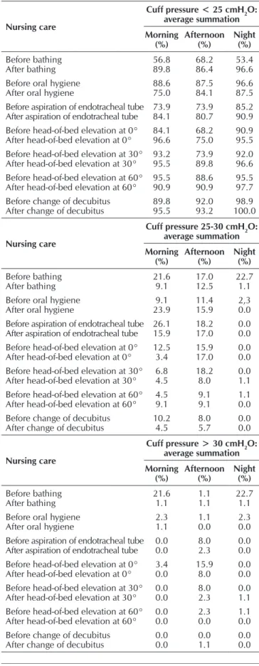
Table 4 shows cuff pressure within normal parameters and when the values were higher and lower than that recommend in the three shifts (morning, afternoon and night).
DISCUSSION
The correct endotracheal cuff pressure must be less than the capillary perfusion pressure, i.e., less than 30 cmH2O. The ideal pressure is between 18 to 22 mmHg and 25 to 30 cm H2O to prevent exhaust air or overinflation. The consensus establishes as an ideal practice to check cuff pressure daily and maintain it between 20 to 34 cmH2O (15 to 25 mmHg)(11). Postoperative dysphagia occurs frequently after anterior cervical spine surgery. This may be related to high endotracheal tube cuff pressure. Whether adaptation and maintaining the pressure after place-ment of the retractor will decrease the incidence of dysphagia, has to be determined by this trial(12). Previous studies have shown that maintaining ETT cuff pressure between 15 and 25 mm Hg can reduce endotracheal intubation-related complications in pa-tients undergoing general anesthesia(13-14).
In the present study, in the morning shift, when the nurses changed the angle of the head-of-bed elevation to 0°, 30°, and 60°, it was observed a decrease from 20.53 mmHg to 18.76 mmHg; from 19,00 mmHg to 18,19 mmHg, and from 18.02 mmHg to 18.29 mmHg, respectively. The statistically significant decrease occurred in 0° and 30°(6).
Regarding the change of body positioning (decubitus), there was a significant change in the cuff pressure in the morning shifts. The decrease in pressure ranged from 18.31 mm Hg to 16.53 mm Hg. Of the 88 checks performed, 95.45% were be-low 25 cmH2O, and 4.54% between 25 to 30 cm H2O. In the evening shift, the average change ranged from 18.40 mmHg to 17.26 mmHg. Of the 88 lower pressure checks, 93.18% were lower than 25 cm H2O; 1.13% above 30 cmH2O, and 5.68% within the desirable parameters, i.e., from 25 to 30 cmH2O. Inflation of the cuff in excess of 30 cm H2O damages the tracheal mucosa by compromising capillary perfusion. When pressures are greater than 50 cm H2O, total obstruc-tion of tracheal blood flow occurs(9).
Regarding nursing care, the in-bed bathing presented a significant change the endotracheal cuff pressure in the three shifts (morning, evening, and night) with mean cuff pres-sure ranging from 29.32 mmHg to 19.37 mmHg; from 24.89 mmHg to 19.77 mmHg, and from 30.63 mmHg to 18.50 mmHg, respectively. It was not possible to find in the litera-ture, studies showing relation with in-bed bathing, which re-quires further studies.
In the morning shift only, the oral hygiene showed a change in the endotracheal cuff pressure statistically significant, with a mean cuff pressure ranging from 20.05 mmHg to 20.42 mmHg. Among all nursing care analyzed, this was the one which less influenced in changing the cuff pressure.
We were no able to find data that could prove whether there is a change in the cuff pressure associated with oral hygiene. We have found the need to check the endotracheal cuff pres-sure prior to performing oral hygiene care as a way to prevent pneumonia associated with mechanical ventilation. The oral Table 4 – Cuff pressure: average summation comparing the three
shifts, São José do Rio Preto, São Paulo, Brazil, 2016
Nursing care
Cuff pressure < 25 cmH2O: average summation
Morning (%)
Afternoon (%)
Night (%)
Before bathing
After bathing 56.889.8 68.286.4 53.496.6
Before oral hygiene
After oral hygiene 88.675.0 87.584.1 96.687.5
Before aspiration of endotracheal tube
After aspiration of endotracheal tube 73.984.1 73.980.7 85.290.9
Before head-of-bed elevation at 0°
After head-of-bed elevation at 0° 84.196.6 68.275.0 90.995.5
Before head-of-bed elevation at 30°
After head-of-bed elevation at 30° 93.295.5 73.989.8 92.096.6
Before head-of-bed elevation at 60°
After head-of-bed elevation at 60° 95.590.9 88.690.9 95.597.7
Before change of decubitus
After change of decubitus 89.895.5 92.093.2 100.098.9
Nursing care
Cuff pressure 25-30 cmH2O: average summation
Morning (%)
Afternoon (%)
Night (%)
Before bathing
After bathing 21.69.1 17.012.5 22.71.1
Before oral hygiene
After oral hygiene 23.99.1 11.415.9 2,30.0
Before aspiration of endotracheal tube
After aspiration of endotracheal tube 26.115.9 18.217.0 0.00.0
Before head-of-bed elevation at 0°
After head-of-bed elevation at 0° 12.53.4 15.917.0 0.00.0
Before head-of-bed elevation at 30°
After head-of-bed elevation at 30° 6.84.5 18.28.0 0.01.1
Before head-of-bed elevation at 60°
After head-of-bed elevation at 60° 4.59.1 9.19.1 1.10.0
Before change of decubitus
After change of decubitus 10.24.5 8.05.7 0.00.0
Nursing care
Cuff pressure > 30 cmH2O: average summation
Morning (%)
Afternoon (%)
Night (%)
Before bathing
After bathing 21.61.1 1.11.1 22.71.1
Before oral hygiene
After oral hygiene 2.31.1 1.10.0 2.30.0
Before aspiration of endotracheal tube
After aspiration of endotracheal tube 0.00.0 8.02.3 0.00.0
Before head-of-bed elevation at 0°
After head-of-bed elevation at 0° 3.40.0 15.98.0 0.00.0
Before head-of-bed elevation at 30°
After head-of-bed elevation at 30° 0.00.0 8.02.3 0.01.1
Before head-of-bed elevation at 60°
After head-of-bed elevation at 60° 0.00.0 2.30.0 1.10.0
Before change of decubitus
hygiene care is necessary to reduce plaque formation and ac-cumulation of waste in the oropharynx, preventing the emer-gence of pathogenic micro-organisms and thus to migration of lower airways and possibly respiratory complications(15).
In the aspiration of the endotracheal tube, there was a sig-nificant difference in pressure cuffs before and after measuring the aspiration in the morning shift, with a mean between the pressures ranging from 20.35 cmH2O to 19.32 cmH2O. In the evening shift, we observed a difference ranging from 25.01 cm H2O to 20.19 cm H2O. When comparing the morning, evening, and night shifts, we observed that 86.4% of the mea-surements in the morning shift were lower than 25 cm H2O; in the evening shift, 82.3% were lower than 25 cm H2O; and in the evening shift, 91.2% presented pressure cuff lower than 25 cm H2O, i.e., changes presenting lower pressures relating to the adequate threshold pressure. The night shift presented the largest number of changes compared to the other shifts.
However, by analyzing the three shifts in a stratified way, the morning and evening shifts showed higher statistically significant difference in pressure cuffs when we performed measures before and after the nursing care. The nursing care performed were in the morning shift: in-bed bathing, oral hy-giene, suction of endotracheal tube, head-of-bed elevation at 0°, head-of-bed elevation at 30º, and changes in body posi-tion. In the afternoon, the nursing care delivered was in-bed bathing, suction of endotracheal tube, head-of-bed elevation at 30º, and changes in body position. The only difference among the night shift and the other two shifts was in in-bed bathing and in head-of-bed elevation at 30°.
One study showed that high pressures were found in the three shifts. However, in the morning shift they had a mean of the values of the pressure cuffs of 36.7 cmH2O. The same was seen in another study, in which the cuff pressure measure-ments above 30 cmH2O were found in the three shifts. Nev-ertheless, the night shift presented more cases of changes in the pressure cuff when compared with the other shifts, which presented 14% of cases(16).
Inflation of the cuff in excess of 30 cm H2O damages the tracheal mucosa by compromising capillary perfusion. When pressures are greater than 50 cm H2O, total obstruction of tra-cheal blood flow occurs(12). Check the pressure of the cuff en-dotracheal tubes through a cuff meter three times a day is im-portant because the greater the number of checks, the greater the chance to adjust the values. However, during the measure-ments may occur an air leak in the uncoupling; another role meant of cuff deflation is the air that is necessary to pressurize the device, showing a value less than the pre-existing one.
Maintaining the cuff pressure ranging from 18 to 22 mmHg reduces the risk of a possible injury to the trachea. However, even if you reached the ideal pressure, but keeping it constant-ly for a time above 12 hours, there may be an inflammatory process in place. Therefore, possible complications are also related to the duration of the intubation. Our data indicate that cuff pressure should be measured after each change in a patient’s body position and supports use of continuous moni-toring of cuff pressure with automatic adaptation to a preset pressure.
Despite the obvious risks associated with excessive pres-sure on the tracheal wall, in daily practice, clinicians rarely evaluate cuff pressure to be sure it is correct. Indeed, cuff pressures outside the target range are common, and the fre-quency with which cuff pressure is measured and adjusted varies from never to at most every 8 hours(5). Monitoring cuff pressure via a manometer results in fewer complications after intubation(14). The extent, if any, that cuff shape influences cuff pressure after changes in body position is unknown(5).
Other study results suggest that changing patient position during mechanical ventilation can lead to significant altera-tions in Pcuff. This study concludes that various factors can induce lesions in the respiratory tract of mechanically venti-lated patients. Such factors include the following: inadequate airway humidification; a high fraction of inspired oxygen; insufficient heating of administered gases; frequent tracheal suction; prolonged endotracheal intubation; prolonged me-chanical ventilation; and inappropriate Pcuff values(4). Cuff pressures >30 cmH2O compress mucosal capillaries, impair blood flow, cause mucosal damage and tracheal rupture, with total occlusion occurring at 50 cmH2O(2).
Changes in a patient’s body position resulted in significant deviations in the cuff pressure of endotracheal tubes. In 40.6% of the measurements, the cuff pressure exceeded the upper tar-get level, and thus theoretically was clinically relevant. Because simple and frequent changes in a patient’s body position may result in potentially harmful cuff pressure, our observations sug-gest a need for a strict monitoring of this pressure(5).
Study limitation and contribution to Nursing
The patients’ anatomical differences were not related to the diameter and pressure inside the endotracheal tubes. Another limitation is concerning to the random allocation and find-ings, which were based on a single institution, which compro-mises data extrapolation.
The study intends to allow greater security in the delivery of health care to the intubated patient, as for the optimal pres-sure volume, thus, preventing accidental extubation and re-ducing the necrosis of the trachea.
CONCLUSION
There were differences before and after health care pro-vided, showing changes in cuff pressure. In-bed bathing and head-of-bed elevation at 30° were the ones that most altered pressure values in the three working shifts. Therefore, it is necessary to measure cuff pressure at least twice per working shift, preferably after bathing.
REFERENCES
1. Chopra M, Jones L, Boulanger C, Benger J, Higginson I, Williamson D, et al. Prospective observational measurement of tracheal tube cuff pressures in the emergency department. Emerg Med J [Internet]. 2010 [cited 2016 Jun 30];27(4):270-1. Available from: https://www.ncbi.nlm.nih.gov/pubmed/20385676
2. Jordan P, Van Rooyen D, Venter D. Endotracheal tube cuff pressure management in adult critical care units. S Afr J Crit Care [Internet]. 2012[cited 2016 Jun 30];28(1):13-6. Available from: http://www.sajcc.org.za/index.php/SAJCC/article/view/129/148 3. Abbasi S, Mahjobipoor H, Kashef P, Massumi G, Aghadavoudi O, Farajzadegan Z, et al. The effect of lidocaine on reducing the
tracheal mucosal damage following tracheal intubation. J Res Med Sci[Internet]. 2013[cited 2016 Mar 30]:18(9):733-8. Available from: https://www.ncbi.nlm.nih.gov/pmc/articles/PMC3872578/
4. Godoy ACF, Vieira RJ, Capitani EM. Endotracheal tube cuff pressure alteration after changes in position in patients under mechanical ventilation. J Bras Pneumol[Internet]. 2008[cited 2016 Mar 30];34(5):294-7. Available from: http://www.scielo.br/pdf/ jbpneu/v34n5/en_v34n5a08.pdf
5. D’Aragon F, Beaudet N, Gagnon V, Martin R, Sansoucy Y. The effects of lidocaine spray and intracuff alkalinized lidocaine on the occurrence of cough at extubation: a double-blind randomized controlled trial. Can J Anaesth[Internet]. 2013[cited 2015 Oct 21];60(4):370-6. Available from: https://dx.doi.org/DOI 10.1007/s12630-013-9896-8
6. Barbas CS, Ísola AM, Farias AM, Cavalcanti AB, Gama Am, Duarte AC, et al. Brazilian recommendations of mechanical ventilation 2013. Rev Bras Ter Intens[Internet]. 2014[cited 2015 Nov 30];26(2):89-121. Available from: https://dx.doi. org/10.5935/0103-507X.20140017
7. Sole ML, Su X, Talbert S, Penoyer DA, Kalita S, Jimenez E, et al. Evaluation of an intervention to maintain endotracheal tube cuf pressure within therapeutic range. Am J Crit Care[Internet]. 2011[cited 2016 Oct 15];20(2):109-17. Available from: https://dx.doi. org/10.4037/ajcc2011661
8. Sole ML, Penoyer DA, Su X, Jimenez E, Kalita SJ, Poalillo E, et al. Assessment of endotracheal cuff pressures by continuous monitoring: a pilot study. Am J Crit Care [Internet]. 2009[cited 2016 Oct 15];18(2):133-43. Available from: https://dx.doi. org/10.4037/ajcc2009441
9. Lizy C, Swinnwn W, Labeau S, Poelaert J, Vogelaers D, Vandewoude K, et al. Cuff pressure of endotracheal tubes after changes in body position in critically ill patients treated with mechanical ventilation. Am J Crit Care[Internet]. 2014 [cited 2015 Nov 30];23(1):e1 8. Available from: https://dx.doi.org/10.4037/ajcc2014489
10. Royal College of Anaesthesia Audit Compendium. Critical care services. In: Nightingale P, Griffiths H, Clayton JA, (Eds.). Section 10.3. Tracheal tube cuff pressures[Internet]. 2016 [cited 2016 Oct 15]. https://www.rcoa.ac.uk/system/files/CSQ-ARB-Contents_ intro.pdf
11. Diretrizes Brasileiras de Ventilação Mecânica. Ventilação Mecânica AMIB e SBPT. Versão eletrônica oficial [Internet]. AMIB e SBPT; 2013 [cited 2016 Oct 10]. Available from: http://itarget.com.br/newclients/sbpt.org.br/2011/downloads/arquivos/Dir_ VM_2013/Diretrizes_VM2013_SBPT_AMIB.pdf
12. Arts MP, Rettig TCD, Vries J, Wolfs JFC, Veld BAI. Maintaining endotracheal tube cuff pressure at 20 mm Hg to prevent dysphagia after anterior cervical spine surgery; protocol of a double-blind randomized controlled trial. BMC Musculoskeletal Disord [Internet] 2013[cited 2016 May 18];14:280-1. Available from: http://www.biomedcentral.com/1471-2474/14/280
13. Sole ML, Penoyer DA, Su X, Jimenez E, Kalita SJ, Poalillo E, et al. Assessment of endotracheal cuff pressure by continuous monitoring: a pilot study. Am J Crit Care [Internet]. 2009[cited 2015 Nov 30];18(2):133-43. Available from: http://ajcc.aacnjournals. org/content/18/2/133.long
14. Liu J, Zhang X, Gong W, Li S, Wang F, Fu S, et al. Correlations between controlled endotracheal tube cuff pressure and postprocedural complications: a multicenter study. Anesth Analg [Internet]. 2010 [cited 2015 Nov 30];111(5):1133-7. Available from: https://www.ncbi.nlm.nih.gov/pubmed/20736432
15. Cerqueira NB, Albuquerque CG, Souza VV, Ramos FF, Andrade FMD, Correia Junior MAV. Fatores que alteram a pressão dos balonetes internos de tubos endotraqueais e a necessidade de sua monitorização. ASSOBRAFIR Ciênc [Internet] 2011 [cited 2016 Apr 12];2(1):29-38. Available from: http://www.uel.br/revistas/uel/index.php/rebrafis/article/download/7905/7717