127
Relevant radiological findings in necrotizing enterocolitis
Radiol Bras 2007;40(2):127–130 Review Article
RELEVANT RADIOLOGICAL FINDINGS FOR THE DIAGNOSIS
OF NECROTIZING ENTEROCOLITIS AND ITS COMPLICATIONS*
Beatriz Regina Alvares1
, Daniel Lahan Martins2
, Renato Lopes Roma3
, Inês Minniti Rodrigues Pereira1
Necrotizing enterocolitis is one of the most frequent and severe gastrointestinal emergencies occurring in the neonatal period. Once necrotizing enterocolitis is suspected a simple abdominal x-ray is a routine exami-nation and this film will play an essential role in the diagnosis of the disease and the follow-up care of the patient, as well as in the detection of complications. In the present study we reviewed the pertinent litera-ture and described the radiological findings, illustrated with cases from our institution. We concluded that the radiological diagnosis of necrotizing enterocolitis done at all stages contributes for an immediate thera-peutic management, reducing the complications and improving the patient’s survival.
Keywords: Necrotizing enterocolitis; Newborn; Radiological diagnosis.
Aspectos radiológicos relevantes no diagnóstico da enterocolite necrosante e suas complicações.
A enterocolite necrosante representa uma das emergências gastrintestinais mais freqüentes e graves no período neonatal. Na suspeita clínica dessa doença, o exame radiológico simples de abdome é um procedimento de rotina, desempenhando um papel fundamental no diagnóstico, acompanhamento e detecção de complica-ções. No presente trabalho, realizamos uma revisão da literatura pertinente e descrevemos os achados radio-lógicos da enterocolite necrosante, ilustrados com casos do nosso serviço. Concluímos que o diagnóstico radiológico da enterocolite necrosante realizado em todas as suas etapas, contribui para uma conduta tera-pêutica imediata, reduzindo as complicações e aumentando a sobrevida dos pacientes.
Unitermos: Enterocolite necrosante; Recém-nascido; Diagnóstico radiológico. Abstract
Resumo
* Study developed in the Department of Radiology of Facul-dade de Ciências Médicas da UniversiFacul-dade Estadual de Campi-nas (Unicamp), CampiCampi-nas, SP, Brazil.
1. PhDs, Professors for Department of Radiology of Faculdade de Ciências Médicas da Universidade Estadual de Campinas (Unicamp), Campinas, SP, Brazil.
2. MD, Resident at Department of Radiology of Faculdade de Ciências Médicas da Universidade Estadual de Campinas (Uni-camp), Campinas, SP, Brazil.
3. Physician Assistant at Department of Radiology of Facul-dade de Ciências Médicas da UniversiFacul-dade Estadual de Campi-nas (Unicamp), CampiCampi-nas, SP, Brazil
Mailing address: Profa. Dra. Beatriz Regina Alvares. Rua Al-berto de Salvo, 238, Distrito de Barão Geraldo. Campinas, SP, Brazil, 13084-759. E-mail: [email protected]
Received March 29, 2005. Accepted after revision July 5, 2005.
INTRODUCTION
Necrotizing enterocolitis (NEC) repsents a condition whose etiology still re-mains unclear, predominantly affecting neonates (NN) weighing less than 1,500 g, and is one of the most frequent and severe gastrointestinal emergencies occurring in the neonatal period(1–3).
Most frequently accepted etiological factors for explaining this condition are intestinal ischemia, resulting in alterations of the intestinal mucosa, excessive bacte-rial growth with formation of gas in the intestinal wall and persistent intestinal
ir-ritation because of oral feeding. The most suggestive clinical findings of this disease are abdominal distension, food or bilious emesis and presence of blood in stools(2,3).
The abdominal plain x-ray is a routine procedure in NN with NEC, since it plays an essential role in the diagnosis, follow-up and detection of complications indicat-ing the need for surgery. Durindicat-ing the treat-ment, NN should undergo serial plain x-rays every hour to allow the early diagno-sis of complications(4,5)
.
RADIOLOGICAL ALTERATIONS
1 – Generalized intestinal distension
The earliest radiological alteration in NEC is a diffused and persistent intestinal gas distension(5). Radiologically, an
intes-tinal loop may be considered as dilated when its measure surpasses the width of the first lumbar vertebral body(6,7). This may be
a non-specific radiological finding in NN with other abnormalities, especially those under continuous ventilation by positive pressure(8). Nevertheless, the suspect of
NEC should be always be raised in radio-logical of NN presenting with persistent,
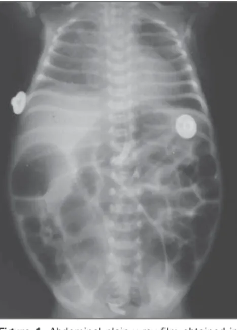
generalized intestinal distension and char-acteristic clinical picture(2,6)(Figure 1).
2 – Localized distension of an intestinal loop
Localized distension o an intestinal loop presenting a tubular configuration and thickened walls was initially described as a radiological sign of imminent perforation. Currently, this radiological sign has not been considered as an imminent risk of perforation anymore, although it is useful to raise the suspicion of NEC at its early phase, since this may mean loop dis-tress(9,10)(Figure 2).
3 – Intestinal pneumatosis
128
Alvares BR et al.
Radiol Bras 2007;40(2):127–130 Figure 2. Abdominal plain x-ray study in dorsal
decubitus demonstrating a single, dilated intesti-nal loop in the right iliac fossa.
Figure 1. Abdominal plain x-ray film obtained in dorsal decubitus demonstrating generalized intes-tinal distension.
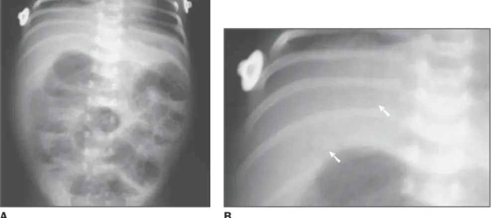
Figure 3(A–E). Abdominal x-ray films of different patients in dorsal decubitus demonstrating intesti-nal pneumatosis characterized by visible radiolucent lines in the walls of the intestinal loops (arrows).
A B
C D
E Intestinal pneumatosis represents the
most pathognomonic radiological finding of NEC, presenting as visible radiolucent, linear, curved-linear or bullous images on the intestinal loop wall. In some circum-stances, the radiological appearance re-sembles the intestinal fecal contents, and the diagnosis is made by means of serial x-rays demonstrating the permanence of
in-tramural gas, contrarily to the feces with present motility(2,11–13) (Figure 3A–E).
4 – Air within the portal system
The intestinal pneumatosis may extend into the venous portal system, and is vis-ible on abdominal plain x-ray films as ra-diolucent, linear images in hepatic projec-tion, and extending to peripheral areas(6,
11,14,15)
(Figures 4A and 4B). This type of distribution allows the differential diagno-sis with air in the biliary tract, with a radio-logical aspect similar to that of air within
➙
➙
➙
➙
➙
➙
➙
➙
129
Relevant radiological findings in necrotizing enterocolitis
Radiol Bras 2007;40(2):127–130 the portal system, but with a more central hepatic localization(6).
5 – Pneumoperitoneum
The term pneumoperitoneum refers to the presence of free air within the perito-neal cavity caused by a perforated hollow, and is a complication from necrotizing enterocolitis(1,2,6,11). The radiological signs
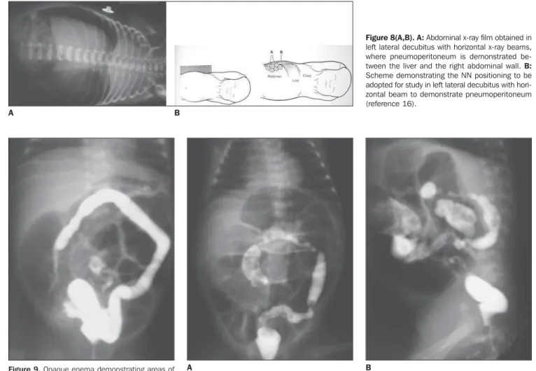
can be seen on abdominal plain x-rays per-formed in dorsal decubitus, with vertical and horizontal x-ray beams, orthostatic po-sition and left lateral decubitus with hori-zontal beams(11,16).
On x-ray films obtained in dorsal decu-bitus with vertical x-ray beams a darkened abdominal cavity is observed, and the in-testinal wall is visualized because of the presence of air both inside and outside the loop (Figure 5). On this view, a falciform ligament may appear, the association of these images being described as “football sign” for resembling the shape of the ball used in the American football(11). On x-ray
films obtained with the patients in orthos-tatic position (Figure 6), dorsal decubitus (Figures 7A and 7B) and left lateral decu-bitus with horizontal x-ray beam (Figures 8A and 8B), the free air movement is ob-served within the abdominal cavity, under the diaphragmatic cupula, anteriorly, or be-tween the liver and the right abdominal wall.
6 – Late complications
Areas of stenosis of the large intestine represent a late complication from necro-tizing enterocolitis, manifesting clinically through obstruction. On plain abdominal x-ray films, a significant intestinal distension is observed with absence of air in the rec-tum(17,18). The diagnosis is confirmed by
opaque enema demonstrating areas of stenosis in the large intestine. In the pres-ence of risk of intestinal rupture, this ex-amination should be performed with di-luted iodinated contrast agent(18,19) (Figures
9, 10A and 10B).
CONCLUSION
The radiological diagnosis of NEC ac-complished in all phases contributes for an immediate therapeutical management, re-ducing the rate of complications and im-proving the patients’ survival.
Figure 5. Abdominal plain x-ray in dorsal decubi-tus showing abdominal hypertransparency because of pneumoperitoneum. Also, the intestinal loop wall is observed, due to the presence of air both inside and outside the loop.
Figure 4(A,B). Abdominal plain x-ray film obtained in dorsal decubitus demonstrating the presence of air within the portal system — radiolucent linear images in hepatic projection, extending to peripheral ar-eas. Also, intestinal pneumatosis is observed (A).
A B
➙ ➙
A B
Figure 7(A,B). A: Abdominal plain x-ray obtained in dorsal decubitus with horizontal x-ray beam. Free air movement is observed anteriorly within the abdominal cavity. B: Scheme demonstrating the NN position-ing to be adopted for study in dorsal decubitus with horizontal beam to demonstrate pneumoperitoneum (reference 16).
Figure 6. Abdominal plain x-ray film obtained in orthostatic position with horizontal x-ray beam (lifted NN) demonstrating pneumoperitoneum under the diaphragmatic cupula (arrows).
130
Alvares BR et al.
Radiol Bras 2007;40(2):127–130 Figure 8(A,B). A: Abdominal x-ray film obtained in left lateral decubitus with horizontal x-ray beams, where pneumoperitoneum is demonstrated be-tween the liver and the right abdominal wall. B:
Scheme demonstrating the NN positioning to be adopted for study in left lateral decubitus with hori-zontal beam to demonstrate pneumoperitoneum (reference 16).
Figure 10(A,B). Anteroposterior (A) and lateral (B) views of opaque enema demonstrating areas of stenosis in the transverse and sigmoid colon.
Figure 9. Opaque enema demonstrating areas of stenosis in the ascending colon.
A B
A B
REFERENCES
1. Kosloske AM, Musemeche A. Necrotizing entero-colitis of the neonate. Clin Perinatol 1989;16:97– 111.
2. Buonomo C. The radiology of necrotizing entero-colitis. Radiol Clin North Am 1999;37:1187–1198. 3. Ostlie DJ, Spilde TL, St Peter SD, et al. Necro-tizing enterocolitis in full-term infants. J Pediatr Surg 2003;38:1039–1042.
4. Kanto WP Jr, Hunter JE, Stoll BJ. Recognition and medical management of necrotizing entero-colitis. Clin Perinatol 1994;21:335–346. 5. Daneman A, Woodward S, da Silva M. The
radi-ology of neonatal necrotizing enterocolitis (NEC). A review of 47 cases and the literature. Pediatr Radiol 1978;7:70–77.
6. Morrison SC, Jacobson JM. The radiology of ne-crotizing enterocolitis. Clin Perinatol 1994;21: 347–363.
7. Edwards DK. Size of gas-filled bowel loops in
in-fants. AJR Am J Roentgenol 1980;135:331–334. 8. Jaile JC, Levin T, Wung JT, Abramson SJ, Ruzal-Shapiro C, Berdon WE. Benign gaseous disten-tion of the bowel in premature infants treated with nasal continuous airway pressure: a study of con-tributing factors. AJR Am J Roentgenol 1992; 158:125–127.
9. Wexler H. The persistent loop sign in neonatal ne-crotizing enterocolitis: a new indication for sur-gical intervention? Pediatr Radiol 1978;126:201– 204.
10. Leonard T Jr, Johnson JF, Pettett PG. Critical evaluation of the persistent loop sign in necrotiz-ing enterocolitis. Radiology 1982;142:385–386. 11. Swischuk LE. Alimentary tract. In: Swischuk LE, editor. Imaging of the newborn, infant, and young child. 5th ed. Philadelphia: Lippincott Williams & Wilkins, 2004;341–589.
12. Heng Y, Schuffler MD, Haggitt RC, Rohrmann CA. Pneumatosis intestinalis: a review. Am J Gastroenterol 1995;90:1747–1758.
13. Pear BL. Pneumatosis intestinalis: a review. Ra-diology 1998;207:13–19.
14. Molik KA, West KW, Rescorla FJ, Scherer LR, Engum SA, Grosfeld JL. Portal venous air: the poor prognosis persists. J Pediatr Surg 2001;36: 1143–1145.
15. Briski LE, Von Berg V, Humes JJ. Necrotizing enterocolitis of the newborn. Ann Clin Lab Sci 1982;12:186–193.
16. Gyll C. The abdomen. In: Meerstadt PWD, Gyll C, editors. Manual of neonatal emergency x-ray interpretation. London: Saunders, 1994;152–237. 17. Virgee JP, Gill JG, Desa D, Somers S, Stevenson GW. Stricures and other late complications of neonatal necrotizing enterocolitis. Clin Radiol 1979;30:25–31.