Knockout Studies Reveal an Important Role of
Plasmodium
Lipoic Acid Protein Ligase A1 for Asexual
Blood Stage Parasite Survival
Svenja Gu¨nther1¤a, Kai Matuschewski2¤b, Sylke Mu¨ller1*
1Division of Infection & Immunity and Wellcome Centre for Parasitology, Faculty of Biomedical and Life Sciences, University of Glasgow, Glasgow, United Kingdom, 2Department of Parasitology, Heidelberg University, School of Medicine, Im Neuenheimer Feld, Heidelberg, Germany
Abstract
Lipoic acid (LA) is a dithiol-containing cofactor that is essential for the function ofa-keto acid dehydrogenase complexes. LA acts as a reversible acyl group acceptor and ‘swinging arm’ during acyl-coenzyme A formation. The cofactor is post-translationally attached to the acyl-transferase subunits of the multienzyme complexes through the action of octanoyl (lipoyl): N-octanoyl (lipoyl) transferase (LipB) or lipoic acid protein ligases (LplA). Remarkably, apicomplexan parasites possess LA biosynthesis as well as scavenging pathways and the two pathways are distributed between mitochondrion and a vestigial organelle, the apicoplast. The apicoplast-specific LipB is dispensable for parasite growth due to functional redundancy of the parasite’s lipoic acid/octanoic acid ligases/transferases. In this study, we show thatLplA1plays a pivotal role during the development of the erythrocytic stages of the malaria parasite. Gene disruptions in the human malaria parasiteP. falciparumconsistently were unsuccessful while in the rodent malaria model parasiteP. bergheitheLplA1gene locus was targeted by knock-in and knockout constructs. However, theLplA1(2)mutant could not be cloned suggesting a critical role of LplA1 for asexual parasite growth in vitro and in vivo. These experimental genetics data suggest that lipoylation during expansion in red blood cells largely occurs through salvage from the host erythrocytes and subsequent ligation of LA to the target proteins of the malaria parasite.
Citation:Gu¨nther S, Matuschewski K, Mu¨ller S (2009) Knockout Studies Reveal an Important Role ofPlasmodiumLipoic Acid Protein Ligase A1 for Asexual Blood Stage Parasite Survival. PLoS ONE 4(5): e5510. doi:10.1371/journal.pone.0005510
Editor:Lisa F. P. Ng, Singapore Immunology Network, Singapore
ReceivedDecember 22, 2008;AcceptedApril 8, 2009;PublishedMay 12, 2009
Copyright:ß2009 Gunther et al. This is an open-access article distributed under the terms of the Creative Commons Attribution License, which permits unrestricted use, distribution, and reproduction in any medium, provided the original author and source are credited.
Funding:This study was supported by The Wellcome Trust (WT061173MA) (SM), a PhD Scholarship funded by Boehringer Ingelheim Fonds (SG) and the European Commission (FP6-funded network of excellence BIOMALPAR program: LSHP-CT-2004-503578. The funders had no role in study design, data collection and analysis, decision to publish, or preparation of the manuscript.
Competing Interests:The authors have declared that no competing interests exist.
* E-mail: [email protected]
¤a Current address: The Walter and Eliza Hall Institute for Medical Research, Melbourne, Australia ¤b Current address: Parasitology Unit, Max Planck Institute for Infection Biology, Berlin, Germany
Introduction
Lipoic acid (6,8-thioctic acid, LA) is an essential cofactor for the multienzyme complexes pyruvate dehydrogenase (PDH), a -ketoglutarate dehydrogenase (KGDH), branched chain a-keto acid dehydrogenase (BCDH) and the glycine cleavage system (GCS). These enzyme complexes are composed of three or four subunits which themselves are oligomers [1,2]. The three subunits that comprise thea-keto acid dehydrogenase complexes (KADH) are thea-keto acid decarboxylase (or E1), the acyltransferase (or E2), which uses lipoamide as a cofactor, and the dihydrolipoamide dehydrogenase (or E3). LA is post-translationally attached to E2 and functions as the so-called ‘‘swinging arm’’ in the reaction catalysed by KADH complexes, accepting the acyl-moiety from E1 and transferring it to coenzyme A to form acyl-CoA [1]. During this reaction the lipoamide cofactor is reduced and subsequently re-oxidised by E3 to regenerate its ability to accept the next acyl-moiety from E1. The GCS works in a similar way to the KADH, with the H-protein being the lipoylated heart of this complex [3].
Usually these enzyme complexes are found in the mitochon-drion of eukaryotic cells, but plastid-bearing organisms also possess
a PDH in the plastid that provides substrates for type II fatty acid biosynthesis operating in the organelle [4]. This situation requires lipoylation machineries in both the mitochondrion and plastids [5].
In most microorganisms, lipoylation occurs through salvage and subsequent ligation of LA to the target protein employing lipoic acid protein ligases (LplA) [6]. LplA attach scavenged LA to the substrate protein in an ATP-dependent reaction. The first step of the reaction leads to the formation of a LA-AMP intermediate and the release of pyrophosphate. The activated lipoyl-moiety is then attached to thee-amino group of a specific lysine residue in the lipoyl-domain of the E2-subunit or the H-protein while AMP is released [6]. In the absence of exogenous LA, bacteria synthesise LAde novo. They ligate the octanoyl-moiety of octanoyl-acyl carrier protein (ACP), an intermediate of fatty acid biosynthesis, to the E2-apo-proteins or apo-H-protein using octanoyl (lipoyl) : N -octanoyl (lipoyl) transferase (LipB). Subsequently, lipoic acid synthase (LipA) catalyses the insertion of two sulphurs into position C6 and C8 of the octanoyl-moiety to form the lipoyl-arm required for KADH and GCS activities [6].
indeed knockout of the apicoplast targeted LipB gene was not lethal forP. falciparumintraerythrocytic stages suggesting that the dually targeted LplA2 compensates for the loss of LipB function [16]. Down-regulation of acyl carrier protein (ACP) expression in T. gondiirevealed that lipoylation levels of the apicoplast PDH was ablated suggesting thatde novofatty acid biosynthesis operating in the organelle is the major source for the LA precursor octanoyl-ACP [15]. This is similar to a study by Witkowski and colleagues [17] who found that one of the major products of mitochondrial fatty acid biosynthesis in mammalian cells is octanoyl-ACP, which is efficiently ligated to the mitochondrial H-protein. Similarly, it was reported that one of the main roles for mitochondrial fatty acid biosynthesis in plants and the protozoan parasiteTrypanosoma bruceiis the generation of octanoyl-ACP to serve as substrate for LA biosynthesis [18,19]. In the light of these studies, it appears imperative to obtain further insights into the importance of the mitochondrial LA salvage pathways present inPlasmodiumwhich clearly is essential to provide LA to the KADH and GCS with the absence of LA biosynthesis in the mitochondrion. This study aimed to elucidate the role of LplA1 in bothP. falciparumandP. bergheiusing reverse genetics approaches.
Results
Knockout studies of LplA1 in P. falciparum
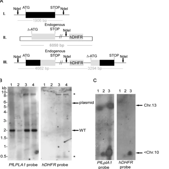
In order to obtain insights into the importance of LA salvage by LplA1 we decided to attempt to knockout theLplA1gene in P. falciparum. We first targeted theP. falciparum LplA1gene by single cross-over recombination using the construct pHH1-LplA1-KO. The KO-construct lacks the ATG start codon and is truncated at the 39 end so that upon recombination of the plasmid by single cross-over two non-functional gene copies are generated. The first copy retains the endogenous promoter and the start codon but is truncated at the 39end through the integration of a premature stop codon into the construct and the second copy is without promoter and start codon (Fig. 1A). Two independently transfected parasite lines were generated and each was taken through three drug selection cycles in order to select for parasites where thePfLplA1 locus had been disrupted by the transfection plasmid. Genomic DNA was isolated and analysed by diagnostic Southern blotting (Fig. 1B). This revealed the presence of endogenous PfLplA1 (1.9 kb band) and linearised plasmid (6 kb band), but no integration events into the parasite genome were detected (Fig. 1B). In order to analyse whether this lack of recombination was due to the essential role ofPfLplA1 for parasite survival or whether other reasons prevent targeting of the PfLplA1 gene locus, two approaches were taken. First, parasites were transfected with the knock-in construct pHH1-LplA1-KOkon, which retains the entire 39end. Upon single cross-over recombination the recombinant locus is predicted to contain one functional gene copy whereas the
gene locus was targeted by the transfection plasmid but some of the regulatory elements present in pHH1-LplA1-KOkon poten-tially might lead to recombination with the parasite genome. The suggestion of a stable integration of pHH1-LplA1-KOkon into an unrelated gene locus is not only consistent with the appearance of the prominent ,9 kb band that cross-reacted with both the PfLplA1probe and thehDHFRprobe but also the concomitant loss of the episome from the parasites despite drug selection. Presumably this stable integration into the parasite genome makes particularly difficult to select for a gene knockout especially if disruption of the gene would result in a growth defect of the null mutants. Similarly, the knock-in of a control plasmid could possibly result in a growth phenotype given that the gene locus is altered by the integration of the transfection plasmid. Confirma-tion that an unrelated gene locus was targeted by pHH1-LplA1 -KOkon came from pulsed field gel electrophoresis (PFGE), where thePfLplA1probe detected a signal on chromosome 13, which is presumably the endogenousPfLplA1gene in wild-type parasites. In contrast, in the LplA1-KOkon parasites the probe detected an additional signal that is likely to correspond to the transfection plasmid integrated into an unrelated gene locus present on one of the smaller chromosomes (Fig. 2C; left panel,,Chr.10). The same signal was detected with the hDHFR probe corroborating the conclusion that the transfected control plasmid readily targeted a gene locus other thanPfLplA1(Fig. 2C, right panel).
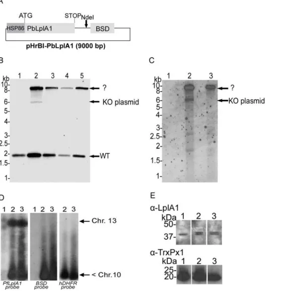
following selection cycles, the 6 kb band diagnostic for the KO-plasmid disappeared and the only band in addition to thePfLplA1 wild-type signal that reacted with both thePfLplA1and thehDHFR probes was a ,9 kb fragment (Fig. 4B,C). This suggests that pHH1-LpA1-KO had recombined with a gene locus other than PfLplA1despite the presence of the episome bearing theP. berghei LplA1 gene. To further analyse the genotype of the double transfected parasites, PFGE was performed and the blot was probed with the PfLplA1-probe, thehDHFR-probe and theBSD -probe. This experiment revealed that the PfLplA1 gene was undisrupted on chromosome 13 and that the two plasmids appeared to be present on a chromosome smaller than chromosome 10, which was not resolved at the bottom of the PFGE gel (Fig. 4D). Further, it should be noted that thePfLplA1 probe also detected the presence of a LplA1gene copy on the smaller chromosome suggesting that at least pHH1-LplA1-KO still carried theLplA1-KOcassette. This suggests that theBSD-positive signal on the smaller chromosome also corresponds to the entire pHrBI-PbLplA1plasmid. It cannot be excluded that pHH1-LplA1 -KO and pHrBI-PbLplA1had actually recombined with each other before they recombined with the non-related gene locus. This possibility was not further analysed. In order to analyse whether the PbLplA1 copy that was introduced into the parasites was actually expressed and thus potentially would compensate for PfLplA1 function, western blots were performed. The results show that parasites co-transfected with knockout and expression plasmid express two proteins that are recognised by theP. falciparumLplA1 antiserum. The two proteins are 70.3% identical (Fig. 3) suggesting that it is likely that the antibody raised against theP. falciparum LplA1 protein cross-reacted with the P. bergheiprotein. This was corroborated by performing a western blot performed onP. berghei blood stage parasite lysate (Fig. 5D). Consistent with this, the blot of the double transfectants revealed the presence of two protein bands of about 45 kDa mass that were barely separated from each other while the antibody only recognised a single protein of similar size in the wild-type parasites (Fig. 4E). A similar result was obtained when aP. falciparumline that was only transfected with pHrBI-PbLplA1 was analysed by western blotting, which also
revealed that the antiserum raised againstPfLplA1 detected two proteins of the expected sizes in these independent transfectants (Fig. 5C). That both proteins appear to migrate faster in the SDS-PAGE than their predicted sizes is likely to be due to the fact that the mitochondrial targeting peptide has been cleaved and the observed sizes correspond to those of the mature proteins. From these experiments we concluded that it is not easily possible to target theP. falciparum LplA1gene locus and we tentatively suggest that LplA1 has an important, if not essential function for P. falciparumduring their erythrocytic development.
Knockout studies of LplA1 inP. berghei
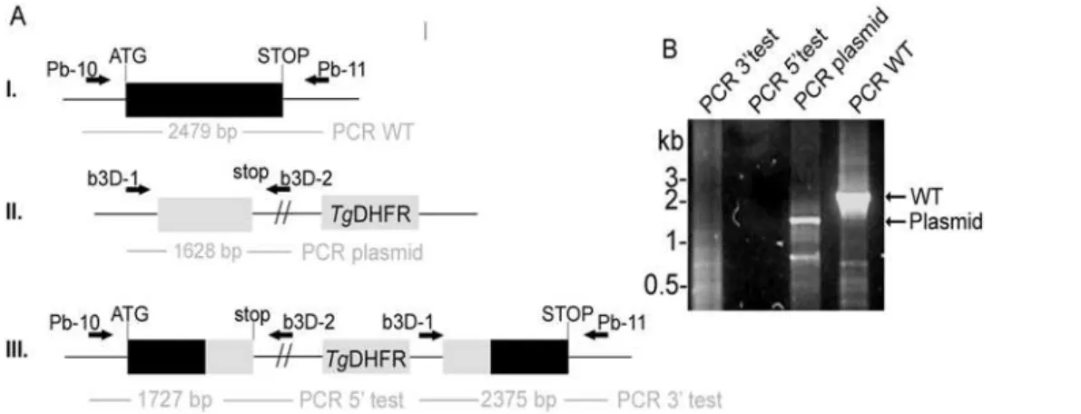
To further investigate the role of LplA1, knockout of theLplA1 gene was attempted in the murine malaria parasiteP. berghei. Two knockout strategies were employed - the first one using single cross-over recombination, similar to the strategy described above for P. falciparum, and the second using double cross-over recombination techniques [21].
The first strategy should result in a disruption of the endogenous gene locus and generate two incomplete copies ofPbLplA1- one truncated at the 39end and one truncated at the 59end (Fig. 6A). This strategy was also used to generate a knock-in construct which should lead to a reconstitution of the endogenous gene upon integration of the plasmid into thePbLplA1gene locus. Parasites were transfected three times with linearised KO-plasmid and after pyrimethamine selection in mice the resistant parasites were subjected to diagnostic PCR analyses (Fig. 6B). According to the PCR results, only endogenous PbLplA1 and plasmid were amplified (lanes 3 and 4 in Fig. 6B), but no PCR product was obtained with the diagnostic primer sets for a disruption of the PbLplA1gene locus (lanes 1 and 2 in Fig. 6B). These data suggest, similar to those described above forP. falciparum, that the LplA1 locus inP. bergheimight either be refractory to recombination or thatLplA1knockout cannot be achieved because of the essential function of the gene/protein. The first conclusion can be excluded because transfection of P. berghei with the control plasmid b3D-LplA-int2 resulted in pyrimethamine resistant parasites that showed integration of the plasmid into the PbLplA1 gene locus Figure 1. The P. falciparum LplA1 locus cannot be disrupted.A. Schematic representation of endogenousPfLplA1gene locus (I.), transfection
plasmid pHH1-LplA-KO (II.) andPfLplA1gene locus after single cross-over recombination (III.). The restriction enzyme used for diagnostic digest is
shown (NdeI) and the expected sizes of diagnostic bands after hybridization withPfLplA1orhDHFR(human dihydrofolate reductase) probes are
indicated.B. Southern blot analyses ofP. falciparumtransfected with pHH1-LplA1-KO. Genomic DNA of wild-type D10 (lane 1) andPfLplA1-KO after 3
selection cycles with WR99210 (lane 2) was digested withNdeI and the Southern blot was probed with theP. falciparum LplA1ORF. The 1.9 kb band
that corresponds to endogenousPfLplA1is visible in both lanes, whereas the transfection plasmid pHH1-LplA1-KO (6 kb band) only is recognised by
the probe in the transfectants. No other bands are visible that are diagnostic for the integration of thePfLplA1gene locus.
doi:10.1371/journal.pone.0005510.g001
(Fig. 7). The authenticity of the PCR products was verified by sequence analysis confirming that theLplA1locus inP. bergheiis not refractory to recombination.
The second approach in theP. bergheisystem was to replace the endogenous PbLplA1 gene with the selectable marker T. gondii DHFR-TSusing the transfection plasmid b3D-LplA-REP. Linear-ised DNA was transfected at 6 independent occasions and pyrimethamine resistant parasites were analysed by PCR (Fig. 8). The results showed that re-circularisation of the linearised plasmid had occurred in 5 out of the 6 occasions, which usually leads to loss
of linearised DNA because the drug pressure applied selects for the presence of the circular plasmid containing the selectable marker. In those parasites, the gene locus was still intact as expected. However, in parasite line 6, it was possible to amplify the diagnostic product for the PbLplA1 gene replacement after two rounds of PCR (Fig. 8B). Again this was verified by subcloning the PCR fragment and analysing its nucleotide sequence. However, this mutant parasite line was lost after transfer into a new animal -a procedure th-at is routinely performed to prop-ag-ate the knockout parasite population and to be able to generate clones (Fig. 8C). Figure 2. Genotypic analysis of P. falciparum transfected with the complementation construct pHH1-LplA1-KOkon.A. Schematic
representation of endogenousPfLplA1gene locus (I.), transfection plasmid pHH1-LplA1-KOkon (II.) andPfLplA1gene locus after single cross-over
recombination (III.). The restriction enzyme used for diagnostic digest is shown (NdeI) and the expected sizes of diagnostic bands after hybridization
withPfLplA1orhDHFRare indicated.B. Southern blot analyses of transfected parasite lines. Genomic DNA of wild-type andLplA1-KOkon parasites was
digested withNdeI and probed with thePfLplA1ORF (left panel). In all parasite lines analysed, the endogenous gene is present (1.9 kb band) and in
lanes 3 and 4 two additional DNA fragments are detected by the probe (in lane 2 less DNA was loaded on the gel so that the plasmid band is hardly
visible). The faint 6 kb band is diagnostic for the presence of the transfection plasmid (see scheme), but the band of,9 kb (*) cannot be assigned to
any specific integration event. Lane 1, wild-type D10; lane 2,PfLplA-KOkon, cycle 1; lane 3,PfLplA-KOkon, cycle 2; lane 4,PfLplA-KOkon, cycle 3. The
same blot was stripped and re-probed with the probe specifically detecting the selectable markerhDHFRand apart from faint plasmid bands at 6 kb
in lanes 2, 3 and 4 two additional bands of,9 kb (*) and 0.5 kb (*) are detected. Lane 1, wild-type D10; lane 2,PfLplA-KOkon, cycle 1; lane 3,PfLplA
-KOkon, cycle 2; lane 4,PfLplA-KOkon, cycle 3.C.Genotyping by pulsed field gel electrophoreses. Chromosomes of wild-typeP. falciparumD10 (lane 1)
and parasites transfected with the pHH1-LplA1-KOkon construct in cycle 0 (lane 2) and cycle 3 (lane 3) were analysed by PFGE and the blots were
probed with thePfLplA1open reading frame (left panel) or with thehDHFRopen reading frame (right panel) present in the transfection plasmid. The
PfLplA1probe detected the endogenousPfLplA1gene locus on chromosome 13 in wild-type and transfected parasites. However, the probe also
detected a strong signal at the bottom of the blot where the non-separated chromosomes,10 are running. ThehDHFRprobe similarly generated
signals on chromosome,10, but there is no sign of integration into theLplA1gene locus on chromosome 13.
These data support that theLplA1locus can be targeted not only by the control plasmid, but also by the replacement plasmid. However, the results suggest that the knockout of theLplA1gene might have severe effects on parasite growth rate and survival during erythrocytic development.
Discussion
The ligation of LA to E2 subunits of the KADH or the apo-H-protein of the GCS is likely to be an essential reaction in most
organisms, unless it can be replaced by the LA biosynthesis pathway like in most bacteria [6]. InPlasmodiumLA biosynthesis and salvage are located in two different organelles, the mitochondrion and the apicoplast [8,10–12]. It further appears that LA cannot be exchanged between the two organelles suggesting that knockout of the mitochondrial salvage pathway might be lethal for the parasites [13–15]. However,Plasmodium possesses two LplA-like proteins with LplA1 present in the mitochondrion and LplA2 present in both mitochondrion and apicoplast [11,12,16]. This situation raises the question whether Figure 3. Alignment ofP. falciparumLplA1 andP. bergheiLplA1.The deduced amino acid sequences of PF13_0083 and PB 000283.02.0 were aligned using ClustalW. The identity between the two sequences was determined to be 70.3% while the similarity is 78%. The predicted sizes of the
two proteins are almost identical with 47.94 kDa for PfLplA1 and 47.97 kDa forPbLplA1. Identical residues are labelled in yellow; homologous
residues are labelled in green. Consensus: gives the consensus amino acid sequence of both proteins. doi:10.1371/journal.pone.0005510.g003
there is redundancy between the two ligases as it was shown that LplA2 can partially compensate for LipB function in the apicoplast [16]. However, our data do not give conclusive information about potential redundancy between LplA1 and LplA2 inP. falciparum, because it was impossible to target theLplA1gene locus, even in
the presence of an expressing episomal copy of the gene; neither was it possible to target theP. falciparum LplA1locus with a knock-in construct (see Fig. 2 and 4).
Therefore, we decided to expand our efforts and attempt a knockout of the LplA1 gene in the murine malaria parasite P.
Figure 4. Genotyping of the double transfection inP. falciparumwith the targeting construct pHH1-LplA1-KO and the trans-species expression construct pHrBI-PbLplA1.The expression plasmid was transfected into theP. falciparumline already carrying pHH1-LplA1-KO in cycle
0.A. Schematic representation of the plasmid pHrBI-PbLplA1carrying the expression cassette ofP. berghei LplA1which is under the control of the
PfHSP86promoter.B.Genotyping of co-transfected parasite lines. Genomic DNA of wild-type (lane 1) and co-transfected parasites isolated after
selection cycle 0 to 3 following transfection (lanes 2 to 5) was digested withNdeI, and the blot was probed with thePfLplA1open reading frame. The
blot showed the endogenousPfLplA1gene specific band at 1.9 kb in lane 2 (cycle 0 after transfection). The pHH1-LplA1-KO plasmid (6 kb) was visible
in cycle 0 (lane 2) and an additional DNA fragment was recognised by the probe, which is unrelated to any expected fragments. TheP. falciparum
LplA1probe did not recognise the presence of the 9 kb pHrBI-PbLplA1expression plasmid (see Supplementary Figure 2) but the signal on the blot is
due to recombination of pHH1-LplA1-KO into an un-related gene locus. The 6 kb pHH1-LplA1-KO plasmid band disappears in lanes 3 to 5 while the
,9 kb band is prominent in these parasite lines.C.Using ahDHFRprobe it was established that the 9 kb band contains bothP. falciparum LplA1and
the selectable marker suggesting that the plasmid had targeted a gene locus unrelated toPfLplA1.D.Analyses of the co-transfected parasite lines by
PFGE supports that the pHH1-LplA1-KO plasmid had recombined with an unrelated gene locus (probehDHFR, right panel). Reprobing the blot with
theBSDprobe (middle panel) that specifically recognises the pHrBI-PbLplA1expression construct, showed that the expression plasmid was also
present on a chromosome that was not resovled under the conditions of this PFGE. ThePfLplA1locus was recognised by thePfLplA1-specific probe
(left panel). Lanes 1, wild-type; lanes 2,P. falciparumco-transfected withLplA1-KO and pHrBI-PbLplA1, cycle 0; lanes 3,P. falciparumco-transfected
withLplA1-KO and pHrBI-PbLplA1, cycle 3.E. Expression of LplA1 protein in co-transfected parasites. The western blot shows parasite extracts that
were isolated from wild-type (lane 1) and two independentP. falciparumlines co-transfected with pHH1-LplA1-KO and pHrBI-PbLplA1(lanes 2 and 3)
probed with a rabbit anti-P. falciparumLplA1 antibody at 1:1000 dilution. The antibody detects one band in the wild-type parasite extracts that
corresponds to the endogenous LplA1 protein. In the co-transfected parasite lines an additional protein is detected, which presumably corresponds
to theP. bergheiLplA1 protein expressed from pHrBI-PbLplA1. The blot was re-probed with a rabbit antibody raised againstP. falciparum1-Cys
berghei. TheP. berghei LplA1gene locus is targetable by knock-in and also a gene replacement construct, clearly demonstrating that this gene locus is not refractory to recombination inP. berghei. Given that the genetic organisation of the LplA1 gene locus is highly syntenic inP. falciparumandP. berghei, it is possible that the failure to obtain a gene disruption ofLplA1inP. falciparumis due to its important function for the parasites during their erythrocytic development. This was also suggested by Allary and colleagues [13] who showed that inhibition ofP. falciparumLplA1 by the LA analogue 8-bromooctanoic acid had deleterious effects on parasite survival. Thus, it appears that LplA1 function cannot be replaced by LplA2 despite the dual targeting of this protein into apicoplast and mitochondrion. One reason for this lack of redundancy might
be the different expression profiles ofLplA1andLplA2genes inP. falciparum; PfLplA1 mRNA is primarily present throughout the intraerythrocytic life of the parasites whilePfLplA2 seems to be primarily expressed in the sexual stages of the parasites [22]. Another possible explanation for the lack of redundancy between the two proteins might be their differential substrate specificities as has previously been suggested by Allary et al. [13]. This is not unusual and has been described before in Listeria monocytogenes, where the two LplAs present in the LA auxotroph organism act on distinct substrates [23,24]. Thus, substrate specificity and time of expression might regulate the function ofLplA1and LplA2inP. falciparum. Our preliminary data on the effect of aLplA2knockout inP. bergheisuggest, however, that the protein is not essential for Figure 5. Analysis of P. falciparum harbouring pHrBI-PbLplA1. A. Southern blot ofP. falciparum transfected with pHrBI-PbLplA1 before
transfection (lane 1) and after transfection (lanes 2 and 3). The DNA was digested withNdeI and the blot was probed with thePfLplA1ORF. The DNA
fragment recognised by the probe corresponds to the endogenousPfLplA1gene. ThePfLplA1probe did not cross-react with the transfected plasmid
pHrBI-PbLplA1.B. To verify the presence of the transfected plasmid, the blot was re-probed with a probe specifically recognising the blasticidin S
deaminase ORF present on the plasmid as selectable marker. The probe hybridised with a DNA fragment diagnostic for the transfected plasmid
(,9.0 kb).C. Western blot ofP. falciparumexpressing PbLplA1. Lane 1, non-transfected parasites; Lane 2,P. falciparumtransfected with
pHrBI-PbLplA1. Equal loading was verified by re-probing the blot with a polyclonal antibody directed against the 1-Cys peroxiredoxin ofP. falciparum. The
polyclonal antiserum raised againstPfLplA1 recognised a protein of,45 kDa in the wild-type parasites which corresponds roughly to the expected
size ofPfLplA1 (predicted size 47.9 kDa). In the transfected parasite line carrying the pHrBI-PbLplA1plasmid two proteins of very similar size were
detected by the antibody – presumably one corresponds to the endogenousPfLplA1 while the second band corresponds toP. bergheiLplA1. This is
surprising as the predicted sizes of both proteins are virtually identical. However, it is possible that the cleavage sites of the mitochondrial targeting
peptides are distinct generating proteins that are just distinguishable by SDS-PAGE.D. Western blot ofP. bergheilysate (lane 1: 16106parasites and
lane 2: 0.26107parasites) usingP. falciparumrabbit anti-LplA1 polyclonal antibody. A band of,45 kDa (similar size to the protein identified in the
double transfected parasites as well as the wild-type parasites only transfected with pHrBI-PbLplA1) reacts strongly with the heterologous antiserum.
doi:10.1371/journal.pone.0005510.g005
blood stage development and has a function important for the parasites’ development in the insect host, which is consistent with the expression profile of this gene (Gu¨nther, Matuschweski and Mu¨ller, unpublished data).
The P. berghei LplA1 gene locus was not only targeted by a knock-in construct but also by the gene replacement construct, which exchanges the PbLplA1 gene with the selectable marker and consequently deletes the PbLplA1 gene. However, it was impossible to characterise the PbLplA1(2) mutants because the initial population of P. berghei LplA1 null-mutants that was obtained appeared to be out-competed by wild-type parasites carrying an episomal selectable marker. This suggested that the
development/growth of LplA1 null-mutants was severely com-promised implying an important role forP. bergheiduring blood stage development.
In conclusion our data suggest that the LplA1 gene in P. falciparumis refractory to recombination, which excludes conclu-sive information as to whether the gene is essential for the survival of the asexual stages of this human pathogenicPlasmodiumspecies in vitro. Two complementary strategies to select LplA1 loss-of-function mutant parasites ofP. bergheiwere employed. Consistent with a severe defect in a null-mutant we could not obtain viable LplA1(2) parasite clones, indicating that LplA1 has a crucial function inPlasmodiumduring their intraerythrocytic life and that Figure 6.PbLplA1is essential for propagation of asexual stages.A.Insertion strategy to generateLplA1null mutants. ThePbLplA1genomic
locus (I.) is targeted with aHpaI-linearized targeting vector (b3D-LplA1-int1, II.) containing 59and 39truncations of theLplA1open reading frame and
theT. gondii DHFR/TSpositive selectable marker. Upon a single cross-over event, the region of homology is duplicated, resulting in two truncated,
non-expressedLplA1copies in the recombinant locus (III.). Wild-type, plasmid, and integration-specific primer combinations are indicated by arrows
and sizes of expected fragments are shown.B.Genotyping indicates absence of successful integration. While episomal plasmid (primer pair b3D-1
and b3D-2 amplifying a 1.63 kb pair; lane 3) and the endogenousLplA1gene (primer pair Pb-10 and Pb-11 amplifying a 2.48 kb fragment; lane 4) are
amplified, no integration-specific bands (lane 1 and 2: 39- and 59-specific integrations, respectively) using the primer pairs b3D-1 and Pb-11 to amplify
a 2.38 kb 39-fragment and Pb-10 and b3D-2 to amplify a 2.45 kb 59-fragment were obtained from each of 3 transfected parasite populations.
doi:10.1371/journal.pone.0005510.g006
Figure 7.PbLplA1is susceptible to gene targeting.A.Control integration strategy to recover one functionalLplA1gene copy. The targeting
vector (b3D-LplA1-int2, II.) contains the endogenous stop codon and 39untranslated regions of the endogenousPbLplA1but lacks the promoter and
the start codon. Upon a single cross-over event, one functionalLplA1copy that is driven by the endogenous promoter and a 59truncated copy of the
gene are generated (III.). Wild-type, plasmid, and integration-specific primer combinations are indicated by arrows and sizes of expected fragments
are shown.B.Genotyping indicates successful integration of the control targeting construct. Both integration-specific PCRs (lane 1, 39specific
integration using primer pair b3D-1 and Pb-11 and lane 2, 59specific integration using primer pair Pb-10 and b3D-2) amplified fragments of the
expected sizes and their authenticity was verified by nucleotide sequencing. The population still contains episomal plasmid (lane 3; primer pair b3D-1 and b3D-2 amplifying a 2.35 kb band which was subcloned and sequenced) and residual wild-type parasites (lane 4; primer pair Pb-10 and Pb11 fragment was amplified and subcloned and sequenced).
development of LplA1 inhibitors has potential for future drug discovery.
Materials and Methods
Materials
Plasmids pHH1, pHBG and pHrBI-1/2 were kind gifts of Professor A.F. Cowman (The Walter and Eliza Hall Institute, Melbourne, Australia) and Professor G.I. McFadden (University of Melbourne, Melbourne, Australia), respectively. Plasmid b3D.DT‘H.‘D was a kind gift of Professor A. P. Waters (Leiden University, The Netherlands) [25].P. bergheiwas grown in NMRI (Naval Medical Research Institute) outbred mice (Charles River Laboratory, Sulzfeld, Germany), or Sprague-Dawley outbred rats (Charles River Laboratory, Sulzfeld, Germany).
Parasites
P. falciparumD10 (Papua New Guinea) were cultured according to [26] in complete RPMI 1640 medium containing 0.1% Albumax II (Invitrogen) at 5% haematocrit. The cultures were maintained at 37uC in an atmosphere of reduced oxygen (3% CO2, 1% O2, 96% N2). Before transfection with pHH1-LplA1 -KO, pHH1-LplA1-KOkon or pHrBI-PbLplA1, parasites were
synchronised using sorbitol according to [27]. Genomic DNA of parasites was isolated using the QIAamp DNA Mini Kit (Qiagen) after parasites were isolated from the red blood cells through saponin lysis [28]. Chromosome blocks for pulse field gel electrophoresis were generated according to standard procedures. Briefly, a 10 ml culture with approximately 5% parasitemia was saponin-lysed and the parasites were pelleted by centrifugation. The parasite pellet was resuspended in 3 pellet volumes of warm (,50uC) phosphate buffered saline (PBS) and the same volume of 2% (w/v) warm low melting point agarose (Invitrogen) was added and the mixture, transferred to plug-molds (BioRad) and allowed to set. Subsequently, the blocks were transferred into 10 mM Tris/ HCl buffer pH 8.0 containing 0.5 mM EDTA, 1% (v/v) sarkosyl and fresh proteinase K at 2 mg/ml and incubated for 48 h at 37uC to free the nucleic acids. Chromosome blocks were stored for up to 7 months in 10 mM Tris/HCl buffer pH 8.0 containing 50 mM EDTA at 4uC.
Protein extracts were prepared from saponin-isolated parasites by resuspending the pellets in lysis buffer (100 mM HEPES (pH 7.4), 5 mM MgCl2, 10 mM EDTA, 0.5% (v/v) TritonX-100, 5mg/ml RNAse, 1 mM phenylmethylsulphonyl fluoride, 1 mM benzamidine, 2mg/ml leupeptin, 10mM E-64, 2mM 1,10-phenanthroline, 4mM pepstatin A) followed by three cycles of Figure 8.PbLplA1is sublethal for asexual parasite growth.A.Gene replacement strategy to generate potentialLplA1-REPnull mutants. The
wild-typePbLplA1genomic locus (I.) is targeted with aKpnI/SacII-linearized replacement vector (b3D-LplA1-REP; II.) containing 59and 39regions of
theLplA1open reading frame that flank theT. gondii DHFR/TSpositive selectable marker. Upon a double cross-over event, the endogenousLplA1
gene is replaced by the selection marker (III.). Wild-type, plasmid, and replacement-specific primer combinations are indicated by arrows and
expected sizes of diagnostic PCRs are given.B.Genotyping indicates transient replacement parasites (left panel). Lane 1: PCR of 39-replacement
fragment using primer pair b3D-1 and Pb-11 which amplified a PCR fragment of 1.18 kb. The fragment was subcloned and its nucleotide sequence verified. Lane 2: PCR using b3D-1 and b3D-2 to amplify a 1.15 kb fragment diagnostic for the recombined episome. Lane 3: PCR diagnostic for presence of wild-type parasites using primer combination Pb-3 and Pb-12 amplifying a 1.84 kb fragment which was sequenced to verify its
authenticity.C.Diagnostic PCRs after the mixed population was transferred into a fresh animal. Lane 1: PCR with primer pair Pb-10 and b3D-3 to
amplify the 59-integration fragment of 1.02 kb. No clear product was detectable. Lane 2: PCR with primer pair b3D-1 and Pb-11 to amplify the 39
-integration fragment of 1.18 kb. No product was detectable. Lane 3: PCR of episomal recombined plasmid using b3D-1 and b3D-2 to amplify a 1.15 kb band. The fragment was subcloned and its sequence verified. Lane 4: PCR diagnostic for presence of wild-type parasites using primer combination Pb-10 and Pb-11 amplifying a 2.47 kb fragment which was sequenced to verify its authenticity.
doi:10.1371/journal.pone.0005510.g008
cellulose column. The red blood cells were then lysed in 0.2% (w/ v) saponin in PBS and after washing the resulting parasite pellet in PBS, it was resuspended in 200ml of PBS and stored at220uC before genomic DNA was extracted using the QIAamp DNA Mini Kit (Qiagen).
Transfection of parasites
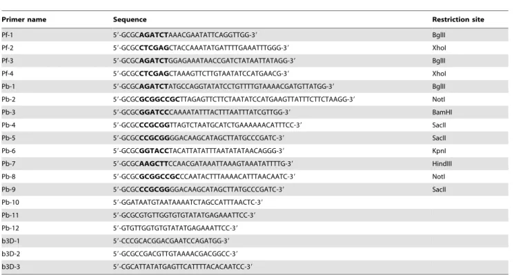
The constructs pHH1-LplA1-KO and pHH1-LplA1-KOkon were generated using the oligonucleotides Pf-1/Pf-2 and Pf-3/Pf-4, respectively (see Table 1). The primers introduced 59 of the PCR products a BglII site and 39 a XhoI site that allowed directional cloning of the products into pHH1 [30]. PCR was performed withPfupolymerase and genomic P. falciparumDNA. The knockout construct is truncated at its 59end lacking the ATG start codon and at the 39 end, a premature stop codon was introduced 237 bp upstream of the natural stop codon. This would result in the formation of two incomplete and inactiveLplA1copies upon single cross-over recombination of the plasmid in theLplA1 gene locus. In contrast, the control construct, corresponding to nucleotides 206–1227, retains the endogenous C-terminus of the LplA1 gene but lack the 59 end and thus should generate a functional copy of LplA1and a non-functional pseudogene upon recombination. It will, however, introduce an artificial 39 UTR downstream of the firstLplA1copy [30]. The expression plasmid pHrBI-PbLplA1encompasses the entire ORF of theP. berghei LplA1 gene, which was amplified fromP. bergheigenomic DNA using the oligonucleotides Pb-1 and Pb-2 (see Table 1). The introduced restriction sitesBglII and NotI allowed directional cloning of the PCR product into the Gateway entry vector pHGB [31]. Through recombination using the single site Gateway cloning system (Invitrogen), thePbLplA1ORF was transferred into the destination vector pHrBI-1/2 which contains the blasticidin S deaminase (BSD) as a selectable marker [31]. All PCR products were initially subcloned into TOPO-Blunt (Invitrogen) and their sequences were verified (The Sequencing Service, University of Dundee, UK, www.dnaseq.co.uk) before cloning into the transfection plasmid pHH1 or entry vector pHGB. Transfection of the constructs was carried out as described before [32,33] and parasites were selected with either 5 nM WR99210 or 2mg/ml blasticidin (Invitrogen). The expression plasmid pHrBI-PbLplA1 was transfected into P. falciparumD10 and theP. falciparumline carrying the pHH1-LplA1 -KO plasmid (cycle 0).
The b3D.DT‘H.‘D-based knockout construct [25] for single homologous recombination in P. bergheiencompasses a 1014 bp PbLplA1fragment corresponding to nucleotides 88 to 1098 (lacking the last 147 bp) which was amplified using the oligonucleotides Pb-3 and Pb-4 (see Table 1),Pfupolymerase and genomicP. berghei DNA. An artificial stop codon was introduced at the 39end of the PCR product and the fragment was cloned into theBamHI and SacII sites of one of the multiple cloning sites of the vector resulting
second strategy PbLplA1 59 UTR and 39 UTR regions were cloned into the two multiple cloning sites of b3D.DT‘H.‘D using the oligonucleotides Pb-6/Pb-7 and Pb-8/Pb-9, respectively (see Table 1). The 59 UTR PCR fragment was 480 bp in length and containedKpnI andHindIII restriction sites; the 39UTR PCR fragment was 516 bp long and hadNotI andSacII restriction sites that allowed directional cloning. The resulting plasmid was called b3D-LplA1-REP. UsingKpnI and SacII, the transfection cassette (containing the two LplA1 UTRs and the selectable marker T. gondiidihydrofolate reductase-thymidylate synthase (TgDHFR-TS) was isolated from the plasmid backbone, and digested plasmid was transfected according to [25]. The transfection was repeated at 6 independent occasions.
Southern blot analyses
For each P. falciparum line that was analysed, 1 to 5mg of genomic DNA was digested withNdeI, separated on a 0.8% (w/v) agarose gel and blotted onto positively charged nylon membrane (GE Healthcare) using standard methods [34]. The blots were prehybridised in 56SSC, 0.1% (w/v) SDS, 5% (w/v) dextrane
sulphate and 1:20 dilution of GI liquid block provided with the Gene Images CDP-Star detection kit from GE Healthcare. After 2 h at 60uC either the fluoresceine-labelledLplAcoding region, the human DHFR(hDHFR) or the blasticidin S deaminase(BSD) probes, generated using the Gene Images Random Prime labelling module according to the manufacturer’s recommendation (GE Health-care), were added and the blots hybridised overnight at the same temperature. After washes with decreasing salt-concentrations, the signals were detected by incubating the blots with an anti-fluoresceine alkaline phosphatase conjugated antibody followed by several washes in 100 mM Tris HCl pH 9.5 containing 300 mM NaCl and 0.3% (v/v) Tween 20. After application of detection solution the blot was exposed to autoradiography film (Kodak).
Pulsed field gel electrophoreses
To separateP. falciparumchromosomes on an agarose gel, pulse field gel electrophoreses were performed using the CHEF-DR III Variable Angle System (BioRad). The conditions used in this study are optimal to separate chromosomes 11 to 14 because theLplA1 gene is located on chromosome 13. The gel consisted of 1% (w/v) agarose in 16TAE and the separation was performed using the
following parameters: 360–800 s pulse, 3 V/cm2 (100 Volts) for 96 hours. The gels were blotted and probed as described above.
Western blot analyses
Briefly, 15mg ofP. falciparumlysates or the protein extract obtained from 16106 or 0.26107 P. berghei were separated on a 4–12%
SDS-PAGE (Invitrogen) and then blotted onto nitrocellulose (Schleicher and Schu¨ll), using standard techniques [34]. The blots were incubated with a rabbit anti-LplA1 antibody (generated against recombinant protein P. falciparum LplA1 by Eurogentec, Belgium) at a dilution of 1:5000 and the secondary anti-rabbit IgG (H+L), HRP conjugate (Promega) at a dilution of 1:10,000 before being developed using the ImmobilonTMWestern Chemilumines-cent HRP Substrate (Millipore).
PCR analyses ofP. bergheitransfectants
The parasite populations that were isolated from infected mice after transfection of the threeP. bergheiconstructs were analysed by PCRs using diagnostic primer combinations that allowed deter-mining whether (i) the gene locus had been targeted by the plasmid, (ii) the endogenous gene was still present and (iii) the plasmid had recombined and was present as an episome. The parasites that were transfected with b3D-LplA1-int1 were analysed with primer pair Pb-10/Pb-11 which should result in the amplification of a 2479 bp fragment if the endogenous gene was still present. In order to analyse whether the recombined transfection plasmid was present in the parasite population, primers b3D-1 and b3D-2 were used, amplifying a 1628 bp fragment. For amplification of PCR products diagnostic for the homologous recombination of the transfected construct with the
gene locus, the two primer sets b3D-1/Pb-11 and Pb-10/b3D-2 were used to analyse the 39 and 59 integration-specific events, respectively. The parasites transfected with the control plasmid b3D-LplA1-int2 were analysed with the same primer sets.
Parasites transfected with the replacement construct b3D-LplA1-REP were analysed using the gene locus specific primer set Pb-3/Pb-11 (amplifying a 1837 bp product) or Pb-10/Pb-12 (amplifying a 2479 bp product). These parasite lines were also analysed for the presence of plasmid using the primer pair b3D-1 and b3D-2, amplifying a 1150 bp fragment. Diagnostic PCRs for the integration event were performed for the 39and 59integration site using the primer combinations b3D-1/Pb-11 (1183 bp) and Pb-10/b3D-3 (1015 bp), respectively (for primer sequences see Table 1).
Acknowledgments
The authors like to thank Dr Janet Storm for critical reading the manuscript and Mrs Anne McIntosh and Mr Andreas Kunze for excellent technical assistance.
Author Contributions
Conceived and designed the experiments: SG KM SM. Performed the experiments: SG. Analyzed the data: SG KM SM. Contributed reagents/ materials/analysis tools: KM. Wrote the paper: KM SM.
References
1. Perham RN (2000) Swinging arms and swinging domains in multifunctional enzymes: catalytic machines for multistep reactions. Annu Rev Biochem 69: 961–1004.
2. Reed LJ, Hackert ML (1990) Structure-function relationships in dihydrolipoa-mide acyltransferases. J Biol Chem 265: 8971–8974.
3. Douce R, Bourguignon J, Neuburger M, Re´beille´ F (2001) The glycine decarboxylase system: a fascinating complex. Trends Plant Sci 6: 167–176.
4. Mooney BP, Miernyk JA, Randall DD (2002) The complex fate of alpha-ketoacids. Annu Rev Plant Biol 53: 357–375.
5. Yasuno R, Wada H (2002) The biosynthetic pathway for lipoic acid is present in plastids and mitochondria inArabidopsis thaliana. FEBS Lett 517: 110–114.
6. Cronan JE, Zhao X, Jiang Y (2005) Function, attachment and synthesis of lipoic acid inEscherichia coli. Adv Microb Physiol 50: 103–146.
Table 1.Oligonucleotides used in this study.
Primer name Sequence Restriction site
Pf-1 59-GCGCAGATCTAAACGAATATTCAGGTTGG-39 BglII
Pf-2 59-GCGCCTCGAGCTACCAAATATGATTTTGAAATTTGGG-39 XhoI
Pf-3 59-GCGCAGATCTGGAGAAATAACCGATCTATAATTATAGG-39 BglII
Pf-4 59-GCGCCTCGAGCTAAAGTTCTTGTAATATCCATGAACG-39 XhoI
Pb-1 59-GCGCAGATCTATGCCAGGTATATCCTGTTTTGTAAAACGATGTTATGG-39 BglII
Pb-2 59-GCGCGCGGCCGCTTAGAGTTCTTCTAATATCCATGAAGTTATTTCTTCTAAGG-39 NotI
Pb-3 59-GCGCGGATCCCAAAATATTTACTTTAATTTATCGTTGG-39 BamHI
Pb-4 59-GCGCCCGCGGTTAGTCTAATGCATCTGAAAAAACATTTCC-39 SacII
Pb-5 59-GCGCCCGCGGGGACAAGCATAGCTTATGCCCGATC-39 SacII
Pb-6 59-GCGCGGTACCTACATTATATTTAATATATAACAGGG-39 KpnI
Pb-7 59-GCGCAAGCTTCCAACGATAAATTAAAGTAAATATTTTG-39 HindIII
Pb-8 59-GCGCGCGGCCGCCCAATACTTTAAAACATTTAACAATC-39 NotI
Pb-9 59-GCGCCCGCGGGGACAAGCATAGCTTATGCCCGATC-39 SacII
Pb-10 59-GGATAATGTAATAAAATCTAGCCATTTAACTC-39
Pb-11 59-GCGCGTGTTGGTGTGTATATGAGAAATTCC-39
Pb-12 59-GTGTTGGTGTGTATATGAGAAATTCC-39
b3D-1 59-CCCGCACGGACGAATCCAGATGG-39
b3D-2 59-GCGCCGACGTTGTAAAACGACGGCC-39
b3D-3 59-CGCATTATATGAGTTCATTTTACACAATCC-39
doi:10.1371/journal.pone.0005510.t001
Toxoplasma gondiiscavenges host-derived lipoic acid despite itsde novosynthesis in the apicoplast. EMBO J 25: 3214–3222.
15. Mazumdar J, Wilson EH, Masek K, Hunter CA, Striepen B (2006) Apicoplast fatty acid synthesis is essential for organelle biogenesis and parasite survival in Toxoplasma gondii. Proc Natl Acad Sci U S A 103: 13192–13197.
16. Gu¨nther S, Wallace L, Patzewitz EM, McMillan PJ, Storm J, et al. (2007) Apicoplast lipoic acid protein ligase B is not essential forPlasmodium falciparum. PLoS Pathog 3: 1938–1949.
17. Witkowski A, Joshi AK, Smith S (2007) Coupling of thede novofatty acid biosynthesis and lipoylation pathways in mammalian mitochondria. J Biol Chem 282: 14178–14185.
18. Gueguen V, Macherel D, Jaquinod M, Douce R, Bourguignon J (2000) Fatty acid and lipoic acid biosynthesis in higher plant mitochondria. J Biol Chem 275: 5016–5025.
19. Stephens JL, Lee SH, Paul KS, Englund PT (2007) Mitochondrial fatty acid synthesis inTrypanosoma brucei. J Biol Chem 282: 4427–4436.
20. Wu Y, Sifri CD, Lei HH, Su XZ, Wellems TE (1995) Transfection ofPlasmodium falciparumwithin human red blood cells. Proc Natl Acad Sci USA 92: 973–977.
29. Bradford MM (1976) A rapid and sensitive method for the quantitation of microgram quantities of protein utilizing the principle of protein-dye binding. Anal Biochem 72: 248–254.
30. Reed MB, Saliba KJ, Caruana SR, Kirk K, Cowman AF (2000) Pgh1 modulates sensitivity and resistance to multiple antimalarials in Plasmodium falciparum. Nature 403: 906–909.
31. Tonkin CJ, van Dooren GG, Spurck TP, Struck NS, Good RT, et al. (2004) Localization of organellar proteins inPlasmodium falciparumusing a novel set of transfection vectors and a new immunofluorescence fixation method. Mol Biochem Parasitol 137: 13–21.
32. Crabb BS, Cowman AF (1996) Characterization of promoters and stable transfection by homologous and nonhomologous recombination inPlasmodium falciparum. Proc Natl Acad Sci U S A 93: 7289–7294.
33. Wu Y, Kirkman LA, Wellems TE (1996) Transformation ofPlasmodium falciparum malaria parasites by homologous integration of plasmids that confer resistance to pyrimethamine. Proc Natl Acad Sci U S A 93: 1130–1134.