Molecular analysis of Salmonella enteritidis
isolates from the Caribbean by pulsed-field
gel electrophoresis
Abiodun Adesiyun,
1Andrew Carson,
2Kelly McAdoo,
2and Craig Bailey
2Using pulsed-field gel electrophoresis (PFGE), between 1987 and 1996 we analyzed Salmo-nella enteritidisisolates from gastroenteritis cases in four Caribbean countries: Barbados, Saint Kitts and Nevis, Saint Lucia, and Trinidad and Tobago. We also determined the resis-tance of the isolates to 12 antimicrobial agents. Of the 129 isolates of S. enteritidisavailable for testing, DNA digested by XbaI revealed 13 distinctive PFGE patterns. The most prevalent
XbaI PFGE patterns were group 1 (88 of 129 isolates, 68.2%) and group 2 (26 of 129, 20.2%). The patterns found among S. enteritidisisolates correlated with the geographical origin of the isolates. Of the 28 isolates from Barbados, 20 of them (71.4%) belonged to XbaI PFGE group 2, and of the 93 isolates from Trinidad and Tobago, 78 of them (83.9%) belonged to group 1.
SpeI digestion of S. enteritidis genome was not as discriminatory as XbaI.
Overall, of the 129 isolates, 67 of them (51.9%) exhibited resistance to one or more of the 12 antimicrobial agents that we tested. The prevalence of resistance was 53.8% for the S. enteri-tidisisolates tested from Trinidad and Tobago, 50.0% for those from Barbados, 28.6% for those from Saint Lucia, and 100.0% for one isolate from the island of Saint Kitts. Resistance was highest to triple sulfur (59 of 129 isolates, 45.7%), followed by furadantoin (10 of 129, 7.8%), ampicillin (7 of 129, 5.4%), and carbamycin (5 of 129, 3.9%).
ABSTRACT
Salmonella enteritidis has become an important cause of food poisoning throughout the world (1–3). For a long time in the Caribbean region S. enteri-tidiswas rarely associated with human
infections (4). In the early 1990s, how-ever, a number of outbreaks and spo-radic cases implicating the microor-ganism were reported (5). There was a subsequent phenomenal increase in the involvement of S. enteritidis in re-ported cases of food poisoning (6).
Various methods have been used for salmonella epidemiological testing, in-cluding antibiotic testing, serotyping, and phage typing (7–9). Recently, var-ious molecular techniques have been applied to characterize S. enteritidis and to conduct epidemiological inves-tigations. Among these techniques are plasmid profile analysis (10), DNA
restriction fragment polymorphisms (11), chromosomal probe fingerprint-ing (12), and pulsed-field gel elec-trophoresis (PFGE) (13, 14).
To date, there have been no reports on the characteristics of S. enteritidis isolated from the Caribbean, nor have there been reports on the relatedness of isolates implicated in foodborne gastroenteritis.
This study utilized PFGE to investi-gate the relationship between S. enteri-tidis isolates recovered from gastro-enteritis cases in various Caribbean countries and to determine the anti-biograms of the isolates.
1 University of the West Indies, Faculty of Medical
Sciences, School of Veterinary Medicine, St. Augus-tine, Trinidad and Tobago. Send correspondence to: Professor A. A. Adesiyun, Faculty of Medical Sciences, School of Veterinary Medicine, The Uni-versity of the West Indies, St. Augustine, Trinidad and Tobago; telephone: (868)-645-8329; fax: (868) 645-9865; e-mail: abiodunadesiyun@hotmail.com
2 University of Missouri, College of Veterinary
MATERIALS AND METHODS
Sources of S. enteritidis isolates
The Caribbean Epidemiology Cen-ter (CAREC), Port of Spain, Trinidad, is the regional reference laboratory for Salmonella, and it served as the source of the S. enteritidis isolates we studied. All Salmonellaclinical isolates from CAREC’s 21 Member Countries (mainly English-speaking) in the Carib-bean sub-region are sent to CAREC for serological typing. Table 1 shows the number of S. enteritidis isolates from gastroenteritis cases (sporadic or out-break) that we obtained from CAREC and studied. All viable isolates of S. enteritidis in the collection of CAREC were selected for study.
Overall, we tested a total of 129 S. enteritidisisolates, with 122 originating from human clinical cases and 7 (from Trinidad and Tobago) from animals. It was not possible to determine which of the isolates originated from out-breaks of gastroenteritis since in the Caribbean region there is no routine investigation or reporting of food-borne outbreaks.
Preparation of bacterial DNA, restriction digests, and running of pulsed-field gel electrophoresis
For our testing all the S. enteritidis strains were plated for isolation on
blood agar. Colonies of pure cultures were then inoculated into 5 mL of brain heart infusion broth and incu-bated overnight at 37 °C to attain log phase growth. An aliquot (1.3 mL) of cell suspension of each isolate was centrifuged at 12 000 ⳯ g for 90 sec-onds. To prepare bacterial DNA we used a procedure described earlier (15).
Agarose plugs were cut into slices to fit gel wells and equilibrated for 15 minutes at room temperature in tubes containing 200 L of restriction
en-zyme buffer (Stratagene, La Jolla, Cali-fornia, United States of America). The buffer was then decanted and replaced with 200 L of fresh restriction
en-zyme buffer for a further 15 minutes of equilibration at room temperature. Restriction digests were done as de-scribed earlier (15).
A 1.0% SeaKem gel (FMC BioProd-ucts, Rockland, Maine, United States) was prepared in 0.5X TBE buffer 60–90 min prior to completion of the restric-tion enzyme digesrestric-tion of bacterial DNA and allowed to solidify at room temperature. DNA digested with ei-ther XbaI or SpeI in gel slices was placed in the wells of SeaKem gel and sealed in place with molten 1% agarose in 0.5X TBE buffer. To wells 1 and 15 of each gel was added standard molecu-lar size markers (lambda ladder). For each run, the sealing agar was allowed to solidify at room temperature and the gel was thereafter transferred into a CHEF-DR III electrophoresis
cham-ber (Bio-Rad Laboratories, Hercules, California, United States) and sub-merged in chilled 0.5X TBE. Elec-trophoresis was performed at 200 V for 20 h with the pulse time ramped from 2 seconds to 40 seconds and the buffer temperature maintained at 14 °C. The gel was subsequently stained in ethid-ium bromide (0.5 g/mL) solution for
30 min at room temperature and then destained in distilled water for approx-imately 1 h prior to photography under ultraviolet light.
Determination of PFGE patterns
Polaroid photographs of PFGE pat-terns were recorded by a desktop scan-ner (Hewlett-Packard, Boise, Idaho, United States) and subsequently sub-jected to a computerized artificial neu-ral network analysis described earlier (15). Grouping was based on location and number of bands in the range of 50 kb to 550 kb.
Comparison of XbaI and SpeI PFGE patterns
Following the identification of the PFGE distinct groups detected by XbaI digestion, a representative of each of the 13 groups subjected to XbaI diges-tion was digested with SpeI restriction enzyme using fresh plugs, and the pat-tern was noted.
TABLE 1. Isolates of S. enteritidissubmitted and studied, by source country and year of isolation, four Caribbean countries, 1987–1996
Trinidad and Tobago Barbados Saint Lucia Saint Kitts and Nevis All sources
Submitted Studied Submitted Studied Submitted Studied Submitted Studied Submitted Studied
Year No. No. % No. No. % No. No. % No. No. % No. No. %
1987 0 0 0.0 2 1 50.0 0 0 0.0 0 0 0.0 2 0 0.0
1989 0 0 0.0 5 1 20.0 6 1 16.7 0 0 0.0 11 2 18.2
1990 1 0 0.0 10 3 30.0 3 2 66.7 0 0 0.0 14 5 35.7
1991 1 1 100.0 20 3 15.0 0 0 0.0 0 0 0.0 21 4 19.0
1992 1 1 100.0 19 5 26.3 0 0 0.0 0 0 0.0 20 6 30.0
1993 0 0 0.0 22 4 18.2 2 0 0.0 1 1 100.0 25 5 20.0
1994 12 8 66.7 18 5 27.8 4 2 50.0 0 0 0.0 34 15 44.1
1995 50 49 98.0 13 5 38.5 2 2 100.0 0 0 0.0 65 56 86.2
1996 70 34 48.6 4 1 25.0 0 0 0.0 0 0 0.0 74 35 47.3
Total 135 93a 68.9 113 28 24.8 17 7 41.2 1 1 100.0 266 129 48.5
Determination of antibiograms of isolates
To determine the antibiograms of the S. enteritidis isolates we used the agar diffusion method of the National Committee for Clinical Laboratory Standards (16). Twelve antimicrobial agents on disks (Difco Laboratories, Inc., Detroit, Michigan, U.S.A.) were used, and their concentrations were: ampicillin (30 g), carbamycin (100 g),
chloramphenicol (30 g), clindamycin
(30 g), furadantoin (300 g),
gen-tamycin (10 units), kanamycin (30 g),
nalidixic acid (30 g), streptomycin
(10 g),
sulphamethoxazole/trimetho-prim (25 g), triple sulfur (300 g),
and tetracycline (30 g). We measured
the zone sizes and used the criteria provided by the disc manufacturer to determine the resistance or sensitivity of the isolates.
RESULTS
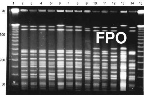
Figure 1 shows the fragment pat-terns of S. enteritidisgenome digested with XbaI; there were 13 distinctive cleavage patterns among the 129 iso-lates of S. enteritidisthat we tested.
Digestion with restriction enzyme SpeI of the same DNA preparations used for the XbaI digestion (shown in Figure 1) is displayed in Figure 2. Only eight distinct fragment patterns were detected (wells 2; 4; 7; 8; 3 and 11; 5, 6, 9, and 10; 12 and 13; and 14).
The sources of the S. enteritidis iso-lates from 1987 through 1996 are shown by country in Table 1; isolates for 1988 were unavailable from CAREC for study. Over the first part of this period, from 1987 through 1993, the four countries submitted a total of 93 S. enteritidis isolates: 3 (3.2%) from Trinidad and Tobago, 78 (83.9%) from Barbados, 11 (11.8%) from Saint Lucia, and 1 (1.2%) from the island of Saint Kitts.
The pattern was different in the sub-sequent period, of 1994 through 1996, with Trinidad and Tobago replacing Barbados as the country from which the largest number of S. enteritidis iso-lates were reported. Of a total of 173
isolates submitted to CAREC over those 3 years, 132 of them (76.3%) were from Trinidad and Tobago, 35 (20.2%) from Barbados, 6 (3.5%) from Saint Lucia, and 0 (0.0%) from Saint Kitts and Nevis.
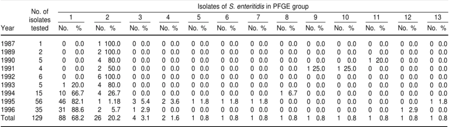
From 1987 through 1996 we tested a total of 129 isolates of S. enteritidis. Of that total, 88 of them (68.2%) belonged to XbaI PFGE group 1, and 26 (20.2%) belonged to XbaI PFGE group 2 (Table 2). Only 4 (3.1%) belonged to group 3,
kb
500
200
50
1 2 3 4 5 6 7 8 9 10 11 12 13 14 15
FIGURE 1. Pulsed-field gel electrophoresis patterns of S. enteritidisisolates generated by enzyme XbaI. Lanes 1 and 15 contained molecular size markers (lambda ladder). Lanes 2–14 show distinct cleavage patterns observed among the 129 S. enteritidisisolates tested
kb
500
200
50
1 2 3 4 5 6 7 8 9 10 11 12 13 14 15
FIGURE 2. Pulsed-field gel electrophoresis separation of restriction fragments of S. enteri-tidisgenome digested with SpeI. Lanes 1 and 15 contained lambda ladder molecular size marker. Lanes 2–14 contained genomes representative of the 13XbaI patterns
FPO
2 (1.6%) belonged to group 4, and the other nine groups had 1 isolate each.
In the 1987–1993 period, of the 23 iso-lates tested, only 1 (4.3%) was classified in group 1, and 19 (82.6%) belonged to group 2. However, for the 1994–1996 period, of a total of 106 isolates tested, 87 of them (82.1%) were in group 1, and only 7 (6.6%) belonged to group 2.
The distribution of XbaI PFGE groups among theS. enteritidisisolates tested from the four countries is shown in Table 3. In Trinidad and To-bago, group 1 was the most prevalent (78 of 93 isolates tested, or 83.9%). In Barbados, group 2 was the most com-mon (20 of 28, or 71.4%). Saint Lucian isolates of S. enteritidis had the same frequency of occurrence, 42.9%, for both group 1 and group 2.
In Trinidad and Tobago, regardless of the year and geographical location of source of S. enteritidisisolates within
the country, PFGE group 1 isolates were the most prevalent. Five (71.4%) of the 7 S. enteritidisof animal origin also belonged to PFGE group 1. In 1994 7 of the 8 (87.5%) isolates that were tested belonged to group 1, in 1995 40 of 49 (81.6%) did so, and in 1996 31 of 34 (91.2%) did so.
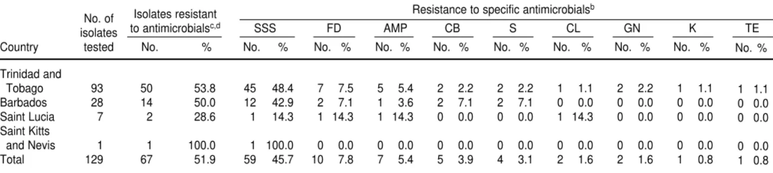
Of the 129 isolates of S. enteritidis tested, 67 of them (51.9%) exhibited re-sistance to one or more antimicrobial agents (Table 4). Overall, resistance was highest to triple sulfur (59 of 129 isolates, or 45.7%), a pattern that was true for all four of the countries. The resistance levels for the other antimi-crobial agents tested were all notice-ably lower, 7.8% or less. The resistance to the various antimicrobial agents was similar in all four of the countries. All of the isolates tested were sensitive to nalidixic acid, chloramphenicol, and sulphamethoxazole/trimethoprim.
A total of 12 resistant patterns were observed, with resistance to triple sul-fur alone most frequent (46 isolates). A total of 59 (45.7%) of the 129 isolates, however, were resistant to either triple sulfur alone or triple sulfur in combi-nation with other antimicrobial agents.
DISCUSSION
Of the four countries from which we studied S. enteritidis strains, only one, Trinidad and Tobago, recorded a signif-icant increase in the absolute number of S. enteritidisisolates from 1994 to 1996. Barbados, on the other hand, had rela-tively high numbers of S. enteritidisfrom as early as 1990. The situation in Barba-dos might be explained in part by the country’s heavy dependence on tour-ism. Worldwide, there has been a surge in the involvement of S. enteritidis in TABLE 2. XbaI pulsed-field gel electrophoresis groups of S. enteritidisisolates, by year, four Caribbean countries, 1987–1996
No. of Isolates of S. enteritidisin PFGE group
isolates 1 2 3 4 5 6 7 8 9 10 11 12 13
Year tested No. % No. % No. % No. % No. % No. % No. % No. %
1987 1 0 0.0 1 100.0 0 0.0 0 0.0 0 0.0 0 0.0 0 0.0 0 0.0
1989 2 0 0.0 2 100.0 0 0.0 0 0.0 0 0.0 0 0.0 0 0.0 0 0.0
1990 5 0 0.0 4 80.0 0 0.0 0 0.0 0 0.0 0 0.0 0 0.0 0 0.0
1991 4 0 0.0 2 50.0 0 0.0 0 0.0 0 0.0 0 0.0 0 0.0 0 0.0
1992 6 0 0.0 6 100.0 0 0.0 0 0.0 0 0.0 0 0.0 0 0.0 0 0.0
1993 5 1 20.0 4 80.0 0 0.0 0 0.0 0 0.0 0 0.0 0 0.0 0 0.0
1994 15 10 66.7 4 26.7 0 0.0 0 0.0 0 0.0 0 0.0 0 0.0 1 6.7
1995 56 46 82.1 1 1.18 3 5.4 2 3.6 1 1.8 1 1.8 1 1.8 0 0.0
1996 35 31 88.6 2 5.7 1 2.9 0 0.0 0 0.0 0 0.0 0 0.0 0 0.0
Total 129 88 68.2 26 20.2 4 3.1 2 1.6 1 0.8 1 0.8 1 0.8 1 0.8
0 0.0 0 0.0 0 0.0 0 0.0 0 0.0
0 0.0 0 0.0 0 0.0 0 0.0 0 0.0
0 0.0 0 0.0 1 20.0 0 0.0 0 0.0
1 25.0 1 25.0 0 0.0 0 0.0 0 0.0
0 0.0 0 0.0 0 0.0 0 0.0 0 0.0
0 0.0 0 0.0 0 0.0 0 0.0 0 0.0
0 0.0 0 0.0 0 0.0 0 0.0 0 0.0
0 0.0 0 0.0 0 0.0 0 0.0 1 1.8
0 0.0 0 0.0 0 0.0 1 2.9 0 0.0
1 0.8 1 0.8 1 0.8 1 0.8 1 0.8
No. % No. % No. % No. % No. %
TABLE 3. XbaI pulsed-field gel electrophoresis groups of S. enteritidisisolates, by country of origin, four Caribbean countries, 1987–1996
No. of Isolates of S. enteritidisbelonging to group
isolates 1 2 3 4 5 6 7 8 9 10 11 12 13
Country tested No. % No. % No. % No. % No. % No. % No. % No. %
Trinidad and
Tobago 93 78 83.9 2 2.2 4 4.3 2 2.2 1 1.1 1 1.1 1 1.1 1 1.1
Barbados 28 7 25.0 20 71.4 0 0.0 0 0.0 0 0.0 0 0.0 0 0.0 0 0.0
Saint Lucia 7 3 42.9 3 42.9 0 0.0 0 0.0 0 0.0 0 0.0 0 0.0 0 0.0
Saint Kitts
and Nevis 1 0 0.0 1 100.0 0 0.0 0 0.0 0 0.0 0 0.0 0 0.0 0 0.0
Total 129 88 68.2 26 20.2 4 3.1 2 1.6 1 0.8 1 0.8 1 0.8 1 0.8
1 1.1 0 0.0 0 0.0 1 1.1 1 1.1
0 0.0 1 3.4 0 0.0 0 0.0 0 0.0
0 0.0 0 0.0 1 14.3 0 0.0 0 0.0
0 0.0 0 0.0 0 0.0 0 0.0 0 0.0
1 0.8 1 0.8 1 0.8 1 0.8 1 0.8
human gastroenteritis (2, 17, 18). It is possible that visitors to Barbados brought in the infections from North America or Europe, two of the areas where S. enteritidishas been increasingly involved in human gastroenteritis.
It was of epidemiological signifi-cance to find that the strains of S. en-teritidisdisplayed a distinct geograph-ical distribution. The XbaI PFGE group 1 strains were predominantly found in Trinidad and Tobago, while group 2 strains were much more prevalent in Barbados. Although there was a slight overlap of XbaI PFGE groups 1 and 2 between the two countries, it was evident that the strains ofS. enteritidisresponsible for gastroenteritis in Trinidad and To-bago and in Barbados differ signifi-cantly. The small number of isolates of S. enteritidis available for study from Saint Lucia and from Saint Kitts and Nevis made it difficult to draw any inferences on the PFGE patterns of isolates from these countries.
In Trinidad and Tobago, regardless of the source of strains (human versus animals, and geographical location) of S. enteritidis and the year of isolation, XbaI PFGE group 1 strains predomi-nated, emphasizing the importance of this strain in gastroenteritis in the country. However, it was difficult to associate the various XbaI groups with outbreaks of gastroenteritis caused by
S. enteritidis. That is because outbreaks of foodborne disease are rarely re-ported or investigated in the Carib-bean. Therefore, a high percentage of the S. enteritidisisolates sent to CAREC for serotyping may have originated from either sporadic cases or out-breaks. PFGE has been employed in various studies for epidemiological investigations in sporadic and large outbreaks of salmonellosis caused by S. enteritidis(3, 14, 19).
If was of zoonotic relevance that in Trinidad and Tobago some of the strains ofS. enteritidisof animal origin belonged to the same PFGE group as those isolated from human gastroen-teritis. S. enteritidis infections in hu-mans have been reported to originate frequently from animals, particularly poultry (20–22).
Our finding that XbaI restriction en-zyme was superior to SpeI restriction enzyme in discriminating the strains of S. enteritidis was hardly a surprise; other researchers have reported simi-lar findings (14, 19).
Regardless of the country of origin or the year of isolation of S. enteritidis,we found that the prevalence of resistance to most of the antimicrobial agents that we tested was low. Resistance to triple sulfur was comparatively high (46%), a finding in agreement with other reports (23, 24). Resistances to furadantoin (7.8%), ampicillin (5.4%), and carbamycin
(3.9%) were also similar to what others have reported (21). Outside the Carib-bean, however, a considerably higher prevalence of resistance to ampicillin has been reported. In Greece, for exam-ple, 30% of S. enteritidis isolates were resistant (18). While the antibiograms of S. enteritidis strains can be used to determine the relatedness of strains (18), they were not helpful in epidemio-logical association in this study.
From our research we can conclude that S. enteritidis has attained some clinical significance in gastroenteritis in the Caribbean, with a particularly dramatic change in Trinidad and To-bago. In addition, the PFGE we per-formed clearly demonstrated that in Barbados and in Trinidad and Tobago there are distinctly different strains of S. enteritidisinvolved in gastroenteritis.
Acknowledgments. We thank the
Inter-American Development Bank for funding the stay of the principal au-thor at the University of Missouri, and for part-sponsorship of the research materials. The assistance rendered by Denise Clarke of the Caribbean Epi-demiology Center in making the iso-lates available is appreciated. Zobaida Khan of the Public Health Laboratory, Port of Spain, kindly assisted in sub-culturing the isolates. Beverly Hartman is thanked for typing the manuscript. TABLE 4. Antibiograms of S. enteritidisisolates from various sources, using 12 antimicrobial agents, four Caribbean countries, 1987–1996a
No. of Isolates resistant Resistance to specific antimicrobials
b
isolates to antimicrobialsc,d SSS FD AMP CB S CL GN K TE
Country tested No. % No. % No. % No. % No. % No. % No. % No. % No. %
Trinidad and
Tobago 93 50 53.8 45 48.4 7 7.5 5 5.4 2 2.2 2 2.2 1 1.1 2 2.2 1 1.1
Barbados 28 14 50.0 12 42.9 2 7.1 1 3.6 2 7.1 2 7.1 0 0.0 0 0.0 0 0.0
Saint Lucia 7 2 28.6 1 14.3 1 14.3 1 14.3 0 0.0 0 0.0 1 14.3 0 0.0 0 0.0
Saint Kitts
and Nevis 1 1 100.0 1 100.0 0 0.0 0 0.0 0 0.0 0 0.0 0 0.0 0 0.0 0 0.0
Total 129 67 51.9 59 45.7 10 7.8 7 5.4 5 3.9 4 3.1 2 1.6 2 1.6 1 0.8
aAll 129 isolates of S. enteritidis were sensitive to chloramphenicol, nalidixic acid, and sulphamethoxazole/trimethoprim.
bSSS = triple sulfur; FD = furadantoin; AMP = ampicillin; CB = carbamycin; S = streptomycin; CL = clindamycin; GN = gentamycin; K = kanamycin; TE = tetracycline. cNumber of isolates resistant to one or more of the antimicrobial agents.
dA total of 12 resistant patterns were observed, namely, SSS (46 isolates), SSS-FD (7 isolates), AMP (2 isolates), FD (2 isolates), CL-AMP-CB (2 isolates), S-GN-SSS (2 isolates), SSS-AMP
(1 isolate), K-SSS (1 isolate), SSS-TE (1 isolate), AMP-CB (1 isolate), S-FD-CB (1 isolate), and S-SSS-AMP-CB (1 isolate). Where only one antimicrobial agent is indicated, it means that re-sistance was exhibited to that agent only.
No. %
1 1.1 0 0.0 0 0.0
1. United States of America, Centers for Disease Control and Prevention. Salmonella surveil-lance report: annual summary. Atlanta: CDC; 1990.
2. Rodrigue DC, Tauxe RV, Rowe B. Interna-tional increase in Salmonella enteritidis: a new pandemic? Epidemiol Infect 1990;105:21–27. 3. Suzuki Y, Ishihara M, Matsumoto M,
Arakawa S, Saito M, Ishikawa M, et al. Mo-lecular epidemiology of Salmonella enteritidis: an outbreak and sporadic cases studied by means of pulsed-field gel electrophoresis. J In-fect 1995; 31:211–217.
4. Hull BP, Spence L, Bassett D, Swanston WH, Tikasingh ES. The relative importance of ro-tavirus and other pathogens in the etiology of gastroenteritis in Trinidadian children. Am J Trop Med Hyg 1982;31:142–148.
5. Caribbean Epidemiology Centre. Surveillance report, 1990. Port of Spain, Trinidad and To-bago: CAREC; 1990.
6. Caribbean Epidemiology Centre. Surveillance reports, 1990–1996. Port of Spain, Trinidad and Tobago: CAREC; 1996.
7. United States of America, Centers for Disease Control and Prevention. Salmonella surveil-lance report, 1989. Atlanta: CDC; 1989. 8. Hickman-Brenner FW, Stubbs AD, Farmer JJ
3d. Phage typing of Salmonella enteritidisin the United States. J Clin Microbiol 1991;29: 2817–2823.
9. Rivera MJ, Rivera N, Castillo J, Rubio MC, Gomez-Lus R. Molecular and epidemiological study of Salmonella clinical isolates. J Clin Mi-crobiol 1991; 29:927–932.
10. Threlfall EJ, Rowe B, Ward LR. Subdivision of Salmonella enteritidisphage types by plasmid profile typing. Epidemiol Infect 1989;102: 459–465.
11. Nastasi A, Mammina C, Villafarte MR. rDNA fingerprinting as a tool in epidemiological analysis of Salmonella typhi infections. Epi-demiol Infect 1991;107:565–576.
12. Bohm H, Karch H. DNA fingerprinting of Es-cherichia coli 0157:47 strains by pulsed-field gel electrophoresis. J Clin Microbiol 1992;30: 2169–2172.
13. Powell NG, Threlfall EJ, Chart H, Schofield SL, Rowe B. Correlation of change in phage type with pulsed field profile and 16S rrn pro-file inSalmonella enteritidisphage types 4, 7 and 9a. Epidemiol Infect 1995;114: 403–411. 14. Thong K, Ngeow Y, Altwegg M, Navaratnam
P, Pang T. Molecular analysis of Salmonella en-teritidis by pulsed-field gel electrophoresis and ribotyping. J Clin Microbiol 1995;33: 1070–1074.
15. Carson CA, Kelly JM, McAdoo KK, Wang D, Higgins B, Bailey CW, et al. Escherichia coli 0157:H7 restriction pattern recognition by ar-tificial neural network. J Clin Microbiol 1995; 33: 2894–2898.
16. National Committee for Clinical Laboratory Standards. Performance standards for antimi-crobial disc susceptibility tests. M2A2. Villa-nora, Pennsylvania, United States: NCCLS; 1992.
17. St Louis ME, Morse DL, Potter ME, DeMelfi TM, Guzewich JJ, Tauxe RV, et al. The emer-gence of grade A eggs as a major source of Sal-monella enteritidis infections. New implica-tions for the control of salmonellosis. JAMA 1988;259:2103–2107.
18. Vatopoulos AC, Mainas E, Balis E, Threlfall EJ, Kanelopoulou M, Kalapothaki V, et al. Molecular epidemiology of ampicillin-resis-tant clinical isolates of Salmonella enteritidis. J Clin Microbiol 1994;32:1322–1325.
19. Murase T, Okitsu T, Suzuki R, Morozumi H, Matsushima A, Nakamura A, et al. Evaluation of DNA fingerprinting by PFGE as an epi-demiologic tool for Salmonellainfections. Mi-crobiol Immunol 1995;39:673–676.
20. Coyle EF, Palmer SR, Ribeiro CD, Jones HI, Howard AJ, Ward L, et al. Salmonella enteri-tidisphage type 4 infection: association with hen’s eggs. Lancet 1988;2(8623):1295–1297. 21. Nair US, Saeed AM, Muriana PM, Kreisle RA,
Barrett B, Sinclair CL, et al. Plasmid profiles and resistance to antimicrobial agents among Salmonella enteritidisisolates from human be-ings and poultry in the midwestern United States. J Am Vet Med Assoc 1995;206(9): 1339–1344.
22. Heffernan HM. Antibiotic resistance among Salmonellafrom human and other sources in New Zealand. Epidemiol Infect 1991;106: 17–23. 23. O’Brien TF, Hopkins JD, Gilleece ES, Medeiros AA, Kent RL, Blackburn BO, et al. Molecular epidemiology of antimicrobial re-sistance in Salmonellafrom animal and human beings in the United States. New Engl J Med 1982; 307:1–6.
24. Frost JA, Ward LR, Rowe B. Acquisition of a drug resistance plasmid which converts Sal-monellaenteritidis phage type 4 to phage type 24. Epidemiol Infect 1989;103:243–248.
Manuscript received 6 July 1999. Revised version ac-cepted for publication on 8 June 2000.
REFERENCES
Mediante electroforesis en gel con pulsos eléctricos (EGPE), se analizaron las Salmo-nella enteritidisaisladas entre 1987 y 1996 en casos de gastroenteritis de cuatro países caribeños: Barbados, Saint Kitts y Nevis, Santa Lucía y Trinidad y Tabago. También se determinó la resistencia de los aislados a 12 antibióticos. La digestión del ADN con la endonucleasa de restricción XbaI reveló 13 patrones distintos de EGPE entre los 129 aislados de S. enteritidisanalizados; los más prevalentes fueron el grupo 1 (88 de 129; 68,2%) y el grupo 2 (26 de 129; 20,2%). Estos patrones se correlacionaron con el origen geográfico de los aislados. Así, de los 28 aislados de Barbados, 20 (71,4%) pertenecían al grupo 2, y de los 93 aislados de Trinidad y Tabago, 78 (83,9%) pertenecían al grupo 1. La digestión del genoma de S. enteritidiscon la endonucleasa de restricción SpeI no fue tan discriminativa como la digestión con XbaI. En general, 67 de los 129 aislados (51,9%) mostraron resistencia a uno o más de los 12 antibióticos probados. La preva-lencia de resistencia fue de 51% en los aislados de Trinidad y Tabago, de 50% en los de Barbados, de 28,6% en los de Santa Lucía y de 100% en el único aislado de la isla de Saint Kitts. La mayor resistencia correspondió a la triple sulfamida (sulfamerazina, sulfadiazina y sulfametazina: 59 de 129; 45,7%), seguida de la nitrofurantoína (10 de 129; 7,8%), la ampicilina (7 de 129; 5,4%) y la carbamicina (5 de 129; 3,9%).
RESUMEN