REVIEW
Molecular mechanisms of RET receptor-mediated
oncogenesis in multiple endocrine neoplasia 2
Simona M. Wagner*, ShuJun Zhu*, Adrian C. Nicolescu, Lois M. Mulligan
Division of Cancer Biology and Genetics, Cancer Research Institute and Department of Pathology & Molecular Medicine, Queen9s University, Kingston, ON, Canada. *contributed equally to the study.
Multiple endocrine neoplasia type 2 is an inherited cancer syndrome characterized by tumors of thyroid and adrenal
tissues. Germline mutations of the
REarranged during Transfection
(RET) proto-oncogene, leading to its
unregulated activation, are the underlying cause of this disease. Multiple endocrine neoplasia type 2 has been a
model in clinical cancer genetics, demonstrating how knowledge of the genetic basis can shape the diagnosis and
treatment of the disease. Here, we discuss the nature and effects of the most common recurrent mutations of
RET
found in multiple endocrine neoplasia type 2. Current understanding of the molecular mechanisms of
RET
mutations and how they alter the structure and function of the RET protein leading to its aberrant activation, and
the effects on
RET
localization and signaling are described.
KEYWORDS:
RET; Multiple Endocrine Neoplasia Type 2; Genotype–Phenotype.
Wagner SM, Zhu SJ, Nicolescu AC, Mulligan LM. Molecular mechanisms of RET receptor-mediated oncogenesis in multiple endocrine neoplasia 2. Clinics. 2012;67(S1):77-84.
E-mail: [email protected] Tel.: 1 613 533 6000 ext.77475
INTRODUCTION
Multiple endocrine neoplasia type 2 (MEN 2) is an
inherited cancer syndrome, characterized by medullary
thyroid carcinoma (MTC). The disease has three clinically
defined subtypes, as described elsewhere in this volume.
Briefly, familial MTC (FMTC), considered the least
aggres-sive form of MEN 2, exhibits MTC without additional
tumors or phenotypes, and frequently shows later onset
than other disease subtypes. MEN 2A is characterized by
MTC with pheochromocytoma, which occurs in
approxi-mately 50% of cases, and parathyroid hyperplasia or
adenoma in 10–35%. Finally, MEN 2B is also characterized
by MTC and pheochromocytoma, but parathyroid
hyper-plasia is rare. This is the most aggressive subtype, with
earliest onset of disease and metastasis, and poorest
prognosis. In MEN 2B, MTC has been documented in
patients as young as 2 months (1). In addition, patients with
MEN 2B frequently present with other non-tumor features
including ganglioneuromatosis of the mouth and gut,
corneal nerve thickening, delayed puberty, and a marfanoid
habitus (2,3).
MEN 2 is dominantly inherited, and its genetic cause,
mutations of the
REarranged during Transfection
(
RET
)
proto-oncogene, was first recognized nearly 20 years ago (4–6).
Since then, the range of mutations identified, their potential
for predicting clinical course, and the underlying functional
effects have been explored. Detection of
RET
mutations in
MEN 2 represents a paradigm for genetically guided patient
management, and genotype–phenotype correlations in this
disease now inform recommended interventions, patient
and family screening, and long-term follow-up (7,8).
Functional characterization of these mutations also has the
potential to define optimal therapeutic regimens, and may
identify additional phenotypic implications that have not
been broadly recognized. Here, we discuss our current
understanding of the molecular mechanisms for the more
common
RET
mutations and their potential significance.
RET RECEPTOR
The
RET
proto-oncogene encodes a receptor tyrosine
kinase that is required for the development of
neural-crest-derived cells, the urogenital system, and the central and
peripheral nervous systems, notably the enteric nervous
system (9,10). The RET protein has a large extracellular
domain containing a cysteine-rich region and a series of
cadherin homology domains, a transmembrane domain, and
an intracellular tyrosine kinase domain, required for RET
phosphorylation and downstream signaling (Figure 1A)
(11,12). The RET kinase is structurally similar to other
tyrosine kinases, sharing many conserved functional motifs
and regulatory residues that have been shown to have
importance for kinase enzyme function (13). RET is activated
by binding of a multi-protein ligand complex. RET binds a
soluble ligand of the glial cell-line-derived neurotrophic
factor (GDNF) family but also requires a co-receptor of the
GDNF family receptors
a
(GFR
a
), which is tethered to
the cell membrane via glycosylphosphatidylinositol linkage
Copyrightß2012CLINICS– This is an Open Access article distributed under the terms of the Creative Commons Attribution Non-Commercial License (http:// creativecommons.org/licenses/by-nc/3.0/) which permits unrestricted non-commercial use, distribution, and reproduction in any medium, provided the original work is properly cited.
No potential conflict of interest was reported.
(Figure 1B) (14,15). Initially, GDNF binds to GFR
a
, and these
complexes are then able to recruit RET to form
heterohex-amers that are concentrated in regions of the cell membrane
called lipid rafts (14,16). These are membrane domains
enriched in glycosylphosphatidylinositol-linked proteins
and signaling molecules that provide a platform not only
for enhanced cell signaling, but also for regulation of receptor
kinase activity and downregulation (17). Activation of RET
leads to stimulation of multiple downstream pathways,
including mitogen-activated protein kinase and extracellular
signal-regulated kinase, phosphoinositide 3-kinase and
pro-tein kinase B, signal transducer and activator of transcription
3, proto-oncogene tyrosine-protein kinase Src1, and focal
adhesion kinase (18,19), that promote cell growth,
prolifera-tion, survival, and/or differentiation.
THE
RET
PROTO-ONCOGENE IN MEN 2
MEN 2 is associated with point mutations of RET,
predictably leading to its activation in the absence of
ligands and co-receptors. Mutations are primarily amino
acid substitutions affecting a very small number of
RET
codons in either the extracellular domain or within the
kinase domain (Table 1; Figure 1A). Mutations are
domi-nant, requiring only a single mutant allele to confer the
disease phenotype. Summaries of MEN 2
RET
mutation
occurrence are well reviewed elsewhere (20–23) or are
available
online
(http://www.arup.utah.edu/database/
MEN2/MEN2_welcome.php). Together, these data suggest
strong overall themes as to functional effects of these
mutations, but also as to their clinical significance.
Strong associations of disease subtype, and also specific
disease phenotypes, with individual RET mutations have
made it possible to stratify risk of MEN 2 by genotype (7,8).
The management guidelines of the American Thyroid
Association (8) base the recommendations for initial
diagnosis, therapeutic intervention, and long-term
follow-up on patient genotype and the current understanding of
the natural history of the disease associated with each
RET
cysteines 609, 611, 618, 620, 630, and 634) in the RET
extracellular domain account for the majority of MEN 2A
cases, and are also common in patients with FMTC.
Intracellular kinase domain mutations are mainly associated
with FMTC and MEN 2B. Mutations in the intracellular
codons 768, 790, 791, 804, and 891 underlie FMTC, and occur
less commonly in patients with MEN 2A (20,24), while
specific mutations of codon 918 (M918T) or 883 (A883F)
account for the vast majority of MEN 2B cases, and are
exclusive to the subtype (3,25). In addition to association
with disease subtype, significant correlations of specific
mutations with disease features are reported. For example,
RET codon 634 mutations carry a greater patient risk for
pheochromocytoma and parathyroid hyperplasia (4,26–28),
and are associated with a higher frequency of detection of
MTC at the time of early thyroidectomy (29). Variation in
clinical presentation has even been observed with different
codon 634 substitutions. The specific substitution of an
arginine at codon 634 (C634R) is strongly associated with
increased risk of parathyroid hyperplasia (4,26–28),
in-creased frequency of distant metastases, earlier onset of
both lymph node and distant metastases, and bilaterality of
pheochromocytoma (30,31).
MOLECULAR MECHANISMS OF RET MUTATIONS
Evidence-based assessment of MEN 2 genotypic data
demonstrate that not all RET mutations have equivalent
clinical significance, although all reported mutations
are thought to lead to ligand-independent constitutive
activation of the RET receptor, autophosphorylation of
RET, and aberrant stimulation of downstream signaling
pathways. It follows that the molecular mechanisms of
mutations associated with these different phenotypes may
also be distinct and that these mechanisms may provide
clues to disease origin and, potentially, treatment for
patients with these mutations. Here, we discuss some of
the current understanding of the mechanisms of RET
dysfunction seen in MEN 2, and explore the potential
implications of these mechanisms.
RET Extracellular Domain Cysteine Residues
The most frequently identified
RET
mutations in MEN
2 affect cysteines in the extracellular cysteine-rich region
(primarily residues between Cys515 and Cys634) (Figure 1A;
Table 1). In the normal protein, intramolecular cysteine–
cysteine disulfide bonds contribute to the tertiary structure
of the RET extracellular domain. Correct positioning of
residues in this region is critical to interactions with GDNF–
GFR
a
ligand complexes (14,15,32). Amino acid
substitu-tions, resulting in replacement of a normal cysteine with any
amino acid, lead to loss of intramolecular bonds and to an
unpaired cysteine that is available for intermolecular
interactions with other mutant RET proteins (Figure 2A)
(33–35). These mutant RET dimers are constitutively active
in the absence of ligands. Furthermore, mutant dimers are
not recruited to lipid rafts through GFR
a
interactions, and
may be activated in other membrane compartments, which
can affect the nature and intensity of the resultant
down-stream signals (36,37). Downdown-stream signaling regulation, via
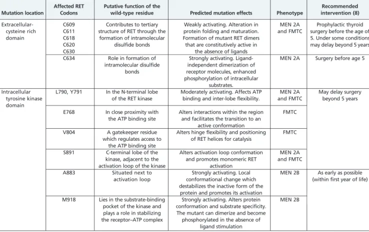
Table 1 -
Molecular effects of RET mutations in multiple endocrine neoplasia 2.
Mutation location
Affected RET Codons
Putative function of the
wild-type residue Predicted mutation effects Phenotype
Recommended intervention (8)
Extracellular-cysteine rich domain
C609 C611 C618 C620 C630
Contributes to tertiary structure of RET through the
formation of intramolecular disulfide bonds
Weakly activating. Alteration in protein folding and maturation. Formation of mutant RET dimers that are constitutively active in
the absence of ligands
MEN 2A and FMTC
Prophylactic thyroid surgery before the age of 5. Under some conditions may delay beyond 5 years C634 Role in formation of
intramolecular disulfide bonds
Strongly activating. Ligand-independent dimerization of receptor molecules, enhanced phosphorylation of intracellular
substrates.
MEN 2A Surgery before age 5
Intracellular tyrosine kinase domain
L790, Y791 In the N-terminal lobe of the RET kinase
Moderately activating. Affects ATP binding and inter-lobe flexibility.
MEN 2A and FMTC
May delay surgery beyond 5 years E768 In close proximity with
the ATP binding site
Alters interactions within the region and facilitates the transition to an
active conformation
FMTC
V804 A gatekeeper residue which regulates access to
the ATP binding site
Alters hinge flexibility and positioning of RET helices for catalysis
FMTC
S891 C-terminal lobe of the kinase, adjacent to the activation loop of the kinase
Alters activation loop conformation and promotes monomeric RET
activation
MEN 2A and FMTC
A883 Situated next to
activation loop
Strongly activating. Local conformational change which destabilizes the inactive form of the
protein and promotes its activation
MEN 2B As early as possible (within first year of life)
M918 Lies in the substrate-binding pocket of the kinase and plays a role in stabilizing the receptor–ATP complex
Strongly activating. Alters protein conformation and substrate specificity. The mutant can dimerize and become
phosphorylated in the absence of ligand stimulation
MEN 2B
FMTC, familial medullary thyroid carcinoma; MEN 2, multiple endocrine neoplasia 2; RET, REarranged during Transfection.
CLINICS 2012;67(S1):77-84 RET receptor-mediated oncogenesis in MEN 2
interactions with ubiquitin ligases such as CasitasB-lineage
lymphoma proto-oncogene (CBL) (38), or with cellular
phosphatases such as SHP1 and SHP2 (39,40) that are
involved in limiting or terminating signals, differ from that
of the raft-associated wild-type receptor, enhancing the
effect of the oncogenic mutation.
Although the molecular mechanisms of activation are
similar, cysteine RET mutations also vary in impact. In
general, mutations located closer to the RET transmembrane
domain have greater transforming ability and are linked to
increased risks of more aggressive MEN 2 disease (41)
(Table 1). Codon 634 mutations confer the greatest degree of
RET activation, with higher levels of autophosphorylation
and transforming ability than the other cysteine mutations,
and are linked to broader phenotypes and more severe
disease, as described above. Interestingly, mutations of
other cysteine residues are believed to affect the efficiency of
RET protein folding and maturation, and to impair
transport to the cell membrane, resulting in decreased
levels of cell surface protein and weaker signaling capability
(33–35,42,43). In fact, a subset of RET cysteine mutations,
sometimes referred to as Janus mutations, can lead to a
partial loss-of-function phenotype, as well as to oncogenic
effects. These mutations, generally affecting codons 609, 611,
618 or 620, are thought to confer cell-type-specific decreases
in functional protein on the cell surface. Inactivating
mutations of RET can lead to the congenital abnormality
Hirschsprung disease, which is characterized by the absence
of the enteric neurons from the distal colon (44). Janus
mutations have been linked to an insufficiency of mature
RET protein in the gut, resulting in the Hirschsprung
phenotype, yet at the same time, risks remain high for MEN
2 phenotypes, as sufficient mature protein is expressed in
the thyroid for development of MTC (45–47).
RET Intracellular Domain Mutations
Intracellular RET kinase mutations fall into two groups:
high-penetrance mutations causing MEN 2B, and less
aggressive mutations that lead to FMTC or, more rarely,
MEN 2A (Table 1). These RET mutations fall within the
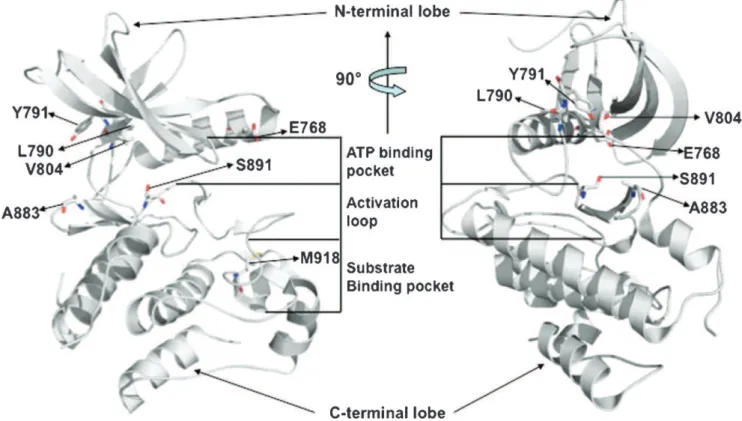
N-terminal and C-N-terminal lobes of the kinase (Figure 3).
Although the mutations are spread out along the linear
protein sequence (Figure 1A), they appear to cluster on
either the ATP-binding face or
substrate-binding/autoinhi-bitory face of the protein tertiary structure, suggesting some
common themes in their functional effects (Figure 3;
Table 1). The precise mechanisms by which these
intracel-lular mutations activate RET are various, but it is suggested
that they all do so through destabilizing the inactive form of
RET, and shifting the equilibrium of RET receptors towards
the active state (48).
MEN 2B Mutations
making RET more active, more of the time (48–51). The
M918T mutation appears to increase the stability of
monomeric active forms of RET, but activation of these
mutants can also be further enhanced by binding of
GDNF–GFR
a
complexes, suggesting that these mutant
RET forms may induce signal transduction from both
within and outside the lipid rafts, perhaps via distinct
signaling complexes (Figure 2B). As a result, RET
down-stream signals are enhanced, and activation of targets is
increased, notably including upregulation of gene
tran-scripts that contribute to cell proliferation or to
metastasis-promoting cell behaviors (52,53). Although it has been
postulated that the M918T mutation alters the preferred
substrates of the mutant RET protein with respect to both
autophosphorylation of RET tyrosine residues, and
phos-phorylation of downstream signaling molecules (54,55),
novel downstream targets that cannot also be stimulated at
lower levels by other less active RET mutants or by ligand
activation of wild-type RET have not been broadly
identified (48–51,53).
An intriguing finding has been that activation of M918T
RET begins before the receptor arrives at the cell surface,
stimulating signaling pathways from the endoplasmic
reticulum before the receptor reaches its fully glycosylated
mature form (56), which has not been observed for other
mutants. RET signaling from intracellular compartments
may differ (in intensity or otherwise) from that at the
plasma membrane, which has been shown to be the case
when wild-type RET is internalized into endosomes
following ligand stimulation (57) and for cytosolic RET
mutants found in papillary thyroid carcinoma (37).
An alanine to phenylalanine substitution at codon 883
(A883F) is the only other recurring MEN 2B mutation
(58,59). Structurally, this residue lies between the activation
and catalytic loops of the kinase (Figure 3), and would be
predicted to increase the flexibility of these domains, so
destabilizing the inactive form of the protein and promoting
its activation. Although generally considered a high-risk
mutation, some studies suggest that it may have a lesser
effect than the M918T mutation (60).
A handful of instances of double mutations in MEN 2B
have also been reported: V804M/E805K (51), V804M/Y806C
(61), and V804M/S904C (62). It appears that the
combina-tion of two mild intracellular mutacombina-tions can cooperate to
produce a more severe mutant. Each mutation alone
(V804M, E805K, Y806C, S904C) has low or no transforming
ability, consistent with the observation that V804M
gener-ally leads to FMTC (discussed below), but when coupled
together, they exert a synergistic effect on the transforming
ability of mutated RET (51,63).
Lower Risk Intracellular Domain Mutations
Recurrent mutations in the intracellular codons 768, 790,
791, 804, and 891 are found in patients with FMTC and, less
commonly, MEN 2A (20,64). This group of mutations is the
most diverse in functional effects, phenotypic variability,
and long-term clinical implications.
Figure 3 - Structure of the RET tyrosine kinase domain. Ribbon diagrams of the intracellular regions of activated RET, in two orientations, showing the positions of key functional features of the kinase: the ATP binding pocket; the activation or autoinhibitory loop; and the substrate binding pocket. Two orientations of the model, displaying the autoinhibitory/substrate binding face (left) and the ATP-binding face (right), are shown. Amino acid residues that are mutated in patients with multiple endocrine neoplasia type 2 (MEN 2) are represented in the stick form. The three-dimensional representation was based on the crystal structure of the phosphorylated (activated) RET tyrosine kinase domain (residues 709–990).
CLINICS 2012;67(S1):77-84 RET receptor-mediated oncogenesis in MEN 2
Mutations of glutamic acid 768 occur almost exclusively
in patients with FMTC, whereas leucine 790 mutations
have been recognized in both FMTC and MEN 2A families
(20–23). These are considered lower penetrance mutations,
associated with later-onset disease, as reflected by
evidence-based clinical management recommendations suggesting
that delayed prophylactic surgery may be acceptable
(Table 1) (8). The E768 and L790 residues lie close to the
ATP binding site (Figure 3) and may alter interactions in
this region, and/or increase flexibility of domains, making
the transition to an active conformation relatively easy.
Mutation of serine 891 to an alanine was initially
recognized as an FMTC mutation, but more recently has
been linked to MEN 2A features (65). Codon 891 lies in a
conserved region of the RET protein, and its mutation
appears to alter protein autoinhibition and ATP binding,
favoring an active conformation. Interestingly, S891A and
Y791F mutations are functionally unique in that they do not
require RET dimerization for full activation, and so RET
autophosphorylation and downstream signaling are not
further enhanced by ligand binding (Figure 2C) (66). As for
other RET mutants, this means that RET is not recruited into
lipid rafts by GFR
a
, and hence it is likely that the nature,
intensity, and duration of signaling is altered for these
mutants (36–40).
The most common of these lower-risk mutations is
substitution of valine 804 (67). This residue lies in the
sequence linking the N-terminal and C-terminal lobes of the
kinase domain, in a conserved region critical for RET–ATP
binding, which is required for activation of the kinase.
Substitution of valine 804 for a leucine (V804L) or methionine
(V804M) changes the conformation of the ATP binding
pocket, making it more permissive for binding ATP, and thus
enhancing RET activation (51,68,69). Residue 804 represents a
classical gatekeeper residue (51), positioned so as to regulate
access to the ATP-binding site. Competitive binding to this
region is the mechanism of action of multikinase inhibitors
such as vandetanib, which has recently been approved for
treatment of advanced MTC (70). Although vandetanib
effectively inhibits wild-type and other mutant RET forms,
the V804L or V804M mutations confer resistance to the drug
(48,68). As other kinase inhibitors (such as sorafenib, which is
currently under review for managing advanced thyroid
carcinoma (71,72)) are not affected by codon 804 mutations
(68),
RET
mutation status can have profound clinical
importance for optimizing treatment regimens.
The molecular effects of substitution of phenylalanine for
tyrosine at codon 791 (Y791F) of RET are not clearly defined.
This residue is not a known site of tyrosine phosphorylation
(73), so direct protein interactions of RET with other
molecules are unlikely to be altered by this mutation. The
position of the residue, close to the ATP binding pocket,
may enhance ATP access, or may again alter protein
flexibility, favoring the active conformation.
In vitro
, Y791F
mutations have been shown to enhance signal transducer
and activator of transcription 3 (STAT3) signaling (74). Like
S891A mutants, the Y791F form of RET appears to exist as
an active monomer as it does not require dimerization to be
activated, and ligand binding does not further enhance
autophosphorylation or downstream signaling (66). The
significance of the Y791F mutation remains somewhat
controversial. Reports have identified this mutation, alone
or in combination with other mutations, in MEN 2A and
FMTC, and in sporadic MTC and pheochromocytoma
tumors (24,75). Co-occurrence of Y791F and codon 634
mutations has been shown to increase the risk of
pheochro-mocytoma in some families (76), whereas other studies have
concluded that this mutation is not pathogenic (77).
Interestingly, a clue is perhaps provided by studies
identifying Y791F and Y791N mutations in patients with
Hirschsprung disease (75,78,79), possibly suggesting that
mutations of tyrosine 791 may act as modifiers of multiple
phenotypes.
CONCLUSIONS AND PERSPECTIVES
The landscape of MEN 2 disease management has been
transformed by the identification and cataloguing of its
underlying genetic causes. Mutation genotype has guided
the evidence-based diagnosis, prediction, and management
of MEN 2 as for few other diseases. However, we are only
beginning to reap the benefits of functional characterization
of
RET
mutations. The new crop of anti-RET therapeutics
being developed has implications not just for MEN 2, but for
thyroid cancer in general, and for other diseases that have
recently been linked to RET activity including pancreatic
and breast cancers (80–82). Conversely, mutations or altered
expression of RET that result in decreased receptor function
have been linked to developmental defects, such as
Hirschsprung disease (44) and kidney anomalies (83,84),
and current research also links GDNF and survival of
dopaminergic neurons in Parkinson disease (85). Together,
these studies clearly indicate that understanding of the
normal functions and physiological role of RET are essential
in assessing the short-term and long-term benefits and
potential harms of novel RET-targeted therapeutics.
AUTHOR CONTRIBUTIONS
Wagner SM prepared the figures and was also responsible for the manuscript writing. Zhu S contributed to the manuscript writing. Nicolescu A prepared the models and figures. Mulligan LM is the senior author who prepared the final manuscript version. Wagner SM and Zhu S contributed equally to the study.
REFERENCES
1. Leboulleux S, Travagli JP, Caillou B, Laplanche A, Bidart JM, Schlumberger M, et al. Medullary thyroid carcinoma as part of a multiple endocrine neoplasia type 2B syndrome: influence of the stage on the clinical course. Cancer. 2002;94(1):44-50, http://dx.doi.org/ 10.1002/cncr.10205.
2. Brauckhoff M, Gimm O, Weiss CL, Ukkat J, Sekulla C, Brauckhoff K, et al. Multiple endocrine neoplasia 2B syndrome due to codon 918 mutation: clinical manifestation and course in early and late onset disease. World J Surg. 2004;28(12):1305-11, http://dx.doi.org/10.1007/s00268-004-7637-4.
3. Brauckhoff M, Machens A, Hess S, Lorenz K, Gimm O, Brauckhoff K, et al. Premonitory symptoms preceding metastatic medullary thyroid cancer in MEN 2B: An exploratory analysis. Surgery. 2008;144(6):1044-50; discussion 50-3, http://dx.doi.org/10.1016/j.surg.2008.08.028. 4. Mulligan LM, Eng C, Healey CS, Clayton D, Kwok JBJ, Gardner E, et al.
Specific mutations of the RET proto-oncogene are related to disease phenotype in MEN 2A and FMTC. Nature Genet. 1994;6:70-4, http:// dx.doi.org/10.1038/ng0194-70.
5. Mulligan LM, Kwok JBJ, Healey CS, Elsdon MJ, Eng C, Gardner E, et al. Germ-line mutations of the RET proto-oncogene in multiple endocrine neoplasia type 2A. Nature. 1993;363:458-60, http://dx.doi.org/10.1038/ 363458a0.
6. Donis-Keller H, Dou S, Chi D, Carlson KM, Toshima K, Lairmore TC, et al. Mutations in the RET proto-oncogene are associated with MEN 2A and FMTC. Hum Mol Genet. 1993;2(7):851-6, http://dx.doi.org/10.1093/ hmg/2.7.851.
8. Kloos RT, Eng C, Evans DB, Francis GL, Gagel RF, Gharib H, et al. Medullary thyroid cancer: management guidelines of the American Thyroid Association. Thyroid. 2009;19(6):565-612, http://dx.doi.org/ 10.1089/thy.2008.0403.
9. Pachnis V, Mankoo B, Costantini F. Expression of the c-ret proto-oncogene during mouse embryogenesis. Development. 1993;119:1005-17. 10. Attie-Bitach T, Abitbol M, Gerard M, Delezoide AL, Auge J, Pelet A, et al. Expression of the RET proto-oncogene in human embryos. Am J Med Genet. 1998;80(5):481-6, http://dx.doi.org/10.1002/(SICI)1096-8628(19981228)80:5,481::AID-AJMG8.3.0.CO;2-6.
11. Takahashi M, Cooper G. ret transforming gene encodes a fusion protein homologous to tyrosine kinases. Mol Cell Biol. 1987;7:1378-85. 12. Takahashi M, Buma Y, Iwamoto T, Inaguma Y, Ikeda H, Hiai H. Cloning
and expression of the ret proto-oncogene encoding a tyrosine kinase with two potential transmembrane domains. Oncogene. 1988;3:571-8. 13. Hanks SK, Quinn AM, Hunter T. The protein kinase family: conserved
features and deduced phylogeny of the catalytic domains. Science. 1988;241(4861):42-52, http://dx.doi.org/10.1126/science.3291115. 14. Parkash V, Leppanen VM, Virtanen H, Jurvansuu JM, Bespalov MM,
Sidorova YA, et al. The structure of the glial cell line-derived neurotrophic factor-coreceptor complex: insights into RET signaling and heparin binding. J Biol Chem. 2008;283(50):35164-72, http:// dx.doi.org/10.1074/jbc.M802543200.
15. Schlee S, Carmillo P, Whitty A. Quantitative analysis of the activation mechanism of the multicomponent growth-factor receptor Ret. Nat Chem Biol. 2006;2(11):636-44, http://dx.doi.org/10.1038/nchembio823. 16. Paratcha G, Ledda F, Baars L, Coulpier M, Besset V, Anders J, et al.
Released GFRalpha1 potentiates downstream signaling, neuronal survi-val, and differentiation via a novel mechanism of recruitment of c-Ret to lipid rafts. Neuron. 2001;29(1):171-84, http://dx.doi.org/10.1016/S0896-6273(01)00188-X.
17. Staubach S, Hanisch FG. Lipid rafts: signaling and sorting platforms of cells and their roles in cancer. Expert Rev Proteomics. 2011;8(2):263-77, http://dx.doi.org/10.1586/epr.11.2.
18. Arighi E, Borrello MG, Sariola H. RET tyrosine kinase signaling in development and cancer. Cytokine and Growth Factor Reviews. 2005;16:441-67, http://dx.doi.org/10.1016/j.cytogfr.2005.05.010. 19. Plaza-Menacho I, Morandi A, Mologni L, Boender P,
Gambacorti-Passerini C, Magee AI, et al. Focal adhesion kinase (FAK) binds RET kinase via its FERM domain, priming a direct and reciprocal RET-FAK transactivation mechanism. J Biol Chem. 2011;286(19):17292-302, http:// dx.doi.org/10.1074/jbc.M110.168500.
20. Margraf RL, Crockett DK, Krautscheid PM, Seamons R, Calderon FR, Wittwer CT, et al. Multiple endocrine neoplasia type 2 RET proto-oncogene database: repository of MEN2-associated RET sequence variation and reference for genotype/phenotype correlations. Hum Mutat. 2009;30(4):548-56, http://dx.doi.org/10.1002/humu.20928. 21. Machens A, Dralle H. Familial prevalence and age of RET germline
mutations: implications for screening. Clin Endocrinol (Oxf). 2008;69(1):81-7, http://dx.doi.org/10.1111/j.1365-2265.2007.03153.x. 22. Raue F, Frank-Raue K. Update multiple endocrine neoplasia type 2. Fam
Cancer. 2010;9(3):449-57, http://dx.doi.org/10.1007/s10689-010-9320-2. 23. Richards ML. Thyroid cancer genetics: multiple endocrine neoplasia type
2, non-medullary familial thyroid cancer, and familial syndromes associated with thyroid cancer. Surg Oncol Clin N Am. 2009;18(1):39-52, viii, http://dx.doi.org/10.1016/j.soc.2008.08.002.
24. Berndt I, Reuter M, Saller B, Frank-Raue K, Groth P, Grussendorf M, et al. A new hot spot for mutations in the ret protooncogene causing familial medullary thyroid carcinoma and multiple endocrine neoplasia type 2A. J Clin Endocrinol Metab. 1998;83(3):770-4, http://dx.doi.org/10.1210/ jc.83.3.770.
25. Machens A, Dralle H. Pheochromocytoma penetrance varies by RET mutation in MEN 2A. Surgery. 2008;143(5):696.
26. Eng C, Clayton D, Schuffenecker I, Lenoir G, Cote G, Gagel RF, et al. The relationship between specific RET proto-oncogene mutations and disease phenotype in multiple endocrine neoplasia type 2: International RET Mutation Consortium. JAMA. 1996;276:1575-9, http://dx.doi.org/ 10.1001/jama.1996.03540190047028.
27. Yip L, Cote GJ, Shapiro SE, Ayers GD, Herzog CE, Sellin RV, et al. Multiple endocrine neoplasia type 2: evaluation of the genotype-phenotype relationship. Arch Surg. 2003;138(4):409-16.
28. Quayle FJ, Fialkowski EA, Benveniste R, Moley JF. Pheochromocytoma penetrance varies by RET mutation in MEN 2A. Surgery. 2007;142(6):800-5; discussion 5 e1, http://dx.doi.org/10.1016/j.surg.2007.09.013. 29. Szinnai G, Meier C, Komminoth P, Zumsteg UW. Review of multiple
endocrine neoplasia type 2A in children: therapeutic results of early thyroidectomy and prognostic value of codon analysis. Pediatrics. 2003;111(2):E132-9, http://dx.doi.org/10.1542/peds.111.2.e132. 30. Punales MK, Graf H, Gross JL, Maia AL. RET codon 634 mutations in
multiple endocrine neoplasia type 2: variable clinical features and clinical outcome. J Clin Endocrinol Metab. 2003;88(6):2644-9, http:// dx.doi.org/10.1210/jc.2002-021422.
31. Rodriguez JM, Balsalobre M, Ponce JL, Rios A, Torregrosa NM, Tebar J, et al. Pheochromocytoma in MEN 2A syndrome. Study of 54 patients.
World J Surg. 2008;32(11):2520-6, http://dx.doi.org/10.1007/s00268-008-9734-2.
32. Amoresano A, Incoronato M, Monti G, Pucci P, de Franciscis V, Cerchia L. Direct interactions among Ret, GDNF and GFRalpha1 molecules reveal new insights into the assembly of a functional three-protein complex. Cell Signal. 2005;17(6):717-27, http://dx.doi.org/10.1016/j.cell-sig.2004.10.012.
33. Carlomagno F, Salvatore G, Cirafici AM, De Vita G, Melillo RM, de Franciscis V, et al. The different RET-activating capability of mutations of cysteine 620 or cysteine 634 correlates with the multiple endocrine neoplasia type 2 disease phenotype. Cancer Res. 1997;57:391-5. 34. Chappuis-Flament S, Pasini A, De Vita G, Segouffin-Cariou C, Fusco A,
Attie T, et al. Dual effect on the RET receptor of MEN 2 mutations affecting specific extracytoplasmic cysteines. Oncogene. 1998;17:2851-61, http://dx.doi.org/10.1038/sj.onc.1202202.
35. Ito S, Iwashita T, Asai N, Murakami H, Iwata Y, Sobue G, et al. Biological properties of Ret with cysteine mutations correlate with multiple endocrine neoplasia type 2A, familial medullary thyroid carcinoma, and Hirschsprung’s disease phenotype. Cancer Res. 1997;57:2870-2. 36. Freche B, Guillaumot P, Charmetant J, Pelletier L, Luquain C,
Christiansen D, et al. Inducible dimerization of RET reveals a specific AKT deregulation in oncogenic signalling. J Biol Chem. 2005;280(44):36584-91, http://dx.doi.org/10.1074/jbc.M505707200. 37. Richardson DS, Gujral TS, Peng S, Asa SL, Mulligan LM. Transcript level
modulates the inherent oncogenicity of RET/PTC oncoproteins. Cancer Res. 2009;69(11):4861-9, http://dx.doi.org/10.1158/0008-5472.CAN-08-4425.
38. Scott RP, Eketjall S, Aineskog H, Ibanez CF. Distinct turnover of alternatively-spliced isoforms of the RET kinase receptor mediated by differential recruitment of the Cbl ubiquitin ligase. J Biol Chem. 2005;280(14):13442-9, http://dx.doi.org/10.1074/jbc.M500507200. 39. Incoronato M, D’Alessio A, Paladino S, Zurzolo C, Carlomagno MS,
Cerchia L, et al. The Shp-1 and Shp-2, tyrosine phosphatases, are recruited on cell membrane in two distinct molecular complexes including Ret oncogenes. Cell Signal. 2004;16(7):847-56, http:// dx.doi.org/10.1016/j.cellsig.2004.01.002.
40. Perrinjaquet M, Vilar M, Ibanez CF. Protein-tyrosine phosphatase SHP2 contributes to GDNF neurotrophic activity through direct binding to phospho-Tyr687 in the RET receptor tyrosine kinase. J Biol Chem. 2010;285(41):31867-75, http://dx.doi.org/10.1074/jbc.M110.144923. 41. Machens A, Hauptmann S, Dralle H. Modification of multiple endocrine
neoplasia 2A phenotype by cell membrane proximity of RET mutations in exon 10. Endocr Relat Cancer. 2009;16(1):171-7, http://dx.doi.org/ 10.1677/ERC7-08-0096.
42. Mograbi B, Bocciardi R, Bourget I, Juhel T, Farahi-Far D, Romeo G, et al. The sensitivity of activated Cys Ret mutants to glial cell line-derived neurotrophic factor is mandatory to rescue neuroectodermic cells from apoptosis. Mol Cell Biol. 2001;21(20):6719-30, http://dx.doi.org/ 10.1128/MCB.21.20.6719-6730.2001.
43. Kjaer S, Hanrahan S, Totty N, McDonald NQ. Mammal-restricted elements predispose human RET to folding impairment by HSCR mutations. Nat Struct Mol Biol. 2010;17(6):726-31, http://dx.doi.org/ 10.1038/nsmb.1808.
44. Amiel J, Sproat-Emison E, Garcia-Barcelo M, Lantieri F, Burzynski G, Borrego S, et al. Hirschsprung disease, associated syndromes and genetics: a review. J Med Genet. 2008;45(1):1-14, http://dx.doi.org/10.1136/ jmg.2007.053959.
45. Arighi E, Popsueva A, Degl’Innocenti D, Borrello MG, Carniti C, Perala NM, et al. Biological effects of the dual phenotypic Janus mutation of ret cosegregating with both multiple endocrine neoplasia type 2 and Hirschsprung’s disease. Mol Endocrinol. 2004;18(4):1004-17, http:// dx.doi.org/10.1210/me.2003-0173.
46. Mulligan LM, Eng C, Attie´ T, Lyonnet S, Marsh DJ, Hyland VJ, et al. Diverse phenotypes associated with exon 10 mutations of the RET proto-oncogene. Hum Mol Genet. 1994;3:2163-7, http://dx.doi.org/10.1093/ hmg/3.12.2163.
47. Moore SW, Zaahl M. Familial associations in medullary thyroid carcinoma with Hirschsprung disease: the role of the RET-C620 ‘‘Janus’’ genetic variation. J Pediatr Surg. 2010; 45(2):393-6, http://dx.doi.org/10.1016/ j.jpedsurg.2009.10.080.
48. Dixit A, Torkamani A, Schork NJ, Verkhivker G. Computational modeling of structurally conserved cancer mutations in the RET and MET kinases: the impact on protein structure, dynamics, and stability. Biophys J. 2009;96(3):858-74, http://dx.doi.org/10.1016/j.bpj.2008. 10.041.
49. Gujral TS, Singh VK, Jia Z, Mulligan LM. Molecular Mechanisms of RET Receptor-Mediated Oncogenesis in Multiple Endocrine Neoplasia 2B. Cancer Res. 2006;66(22):10741-9, http://dx.doi.org/10.1158/0008-5472.CAN-06-3329.
50. Knowles PP, Murray-Rust J, Kjaer S, Scott RP, Hanrahan S, Santoro M, et al. Structure and chemical inhibition of the RET tyrosine kinase domain. J Biol Chem. 2006;281(44):33577-87, http://dx.doi.org/10.1074/jbc.M605604200. 51. Cranston AN, Carniti C, Oakhill K, Radzio-Andzelm E, Stone EA, McCallion AS, et al. RET is constitutively activated by novel tandem
CLINICS 2012;67(S1):77-84 RET receptor-mediated oncogenesis in MEN 2
mutations that alter the active site resulting in multiple endocrine neoplasia type 2B. Cancer Res. 2006;66(20):10179-87, http://dx.doi.org/ 10.1158/0008-5472.CAN-06-0884.
52. Jain S, Watson MA, DeBenedetti MK, Hiraki Y, Moley JF, Milbrandt J. Expression profiles provide insights into early malignant potential and skeletal abnormalities in multiple endocrine neoplasia type 2B syndrome tumors. Cancer Res. 2004;64(11):3907-13, http://dx.doi.org/10.1158/ 0008-5472.CAN-03-3801.
53. Hickey JG, Myers SM, Tian X, Zhu SJ, JL VS, Andrew SD, et al. RET-mediated gene expression pattern is affected by isoform but not oncogenic mutation. Genes Chromosomes Cancer. 2009;48(5):429-40, http://dx.doi.org/10.1002/gcc.20653.
54. Salvatore D, Melillo RM, Monaco C, Visconti R, Fenzi G, Vecchio G, et al. Increased in vivo phosphorylation of ret tyrosine 1062 is a potential pathogenetic mechanism of multiple endocrine neoplasia type 2B. Cancer Res. 2001;61(4):1426-31.
55. Songyang Z, Carraway KL, Eck MJ, Harrison SC, Feldman RA, Mohammadi M, et al. Catalytic specificity of protein-tyrosine kinases is critical for selective signalling. Nature. 1995;373:536-9, http:// dx.doi.org/10.1038/373536a0.
56. Runeberg-Roos P, Saarma M. Neurotrophic factor receptor RET: structure, cell biology, and inherited diseases. Ann Med. 2007;39(8):572-80, http:// dx.doi.org/10.1080/07853890701646256.
57. Richardson DS, Lai AZ, Mulligan LM. RET ligand-induced internaliza-tion and its consequences for downstream signaling. Oncogene. 2006;25(22):3206-11, http://dx.doi.org/10.1038/sj.onc.1209349. 58. Gimm O, Marsh DJ, Andrew SD, Frilling A, Dahia PLM, Mulligan LM, et al.
Germline dinucleotide mutation in codon 883 of the RET proto-oncogene in multiple endocrine neoplasia type 2B without codon 918 mutation. J Clin Endocrinol Metab. 1997;82:3902-4, http://dx.doi.org/10.1210/ jc.82.11.3902.
59. Smith DP, Houghton C, Ponder BAJ. Germline mutation of RET codon 883 in two cases of de novo MEN 2B. Oncogene. 1997;15:1213-7, http:// dx.doi.org/10.1038/sj.onc.1201481.
60. Jasim S, Ying AK, Waguespack SG, Rich TA, Grubbs EG, Jimenez C, et al. Multiple Endocrine Neoplasia Type 2B with a RET Proto-Oncogene A883F Mutation Displays a More Indolent Form of Medullary Thyroid Carcinoma Compared with a RET M918T Mutation. Thyroid. 2011;21(2):189-92, http://dx.doi.org/10.1089/thy.2010.0328.
61. Miyauchi A, Futami H, Hai N, Yokozawa T, Kuma K, Aoki N, et al. Two germline missense mutations at codons 804 and 806 of the RET proto-oncogene in the same allele in a patient with multiple endocrine neoplasia type 2B without codon 918 mutation. Jpn J Cancer Res. 1999;90(1):1-5.
62. Menko FH, van der Luijt RB, de Valk IA, Toorians AW, Sepers JM, van Diest PJ, et al. Atypical MEN type 2B associated with two germline RET mutations on the same allele not involving codon 918. J Clin Endocrinol Metab. 2002;87(1):393-7, http://dx.doi.org/10.1210/jc.87.1.393. 63. Iwashita T, Murakami H, Kurokawa K, Kawai K, Miyauchi A, Futami H,
et al. A two-hit model for development of multiple endocrine neoplasia type 2B by RET mutations. Biochem Biophys Res Commun. 2000;268(3):804-8, http://dx.doi.org/10.1006/bbrc.2000.2227.
64. Romei C, Mariotti S, Fugazzola L, Taccaliti A, Pacini F, Opocher G, et al. Multiple endocrine neoplasia type 2 syndromes (MEN 2): results from the ItaMEN network analysis on the prevalence of different genotypes and phenotypes. Eur J Endocrinol. 2010;163(2):301-8, http://dx.doi.org/ 10.1530/EJE-10-0333.
65. Schulte KM, Machens A, Fugazzola L, McGregor A, Diaz-Cano S, Izatt L, et al. The clinical spectrum of multiple endocrine neoplasia type 2a caused by the rare intracellular RET mutation S891A. J Clin Endocrinol Metab. 2010;95(9):E92-7, http://dx.doi.org/10.1210/jc.2010-0375. 66. Plaza Menacho I, Koster R, van der Sloot AM, Quax WJ, Osinga J, van
der Sluis T, et al. RET-familial medullary thyroid carcinoma mutants Y791F and S891A activate a Src/JAK/STAT3 pathway, independent of glial cell line-derived neurotrophic factor. Cancer Res. 2005;65(5):1729-37, http://dx.doi.org/10.1158/0008-5472.CAN-04-2363.
67. Mukherjee S, Zakalik D. RET codon 804 mutations in multiple endocrine neoplasia 2: genotype-phenotype correlations and implications in clinical management. Clin Genet. 2011;79(1):1-16, http://dx.doi.org/10.1111/ j.1399-0004.2010.01453.x.
68. Carlomagno F, Guida T, Anaganti S, Vecchio G, Fusco A, Ryan AJ, et al. Disease associated mutations at valine 804 in the RET receptor tyrosine
kinase confer resistance to selective kinase inhibitors. Oncogene. 2004;23(36):6056-63, http://dx.doi.org/10.1038/sj.onc.1207810. 69. Carlomagno F, Guida T, Anaganti S, Provitera L, Kjaer S, McDonald NQ,
et al. Identification of tyrosine 806 as a molecular determinant of RET kinase sensitivity to ZD6474. Endocr Relat Cancer. 2009;16(1):233-41, http://dx.doi.org/10.1677/ERC-08-0213.
70. Wells SA, Jr., Gosnell JE, Gagel RF, Moley J, Pfister D, Sosa JA, et al. Vandetanib for the treatment of patients with locally advanced or metastatic hereditary medullary thyroid cancer. J Clin Oncol. 2010;28(5):767-72, http://dx.doi.org/10.1200/JCO.2009.23.6604. 71. Gupta-Abramson V, Troxel AB, Nellore A, Puttaswamy K, Redlinger M,
Ransone K, et al. Phase II trial of sorafenib in advanced thyroid cancer. J Clin Oncol. 2008;26(29):4714-9, http://dx.doi.org/10.1200/ JCO.2008.16.3279.
72. Kloos RT, Ringel MD, Knopp MV, Hall NC, King M, Stevens R, et al. Phase II trial of sorafenib in metastatic thyroid cancer. J Clin Oncol. 2009;27(10):1675-84. Epub 2009/03/04, http://dx.doi.org/10.1200/ JCO.2008.18.2717.
73. Kawamoto Y, Takeda K, Okuno Y, Yamakawa Y, Ito Y, Taguchi R, et al. Identification of RET autophosphorylation sites by mass spectrometry. J Biol Chem. 2004;279:14213-24, http://dx.doi.org/10.1074/ jbc.M312600200.
74. Plaza-Menacho I, van der Sluis T, Hollema H, Gimm O, Buys CH, Magee AI, et al. Ras/ERK1/2-mediated STAT3 Serine 727 phosphorylation by familiar medullary thyroid carcinoma-associated RET mutants induces full activation of STAT3 and is required for c-fos promoter activation, cell mitogenicity and transformation. J Biol Chem. 2007;282(9):6415-24, http:// dx.doi.org/10.1074/jbc.M608952200.
75. Vaclavikova E, Dvorakova S, Sykorova V, Bilek R, Dvorakova K, Vlcek P, et al. RET mutation Tyr791Phe: the genetic cause of different diseases derived from neural crest. Endocrine. 2009;36(3):419-24, http:// dx.doi.org/10.1007/s12020-009-9242-7.
76. Toledo RA, Wagner SM, Coutinho FL, Lourenco DM, Jr., Azevedo JA, Longuini VC, et al. High penetrance of pheochromocytoma associated with the novel C634Y/Y791F double germline mutation in the RET protooncogene. J Clin Endocrinol Metab. 2010;95(3):1318-27, http:// dx.doi.org/10.1210/jc.2009-1355.
77. Erlic Z, Hoffmann MM, Sullivan M, Franke G, Peczkowska M, Harsch I, et al. Pathogenicity of DNA variants and double mutations in multiple endocrine neoplasia type 2 and von Hippel-Lindau syndrome. J Clin Endocrinol Metab. 2010;95(1):308-13, http://dx.doi.org/10.1210/jc.2009-1728.
78. Seri M, Yin L, Barone V, Bolino A, Celli I, Bocciardi R, et al. Frequency of RET mutations in long- and short-segment Hirschsprung disease. Hum Mutat. 1997;9(3):243-9, http://dx.doi.org/10.1002/(SICI)1098-1004(1997)9:3,243::AID-HUMU5.3.0.CO;2-8.
79. Fitze G, Paditz E, Schlafke M, Kuhlisch E, Roesner D, Schackert HK. Association of germline mutations and polymorphisms of the RET proto-oncogene with idiopathic congenital central hypoventilation syndrome in 33 patients. J Med Genet. 2003;40(2):E10, http://dx.doi.org/10.1136/ jmg.40.2.e10.
80. Funahashi H, Okada Y, Sawai H, Takahashi H, Matsuo Y, Takeyama H, et al. The role of glial cell line-derived neurotrophic factor (GDNF) and integrins for invasion and metastasis in human pancreatic cancer cells. J Surg Oncol. 2005;91(1):77-83, http://dx.doi.org/10.1002/jso.20277. 81. Esseghir S, Todd SK, Hunt T, Poulsom R, Plaza-Menacho I, Reis-Filho JS,
et al. A role for glial cell derived neurotrophic factor induced expression by inflammatory cytokines and RET/GFR alpha 1 receptor up-regulation in breast cancer. Cancer Res. 2007;67(24):11732-41, http://dx.doi.org/ 10.1158/0008-5472.CAN-07-2343.
82. Gil Z, Cavel O, Kelly K, Brader P, Rein A, Gao SP, et al. Paracrine regulation of pancreatic cancer cell invasion by peripheral nerves. J Natl Cancer Inst. 2010;102(2):107-18, http://dx.doi.org/10.1093/jnci/djp456. 83. Skinner MA, Safford SD, Reeves JG, Jackson ME, Freemerman AJ. Renal
aplasia in humans is associated with RET mutations. Am J Hum Genet. 2008;82(2):344-51, http://dx.doi.org/10.1016/j.ajhg.2007.10.008. 84. Zhang Z, Quinlan J, Hoy W, Hughson MD, Lemire M, Hudson T, et al. A
common RET variant is associated with reduced newborn kidney size and function. J Am Soc Nephrol. 2008;19(10):2027-34, http://dx.doi.org/ 10.1681/ASN.2007101098.