Cop
yright
© ABE&M t
odos os dir
eit
os r
eser
vados
.
original article
Mutation analysis in two
Chinese families with multiple
endocrine neoplasia type 1
Análise de mutações em duas famílias chinesas com neoplasia endócrina múltipla tipo 1
Zhang Wen1, Quan Liao1, Ya Hu1, Yupei Zhao1
ABSTRACT
Objective: This study aimed at identiing mutations in two Chinese genealogies with MEN1. Subjects and methods: Three members of two Chinese families with MEN1 were enrolled in this study, and all of the coding regions and adjacent sequences of the MEN1 gene were ampli-ied and sequenced. Results: A recurrent mutation of heterozygous change T>A at IVS 4+1 was found in family I, and a novel insGAGGTGG mutation (c.703-709dup7bp) resulted in a frame-shift (p.A237Gfsx13) in family II. Conclusion: We are able to add a new mutation of MEN1 gene in Chinese patients with MEN1 that will be useful for the diagnosis and treatment of the disease.
Arq Bras Endocrinol Metab. 2012;56(3):184-9
Keywords
Multiple endocrine neoplasia type 1; MEN1 gene; germline mutation; menin
RESUMO
Objetivo: O objetivo deste estudo foi identiicar as mutações em duas famílias chinesas com NEM1. Sujeitos e métodos: Três membros das duas famílias chinesas foram estudados. Em todos eles, as regiões codiicadoras e sequências adjacentes do gene MEN1 foram ampliica-das e sequenciaampliica-das. Resultados: Uma alteração heterozigota recorrente de T>A em IVS 4+1 foi encontrada na família I, e uma nova mutação insGAGGTGG (c.703-709dup7bp) levou a um frameshift (p.A237Gfsx13) na família II. Conclusão: Adicionou-se uma nova mutação ao gene MEN1 em pacientes chineses com diagnóstico de NEM1 que vai ser útil no diagnóstico e trata-mento da doença. Arq Bras Endocrinol Metab. 2012;56(3):184-9
Descritores
Neoplasia endócrina múltipla tipo 1; gene MEN1; mutação germinal
Correspondence to:
Quan Liao/Yupei Zhao Department of General Surgery, Peking Union Medical College Hospital, Chinese Academy of Medical Science & Peking Union Medical College, Tsinghua University, 1 Shuai Fu Yuan Hu Tong, 100730 – Beijing, China [email protected] [email protected]
Received on 12/Sept/2011 Accepted on 27/Feb/2012
1 Department of General Surgery, Peking Union Medical College Hospital, Chinese Academy of Medical Science & Peking Union Medical College, Tsinghua University, Beijing 100730, China
INTRODUCTION
M
ultiple endocrine neoplasia type 1 (MEN1), irst reported by Wermer (1) in 1954, is an autosomal dominant disorder characterized by varying combina-tions of tumors involving the parathyroid (90%-100%), enteropancreatic neuroendocrine tissues (30%-75%) and the anterior pituitary (50%-65%), with penetrance higher than 90% by age 50 (2,3). Clinical manifesta-tions of MEN1 are generally related to their products of secretion and, less frequently, to their primary sites or metastasis (4). In the absence of treatment, patients with MEN1 die earlier (5). Nonfunctional pancreaticCop
yright
© ABE&M t
odos os dir
eit
os r
eser
vados
.
localized to chromosome 11q13. The MEN1 gene consists of 10 exons that span approximately 9 kb of ge-nomic DNA. MEN1 germline mutations can be identi-ied in more than 85% of MEN1 families.
In the present study, we analyzed and identiied
MEN1 germline mutations in two Chinese families, supporting the practical importance of early detection and individualized surgical therapy for MEN1 patients.
SUBJECTS AND METHODS
Clinical data
Diagnosis of MEN1 was based on the presence of tu-mors in two or more of the three principal systems, i.e., parathyroid, anterior pituitary, and enteropancreatic neuroendocrine tissues (14). Detailed family histories were obtained from the probands and available family members. Individual tumors were classiied according to clinical features.
Patient 1 of family I visited our hospital due to repeated hypoglycemic attacks and, at age 48, she re-ceived a hypophysectomy at another hospital. She had a history of hypertension, but no deinite history of hereditary disease (Figure 1). Laboratory indings for the patient upon admission are summarized in table 1. Elevated levels of serum Ca2+, PTH, insulin, and
gas-trin, and decreased levels of blood glucose were noted. Parathyroid MIBI imaging showed right parathyroid with hyperparathyroidism (Figure 2A). CT scan of the pancreas revealed multiple nodules with partial calcii-cation, as well as a left adrenal nodule (adenoma; Figu-re 2B~F). MRI images of the skull and brain found an irregular pituitary with homogeneous signal intensity. Accor ding to these indings, the patient was then di-agnosed with MEN1 (pancreatic tumor, hyperpara-thyroidism with right parathyroid tumor, left adrenal ade noma). The right parathyroid tumor was complete-ly removed by surgery, and pathology conirmed the diag nosis of adenoma (Figure 3A). During laparotomy, ultrasonography showed one nodule in the pancreatic head (diameter: 2.5 cm), and one nodule in the pan-creatic tail (diameter: 1.5 cm). Subsequently, these two pancreatic tumors were resected. Pathology and im-munohistochemistry conirmed the diagnosis of insu-linomas for two pancreatic tumors (Figure 3B~E). No treatment for left adrenal adenoma was determined at this time, but the patient underwent regular follow-up.
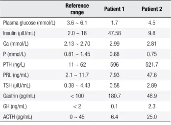
Table 1. Laboratory indings in the two patients
Reference
range Patient 1 Patient 2
Plasma glucose (mmol/L) 3.6 ~ 6.1 1.7 4.5 Insulin (µIU/mL) 2.0 ~ 16 47.58 9.8 Ca (mmol/L) 2.13 ~ 2.70 2.99 2.81 P (mmol/L) 0.81 ~ 1.45 0.68 0.75 PTH (ng/L) 11 ~ 62 596 521.7 PRL (ng/mL) 2.1 ~ 11.7 7.93 47.6 TSH (µIU/mL) 0.38 ~ 4.43 0.58 2.89 Gastrin (pg/mL) < 100 180.7 48.9 GH (ng/mL) < 2 0.1 2.3 ACTH (pg/mL) 0 ~ 45 6.4 25.0
Figure 1. Genealogy chart of family I.
Patient 2 of family II visited our hospital due to re-current diarrhea for about 4 years. At age 32, he recei-ved a hypophysectomy and postoperative radiotherapy at another hospital. His elder brother had undergone enucleation of a pancreatic insulinoma, and resection of a parathyroid adenoma (Figure 4). Laboratory indings for the patient upon admission are summarized in table 1. Elevated levels of serum Ca2+, PTH, PRL, and GH, and
Cop
yright
© ABE&M t
odos os dir
eit
os r
eser
vados
.
Figure 2. Imaging data of patient 1. 99mTc-MIBI scan (A) showed enlargement of a right parathyroid gland. CT scan showed nodule in pancreatic head
with partial calciication (B), nodule in pancreatic tail (C, D), and nodule in left adrenal (E, F).
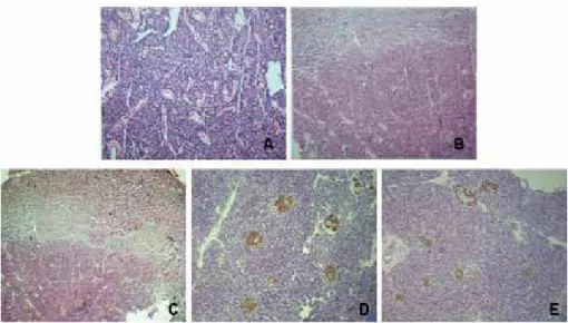
Figure 3. Histopathological section A shows parathyroid adenoma (HE, original magniication 100x), and sections B, C show an insulinoma in the
pancreatic head and tail, respectively (HE, original magniication 100x). Immunohistochemistry sections D, E show positive immunohistochemical analysis for insulin of pancreatic head and tail tumor, respectively (original magniication 100x).
Figure 4. Genealogy chart of family II.
These two patients were diagnosed with MEN1 with all the characteristics of gastrointestinal neuroendocrine tumors, parathyroid adenomas, and pituitary tumors. The elder brother of the patient in family II also had history of insulinoma and parathyroid adenoma.
Cop
yright
© ABE&M t
odos os dir
eit
os r
eser
vados
.
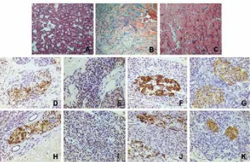
Figure 6. Histopathological section A shows parathyroid adenoma (HE, original magniication, 100x), sections B, C show well differentiated neuroendocrine
tumors in the pancreatic head and tail, respectively (HE, original magniication, 100x). Immunohistochemistry sections D, E, F, G show positive immunohistochemical analysis of pancreatic head tumor for somatostatin, gastrin, glucagon, and insulin, respectively (original magniication, 400x), sections H, I, J, K show positive immunohistochemical analysis of pancreatic tail tumor for somatostatin, gastrin, glucagon, and insulin, respectively (original magniication, 400x).
Figure 5. Imaging data of patient 2. 99mTc-MIBI scan (A) showed enlargement of left parathyroid gland. CT scan showed nodule in the pancreatic tail (B,
C, D) and a nodule in the pancreatic head (E, F).
DNA extraction and mutation screening
Venous blood samples were obtained from three patients of two families (1 female and 2 males) for genetic analy-ses. Genomic DNA was isolated from peripheral blood. Exons were ampliied by polymerase chain reaction (PCR) (15). Puriied PCR products were then sequenced.
RESULTS
Cop
yright
© ABE&M t
odos os dir
eit
os r
eser
vados
.
DISCUSSION
A total of 1,336 mutations of MEN1 have been re-ported in the irst decade following identiication of this gene. The 1,133 germline mutations are scattered throughout the entire 1,830-bp coding region and spli-ce sites of the MEN1 gene, and consist of 23% nonsen-se mutation, 9% splice site mutations, 41% frameshift deletions or insertions, 6% in-frame deletions or inser-tions, 20% missense mutation, and 1% whole or par-tial gene deletions (16,17). The MEN1 gene encodes a 610-amino acid protein referred to as menin, which is ubiquitously expressed, and is predominantly a nuclear protein in nondividing cells (18). However, in dividing cells, it is mainly found in the cytoplasm (19). Menin has at least three nuclear localization signals (20).
MEN1 germline mutations were identiied in more than 85% of the MEN1 families investigated when full--length sequencing of the open reading frame was per-formed (21-23). Until now, some genes were recently
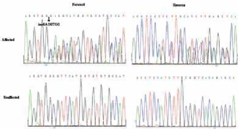
Figure 7. DNA sequence analysis of MEN1 in unaffected and affected individuals of family I. A heterozygous change T > A at IVS4+1 was noted in
affected individuals.
Figure 8. DNA sequence analysis of MEN1 in unaffected and affected individuals in family II. An insGAGGTGG mutation (c.703-709dup7bp) was found,
resulting in a frameshift (p.A237Gfsx13).
identiied as probable causes of MEN1-like condition, such as mixed lineage leukemia (MLL): p15, p18, p21
Cop
yright
© ABE&M t
odos os dir
eit
os r
eser
vados
.
MEN1 should control most features of excess of some hormones (gastrin, PRL etc.). Surgery should control features of excess of other hormones (PTH and insu-lin). However, surgery has not been shown to prevent or cure MEN1-related cancers (14).
In conclusion, we have identiied a novel insGA-GGTGG mutation (c.703-709dup7bp) resulting in a frameshift (p.A237Gfsx13) in a Chinese family. This inding extends our knowledge of the variety of genetic abnormalities associated with familial MEN1. Functio-nal studies are necessary to evaluate and understand the impact of this insertion mutation on the clinical picture.
Disclosure: no potential conlict of interest relevant to this article was reported.
REFERENCES
1. Wermer P. Genetic aspects of adenomatosis of endocrine glands. Am J Med. 1954;16(3):363-71.
2. Trump D, Farren B, Wooding C, Pang JT, Besser GM, Buchanan KD, et al. Clinical studies of multiple endocrine neoplasia type 1 (MEN1). QJM. 1996;89(9):653-69.
3. Skogseid B, Rastad J, Oberg K. Multiple endocrine neoplasia type 1. Clinical features and screening. Endocrinol Metab Clin North Am. 1994;23(1):1-18.
4. Thakker RV. Multiple endocrine neoplasia type 1 (MEN1). Best Pract Res Clin Endocrinol Metab. 2010;24(3):355-70.
5. Dean PG, van Heerden JA, Farley DR, Thompson GB, Grant CS, Har-msen WS, et al. Are patients with multiple endocrine neoplasia type I prone to premature death? World J Surg. 2000;24(11):1437-41. 6. Arnold R. Endocrine tumours of the gastrointestinal tract.
In-troduction: deinition, historical aspects, classiication, staging, prognosis and therapeutic options. Best Pract Res Clin Gastroen-terol. 2005;19(4):491-505.
7. Norton JA, Kivlen M, Li M, Schneider D, Chuter T, Jensen RT. Mor-bidity and mortality of aggressive resection in patients with ad-vanced neuroendocrine tumors. Arch Surg. 2003;138(8):859-66. 8. Shepherd JJ, Challis DR, Davies PF, McArdle JP, Teh BT, Wilkinson
S. Multiple endocrine neoplasm, type 1. Gastrinomas, pancreatic neo plasms, microcarcinoids, the Zollinger-Ellison syndrome, lym-ph nodes, and hepatic metastases. Arch Surg. 1993;128(10):1133-42. 9. Fendrich V, Langer P, Celik I, Bartsch DK, Zielke A, Ramaswamy A,
et al. An aggressive surgical approach leads to long-term sur-vival in patients with pancreatic endocrine tumors. Ann Surg. 2006;244(6):845-51; discussion 852-43.
10. Abe T, Sato M, Okumura T, Shioyama Y, Kiyoshima M, Asato Y, et al. FDG PET/CT indings of thymic carcinoid and bronchial carci-noid in a patient with multiple neuroendocrine neoplasia type 1. Clin Nucl Med. 2008;33(11):778-9.
11. Grama D, Eriksson B, Martensson H, Cedermark B, Ahren B, Kris-toffersson A, et al. Clinical characteristics, treatment and survival in patients with pancreatic tumors causing hormonal syndromes. World J Surg. 1992;16(4):632-9.
12. Garbrecht N, Anlauf M, Schmitt A, Henopp T, Sipos B, Raffel A, et al. Somatostatin-producing neuroendocrine tumors of the duo-denum and pancreas: incidence, types, biological behavior,
asso-ciation with inherited syndromes, and functional activity. Endocr Relat Cancer. 2008;15(1):229-41.
13. Goudet P, Murat A, Binquet C, Cardot-Bauters C, Costa A, Rusz-niewski P, et al. Risk factors and causes of death in MEN1 disease. A GTE (Groupe d’Etude des Tumeurs Endocrines) cohort study among 758 patients. World J Surg. 2010;34(2):249-55.
14. Brandi ML, Gagel RF, Angeli A, Bilezikian JP, Beck-Peccoz P, Bordi C, et al. Guidelines for diagnosis and therapy of MEN type 1 and type 2. J Clin Endocrinol Metab. 2001;86(12):5658-71.
15. Hai N, Aoki N, Matsuda A, Mori T, Kosugi S. Germline MEN1 mu-tations in sixteen Japanese families with multiple endocrine neo-plasia type 1 (MEN1). Eur J Endocrinol. 1999;141(5):475-80. 16. Chandrasekharappa SC, Guru SC, Manickam P, Olufemi SE, Collins
FS, Emmert-Buck MR, et al. Positional cloning of the gene for mul-tiple endocrine neoplasia-type 1. Science. 1997;276(5311):404-7. 17. Lemos MC, Thakker RV. Multiple endocrine neoplasia type 1
(MEN1): analysis of 1336 mutations reported in the irst decade following identiication of the gene. Hum Mutat. 2008;29(1):22-32. 18. Guru SC, Goldsmith PK, Burns AL, Marx SJ, Spiegel AM, Collins
FS, et al. Menin, the product of the MEN1 gene, is a nuclear pro-tein. Proc Natl Acad Sci U S A. 1998;95(4):1630-4.
19. Huang SC, Zhuang Z, Weil RJ, Pack S, Wang C, Krutzsch HC, et al. Nuclear/cytoplasmic localization of the multiple endocrine neo-plasia type 1 gene product, menin. Lab Invest. 1999;79(3):301-10. 20. La P, Desmond A, Hou Z, Silva AC, Schnepp RW, Hua X. Tumor
suppressor menin: the essential role of nuclear localization signal domains in coordinating gene expression. Oncogene. 2006;25(25):3537-46.
21. Agarwal SK, Kester MB, Debelenko LV, Heppner C, Emmert-Buck MR, Skarulis MC, et al. Germline mutations of the MEN1 gene in familial multiple endocrine neoplasia type 1 and related states. Hum Mol Genet. 1997;6(7):1169-75.
22. Bassett JH, Forbes SA, Pannett AA, Lloyd SE, Christie PT, Wooding C, et al. Characterization of mutations in patients with multiple endocrine neoplasia type 1. Am J Hum Genet. 1998;62(2):232-44. 23. Poncin J, Abs R, Velkeniers B, Bonduelle M, Abramowicz M,
Le-gros JJ, et al. Mutation analysis of the MEN1 gene in Belgian patients with multiple endocrine neoplasia type 1 and related di-seases. Hum Mutat. 1999;13(1):54-60.
24. Taguchi R, Yamada M, Horiguchi K, Ozawa A, Shibusawa N, Hashi-moto K, et al. Haploinsuficient and predominant expression of multiple endocrine neoplasia type 1 (MEN1)-related genes, MLL, p27(Kip1) and p18(Ink4C) in endocrine organs. Biochem Biophys Res Commun. 2011;415(2):378-83.
25. Agarwal SK, Mateo CM, Marx SJ. Rare germline mutations in cyclin-dependent kinase inhibitor genes in multiple endocrine neoplasia type 1 and related states. J Clin Endocrinol Metab. 2009;94(5):1826-34.
26. Ishida E, Yamada M, Horiguchi K, Taguchi R, Ozawa A, Shibusa-wa N, et al. Attenuated expression of menin and p27 (Kip1) in an aggressive case of multiple endocrine neoplasia type 1 (MEN1) associated with an atypical prolactinoma and a malignant pan-creatic endocrine tumor. Endocr J. 2011;58(4):287-96.
27. Georgitsi M, Raitila A, Karhu A, van der Luijt RB, Aalfs CM, Sane T, et al. Germline CDKN1B/p27Kip1 mutation in multiple endocrine neoplasia. J Clin Endocrinol Metab. 2007;92(8):3321-5.
28. Pannett AA, Thakker RV. Multiple endocrine neoplasia type 1. En-docr Relat Cancer. 1999;6(4):449-73.
29. Thakker RV. Multiple endocrine neoplasia--syndromes of the twentieth century. J Clin Endocrinol Metab. 1998;83(8):2617-20. 30. Marini F, Falchetti A, Del Monte F, Carbonell Sala S, Gozzini A, Luzi