Which factors are associated with global
cognitive impairment in Wilson’s disease?
Norberto Anízio Ferreira Frota1, Egberto Reis Barbosa2, Claudia Sellitto Porto3, Leandro Tavares Lucato4, Carla Rachel Ono4, Carlos Alberto Buchpiguel4,
Alexandre Aluizio Costa Machado2, Paulo Caramelli5
ABSTRACT. Background: Patients with Wilson’s disease (WD) present cognitive impairment, especially in executive functions. Which other factors might be associated with global cognitive decline in these patients remains unclear.
Objective: To assess which factors are associated with worse performance on a global cognitive test in patients with WD. Methods: Twenty patients with WD underwent cognitive assessment with the following tests: the Mini-Mental State Examination (MMSE), Dementia Rating Scale (DRS), verbal fluency test, brief cognitive battery, clock drawing test, Frontal Assessment Battery, Stroop test, Wisconsin card sorting test, Hopper test, cubes (WAIS) and the Pfeffer questionnaire. MRI changes were quantified. Patients with poor performance on the DRS were compared to patients with normal performance. Results: Nine patients had a poor performance on the DRS. This group had lower educational level (9.11±3.58 × 12.82±3.06) and a greater number of changes on MRI (9.44±2.74 × 6.27±2.45). The presence of hyperintensity in the globus pallidus on MRI was more frequent in this group (66.6% vs 9.0%), with OR=5.38 (95% CI 0.85-33.86). Conclusion: Global cognitive impairment was prevalent in this sample of patients with WD and was associated with low educational level, number of changes on MRI and MRI hyperintensity in the globus pallidus.
Key words: Wilson’s disease, cognitive impairment, neuropsychological tests, magnetic resonance imaging, globus pallidus.
QUAIS FATORES ESTÃO ASSOCIADOS AO COMPROMETIMENTO COGNITIVO GLOBAL NA DOENÇA DE WILSON?
RESUMO. Embasamento: Pacientes com doença de Wilson (DW) apresentam comprometimento cognitivo, principalmente de funções executivas. Existem dúvidas sobre quais outros fatores poderiam estar associados ao declínio cognitivo global nesses pacientes. Objetivo: Avaliar quais fatores estão associados ao pior desempenho em teste cognitivo global em pacientes com DW. Métodos: Vinte pacientes com DW em tratamento regular foram submetidos à avaliação cognitiva com os seguintes testes: Mini-Exame do Estado Mental, escala de demência de Mattis (DRS), fluência verbal, bateria cognitiva breve, desenho do relógio, bacteria de avaliação frontal, Stroop, teste de seleção de cartões de Wisconsin, Hopper, Cubos e ao questionário de Pfeffer. As alterações em RM foram quantificadas. Pacientes com desempenho alterado na DRS foram comparados aos pacientes com desempenho normal. Resultados: Nove pacientes apresentavam desempenho alterado na DRS. Eles apresentavam menor nivel educacional (9,11±3,58 × 12,82±3,06 anos, respectivamente) e maior quantidade de alterações na RM (9,44±2,74 × 6,27±2,45). A presença de hipersinal no globo pálido na RM foi mais frequente nesse grupo (66,6% × 9,0%), com OR=5,38 (IC 95% 0,85-33,86). Conclusão: Alterações cognitivas globais foram frequentes nesta amostra de pacientes com DW e se associaram à baixa escolaridade, quantidade de alterações em RM e a hipersinal no globo pálido à RM.
Palavras-chave: doença de Wilson, deterioração cognitiva, testes neuropsicológicos, imagem de ressonância magnética, globus pallidus.
This study was conducted at the Department of Neurology, University of São Paulo School Medicine, São Paulo,SP, Brazil.
1 MD, PhD. School of Medicine, University of Fortaleza, Fortaleza, CE, Brazil. MD, PhD. 2Department of Neurology, University of São Paulo School of Medicine, São Paulo, SP, Brazil. 3 PhD. Department of Neurology, University of São Paulo School of Medicine, São Paulo, SP Brazil. 4 MD, PhD. Department of Radiology, University of São Paulo School of Medicine, São Paulo, SP, Brazil. 5 MD, PhD. Department of Internal Medicine, Faculty of Medicine, Federal University of Minas Gerais, MG, Brazil. Norberto Anizio Ferreira Frota. University of Fortaleza – Rua Desembargador Floriano Benevides, 221 / Third Floor – 60811-905 Fortaleza CE – Brazil. E-mail: [email protected]
Disclosure: The authors report no conflicts of interest.
Received October 17, 2016. Accepted in final form November 10, 2016.
INTRODUCTION
W
ilson’s disease (WD) is a rare genetic condition described over 100 years ago, characterized by the accumulation of copper in many organs, including the central nervous system.1he cognitive symptoms present in the irst recorded patients were not studied in depth for some time.2 he irst studies assessing cognitive performance described changes in memory and executive functions in patients with neurological symptoms of WD,3,4 rais-ing doubts as to whether the motor symptoms were in fact responsible for the cognitive impairment.3 Patients with only hepatic symptoms did not exhibit cognitive abnormalities.5
In 2002, a study found that patients with lesions restricted to the basal nuclei displayed cognitive changes compared to healthy controls.6 Recently, it has been noted that patients with neurological symptoms but no depressive or anxiety disorders, present cognitive impairment, mainly in executive functions. Further-more, the number of deicits on cognitive tests was asso-ciated with the intensity of brain Magnetic Resonance Imaging (MRI) changes, especially hyperintense signals on T2 images and atrophy.7
A study of 12 patients with WD sought to correlate the topographies of hyperintensities on MRI with cogni-tive proile and suggested that changes in the putamen are related to worse performance on cognitive tests.8 However, the small number of patients and the fact that only descriptive analysis of the data was performed limit this inding.
Nevertheless, it is known that the intensity of changes on MRI and the severity of motor symptoms are related to cognitive changes.7
Not all patients with WD exhibited deicits on the cognitive tests. Normal overall performance on cog-nitive tests can be observed in more than 50% of WD patients.3,7 It is not clear which factors are related to cog-nitive symptoms, such as disease duration, medications used or the speciic topography of T2-hyperintensity changes.
In the present study, data previously published by us was reevaluated, comparing a group of patients with WD that exhibited deicits on global cognitive tests against cognitively unimpaired WD patients.
METHODS
he methodology used to select the patients, as well as the cognitive evaluation and neuroimaging protocols, has been described in detail elsewhere.7 A brief descrip-tion is given below.
Patients. Twenty patients with WD were selected based on clinical history, physical examination, serum cerulo-plasmin levels (<20 mg/dl), 24-h urinary copper excre-tion (>100 mcg/24 h) and ophthalmologic examinaexcre-tion by slit lamp. All patients were followed at a Movement Disorders Clinic from September 2006 to October 2007. he patients were undergoing regular treatment for at least one year, without neurological decline. Patients with severe dysarthria or anarthria that could impair speech comprehension, as well as those with motor disorders which could prevent them from performing written tasks, as well as clinical signs of hepatic enceph-alopathy, depression/anxiety according to the Goldberg scale, were excluded.9 All patients signed a consent form and the study was approved by the institution’s Ethics Committee.
Neurological evaluation. All patients were evaluated by the lead researcher of the study (NAFF) using the motor symptoms scale (MSS)7,10 which assesses 13 items (bradykinesia, rigidity, postural instability, tremor, dystonia, chorea, athetosis, cerebellar disturbances, dysarthria, dysphagia, walking diiculties, psychiatric disturbances and other alterations), each graded from 0 to 3 (absent, slight, moderate and intense). Hence, the total score on the scale ranges from 0 to 39.
Cognitive evaluation. All patients were submitted on diferent days to a two-stage cognitive evaluation, each lasting around 40 minutes.
he irst session was applied by the same examining neurologist (NAFF) and included the following tests: Mini-Mental State Examination;11 a memory test of igures,12 clock drawing;13 verbal luency tests (animals and FAS); CERAD naming test;14 Stroop test15 and the Frontal Assessment Battery (FAB).16 he patients’ rela-tives also answered the Pfefer Functional Activities Questionnaire.17
he second evaluation session was performed by an experienced neuropsychologist (CSP), with a similar duration to that of the irst evaluation, involving the following tests: Mattis Dementia Rating Scale (DRS),18 Wisconsin Card Sorting Test (WCST) and the Hooper and Cubes tests (subscale of Wechsler intelligence scale).
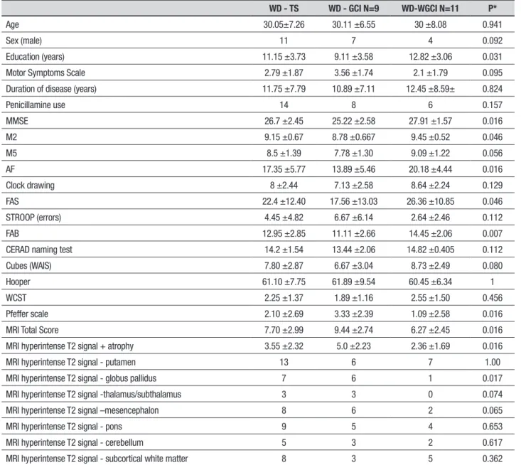
Table 1. Clinical, cognitive and magnetic resonance imaging features.
WD - TS WD - GCI N=9 WD-WGCI N=11 P*
Age 30.05±7.26 30.11 ±6.55 30 ±8.08 0.941
Sex (male) 11 7 4 0.092
Education (years) 11.15 ±3.73 9.11 ±3.58 12.82 ±3.06 0.031
Motor Symptoms Scale 2.79 ±1.87 3.56 ±1.74 2.1 ±1.79 0.095
Duration of disease (years) 11.75 ±7.79 10.89 ±7.11 12.45 ±8.59± 0.824
Penicillamine use 14 8 6 0.157
MMSE 26.7 ±2.45 25.22 ±2.58 27.91 ±1.57 0.016
M2 9.15 ±0.67 8.78 ±0.667 9.45 ±0.52 0.046
M5 8.5 ±1.39 7.78 ±1.30 9.09 ±1.22 0.056
AF 17.35 ±5.77 13.89 ±5.46 20.18 ±4.44 0.016
Clock drawing 8 ±2.44 7.13 ±2.58 8.64 ±2.24 0.129
FAS 22.4 ±12.40 17.56 ±13.03 26.36 ±10.85 0.046
STROOP (errors) 4.45 ±4.82 6.67 ±6.14 2.64 ±2.46 0.112
FAB 12.95 ±2.85 11.11 ±2.66 14.45 ±2.06 0.007
CERAD naming test 14.2 ±1.54 13.44 ±2.06 14.82 ±0.405 0.112
Cubes (WAIS) 7.80 ±2.87 6.67 ±3.04 8.73 ±2.49 0.080
Hooper 61.10 ±7.75 61.89 ±9.54 60.45 ±6.34 1
WCST 2.25 ±1.37 1.89 ±1.16 2.55 ±1.50 0.456
Pfeffer scale 2.10 ±2.69 3.33 ±2.39 1.09 ±2.58 0.016
MRI Total Score 7.70 ±2.99 9.44 ±2.74 6.27 ±2.45 0.016
MRI hyperintense T2 signal + atrophy 3.55 ±2.32 5.0 ±2.23 2.36 ±1.69 0.016
MRI hyperintense T2 signal - putamen 13 6 7 1.00
MRI hyperintense T2 signal - globus pallidus 7 6 1 0.017
MRI hyperintense T2 signal -thalamus/subthalamus 3 3 0 0.074
MRI hyperintense T2 signal –mesencephalon 8 6 2 0.065
MRI hyperintense T2 signal - pons 9 5 4 0.653
MRI hyperintense T2 signal - cerebellum 5 3 2 0.617
MRI hyperintense T2 signal - subcortical white matter 8 3 5 0.362
MMSE: Mini-Mental State Examination; M2: memory (learning); M5: memory (delayed recall); AF: animal fluency; FAS: phonemic fluency; FAB: Frontal Assessment Battery; WCST: Wisconsin Card Sorting Test; WD: Wilson’s disease; WD-GCI: Wilson’s disease with global cognitive Impairment; WD-WGCI+ Wilson’s disease without global cognitive impairment. *WD-GCI × WD-WGCI. rules suggested by Porto et al. in the Brazilian
popula-tion,19 using percentile values ≤10.
MRI. Magnetic resonance imaging examinations were performed using a 1.5 Tesla according to a previously published acquisition protocol.7 he analyses of the images were performed by an experienced neurora-diologist (LTL), blinded to the clinical and cognitive pictures of the patients, using a scale7 for quantitative assessment of the MRI structural changes. On the scale, one point is given in the presence of a hyperintense T2 signal in each of the following structures (maximum 7 points): putamen, globus pallidus, caudate,
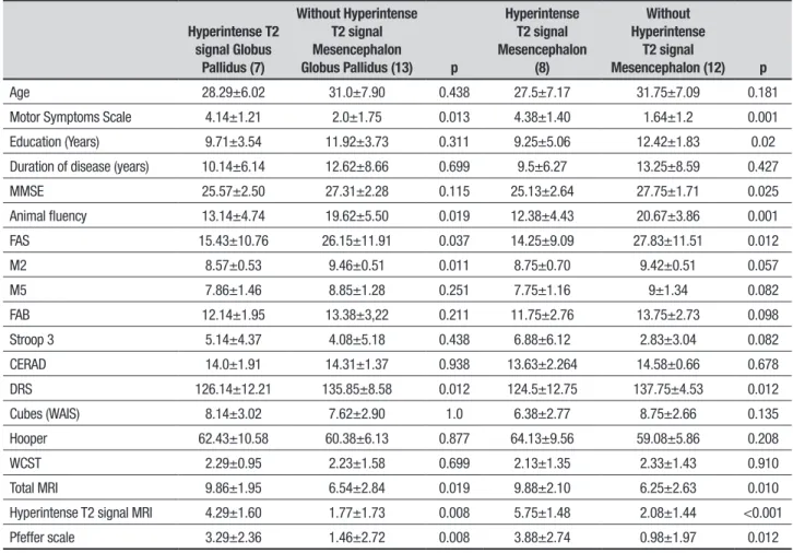
Table 2. Clinical, cognitive and total magnetic resonance imaging in patients with specific topographic MRI changes.
Hyperintense T2 signal Globus
Pallidus (7)
Without Hyperintense T2 signal Mesencephalon
Globus Pallidus (13) p
Hyperintense T2 signal Mesencephalon
(8)
Without Hyperintense
T2 signal
Mesencephalon (12) p
Age 28.29±6.02 31.0±7.90 0.438 27.5±7.17 31.75±7.09 0.181
Motor Symptoms Scale 4.14±1.21 2.0±1.75 0.013 4.38±1.40 1.64±1.2 0.001
Education (Years) 9.71±3.54 11.92±3.73 0.311 9.25±5.06 12.42±1.83 0.02
Duration of disease (years) 10.14±6.14 12.62±8.66 0.699 9.5±6.27 13.25±8.59 0.427
MMSE 25.57±2.50 27.31±2.28 0.115 25.13±2.64 27.75±1.71 0.025
Animal fluency 13.14±4.74 19.62±5.50 0.019 12.38±4.43 20.67±3.86 0.001
FAS 15.43±10.76 26.15±11.91 0.037 14.25±9.09 27.83±11.51 0.012
M2 8.57±0.53 9.46±0.51 0.011 8.75±0.70 9.42±0.51 0.057
M5 7.86±1.46 8.85±1.28 0.251 7.75±1.16 9±1.34 0.082
FAB 12.14±1.95 13.38±3,22 0.211 11.75±2.76 13.75±2.73 0.098
Stroop 3 5.14±4.37 4.08±5.18 0.438 6.88±6.12 2.83±3.04 0.082
CERAD 14.0±1.91 14.31±1.37 0.938 13.63±2.264 14.58±0.66 0.678
DRS 126.14±12.21 135.85±8.58 0.012 124.5±12.75 137.75±4.53 0.012
Cubes (WAIS) 8.14±3.02 7.62±2.90 1.0 6.38±2.77 8.75±2.66 0.135
Hooper 62.43±10.58 60.38±6.13 0.877 64.13±9.56 59.08±5.86 0.208
WCST 2.29±0.95 2.23±1.58 0.699 2.13±1.35 2.33±1.43 0.910
Total MRI 9.86±1.95 6.54±2.84 0.019 9.88±2.10 6.25±2.63 0.010
Hyperintense T2 signal MRI 4.29±1.60 1.77±1.73 0.008 5.75±1.48 2.08±1.44 <0.001
Pfeffer scale 3.29±2.36 1.46±2.72 0.008 3.88±2.74 0.98±1.97 0.012
MMSE: Mini-Mental State Examination; M2: memory (learning); M5: memory (delayed recall); AF: animal fluency; FAS: phonemic fluency; FAB: Frontal Assessment Battery; DRS: Dementia Rating Scale; WCST: Wisconsin Card Sorting Test.
classiied as follows: absent (0), slight (1) or severe (2). Hence, the inal score ranged from 0 to 16, with higher scores indicating greater involvement.
Statistical analyses. An overall descriptive analysis of patients’ DRS performance was irst carried out and the group exhibiting impairmentson the DRS was then compared with the unimpaired group. he Mann-Whitney test was employed for continuous variables and Fisher’s test for categorical variables. In order to investigate associations of categorical variables with global cognitive impairment, odds ratio (OR) values were calculated. Cognitive and motor performances were also compared according to speciic topographies of MRI hyperintensities using the Mann-Whitney test. he associations found on the OR analysis were conirmed or otherwise bylogistic regression models, each with two variables. he irst model was adjusted for education, the second for motor scale scores, while the third model was adjusted for total scores on the
MRI scale. he p-value used for statistical signiicance was set at <0.05.
RESULTS
Impaired scores on the DRS were observed in nine out of the 20 patients evaluated, with a median of 123 points for the impaired group and 140 points for the group with no impairment. Comparison between the clinical, cognitive and neuroimaging features of the overall sample are depicted in Table 1, together with those of the two subgroups of patients.
he group with DRS total score deicits performed signiicantly worsein comparison to the other subgroup on the Initiative/Perseveration and Conceptualization subscales, with medians of 29 vs. 37 and 32 vs. 38, respectively. No statistical diference was found for the other subscales.
to separate assessment of those individuals with hyper-intensity signal changes in the globus pallidus and mesencephalon, and comparisons to patients with no signal change in these topographies. he patients with hyperintensity signal in these regions had higher scores on the MSS, displayed global cognitive deicits (MMSE and DRS), changes in executive function (verbal luency – animals and FAS), as well as changes in the learning phase of the memory test (globus pallidus) (Table 2).
he neuroimaging factors related to poor global cognitive performance were determined by calculating the OR for each feature. he presence of hyperinten-sity signal in the globus pallidus (signiicance) and in the mesencephalon (tendency) were associated with a worse global performance (Table 3). he former associa-tion remained after correcting for educaassocia-tion, but signii-cance was lost when correcting for neurological and MRI scores. he hyperintensity signal in the mesencephal on did not have a statistically signiicant association.
DISCUSSION
We observed that 45% of our sample had low total score on the DRS (below the 10th percentile). his rate is slightly higher to that found by Medalia in 1988.3 When compared to the group with normal performance, there was no diference in relation to age or time of symptom onset. his suggests that after at least one year of follow-up and continuous treatment, the occurrence of cogni-tive decline is not related to the duration of symptoms.
he use of D-Penicillamine also did not difer between the two groups. here are no previous studies assessing the role of speciic treatments on cognitive symptoms in WD. he use of D-Penicillamine was associ-ated with an improvement in the MRI parameters com-pared to Zinc, but without impact on motor changes.20 In order assess the impact of better treatment and dis-ease duration on the occurrence of cognitive symptoms, a prospective study would be required in which patients with no previous treatment and cognitive evaluation prior to the study were selected and retested after a one-year follow up.
he population with worse cognitive performance
tended to have higher MSS scores. he intensity of motor symptoms is associated to the quantity of altered cognitive tests7 and can also inluence performance on some tests.3 We used tests with lower motor perfor-mance demands and excluded patients with serious motor problems. he small number of patients in each group might have prevented statistical signiicance from being reached in this case.
he tendency for a greater number of men with cognitive impairment has not been reported in previ-ous studies. Our inding is probably related to the fact that men were less educated than women in our sample (9.82 vs. 12.78 years, respectively; p=0.031). Indeed, education was the only sociodemographic diference between the two groups. We used education-adjusted cut-of scores for the DRS, nevertheless, the population with lower educational level showed greater impair-menton the test. Low educational level is related to a greater risk for dementia,21 probably due to a smaller cognitive reserve.22 In the present study, no diference was observed in neuroimaging scale score for the group with ≤11years of education compared to those with > 11 years of schooling on the full MRI scale (7.77 vs 7.57 p=0.877), or for hypersignal (2.77 vs 2.43 p=0.877) or hyperintensity signal plus atrophy (3.77 vs 3.14 p=0.757). Furthermore, no correlation was detected between the number of years of education and scores on the MRI full scale (r2: –0.303 p=0.194). hese ind-ings suggest that for the same number of neuroimaging changes, individuals with less education are more sus-ceptible to worse cognitive performance.
Cognitive performance in the population with impairment on the DRS was lower, especially in the executive function tests. he subscales with lowest per-formance were Initiation/Perseveration and Concep-tualization, both associated with executive functions. his observation is in agreement with a previousstudy.23 Changes in response time,23 inhibitory control,7 selective attention24 and working memory23 have been reported in patients with WD presenting neurological symptoms. he absence of diference on the Stroop test might have occurred because this is a common deicit in patients
Table 3. Association between MRI changes and cognitive impairment.
Topography
Hyperintense T2 Signal OR (95% CI)
Model A+
ß (95% CI)
Model B++
ß (95% CI)
Model C+++
ß (95% CI)
Globus pallidus 5.38 (0.85-33.86)* 22.14 (1.19-410.30)* 13.473 (0.82-220.09) 9.06 (0.627-131.16)
Mesencephalon 3.00 (0.86-10.40) 5.04 (0.44-57.87) 5.38 (0.283-102.47) 3.11 (0.287-33.814)
with WD,7 even in cases whose performance on other cognitive tests is normal.
Memory deicits may be seen in patients with WD, more on encoding than in the delayed recall,3,6,7 as was seen in our sample. he normal performances on tests related to praxis and visuospatial abilities conirm that these are not domains afected in patients with WD.3,23
he pattern of preferential involvement of the execu-tive functions and memory (learning) suggests dysfunc-tion in cortico-subcortical circuits of the frontal lobe. In 2002, Seniow et al. reported that patients with lesions restricted to the basal nuclei also showed cognitive changes.6 A previous study seeking to correlated cog-nitive performance with MRI changes suggested that putamen involvement might be associated with a higher number of changes in cognitive tests.8 In the study, only 12 patients were evaluated, three of which had normal MRI, thus limiting the study to nine patients and pre-venting the use of any statistical evaluation. For this reason, the results were only descriptive.
In our sample, all 20 patients presented changes on brain MRI.7 he putamen was the topography most frequently afected (sign changes), but without difer-ence in frequency between the groups with and without cognitive impairment. When we compared the seven patients with no putamen changes to the remaining,13 no statistical diferences were found for any of the tests except the MSS, suggesting that changes in this topog-raphy impact motor more than cognitive performance. In our study we found that the topographic asso-ciation between cognitive and T2 hyperintensity signal changes had a tendency to be associated with involve-ment of the mesencephalon and attained statistical sig-niicance in the globus pallidus. Patients with changes in these topographies had worse performance on executive function tests, such as luency and learning memory, but also had more severe motor symptoms and changes on MRI. Correcting for these factors decreased statistical signiicance, which persisted only after the adjustment-for educational level.
Few studies have evaluated the role of the globus pal-lidus in executive functions or in global cognitive per-formance. One study entailing deep brain stimulation (DBS) in patients with Parkinson’s disease indicated that this would be a safer surgical site than the subthalamic nucleus for preventing cognitive dysfunctions.25 A study of patients with generalized dystonia observed cogni-tive worsening, mainly on digit span, errors on attention tests and worse recall in the Rey Auditory Verbal Learn-ing Test, after placement DBS probes into the internal globus pallidus.26
More recently, observing pathological studies in patients with Huntington’s disease that had a similar cognitive proile to our sample,7 a linear correlation was found between atrophy of the external globus pallidus and its most ventral part with global cognitive dysfunc-tion in these patients.27 his result, taken together with our indings, suggests that the globus pallidus plays more than merely motor roles in the basal nuclei and that changes in this topography are associated with worse cognitive performance.
he small number of patients in our group might have limited the assessment of our data and prevented attainment of statistical signiicance on some tests. he fact that we were unable to deine which parts of the glo-bus pallidus and the mesencephalon were more involved also prevented a better correlation between cognitive performance and neuroimaging changes.
Despite these limitations, our study conirms that global cognitive impairments occur in a considerable proportion of patients with WD. hese changes are asso-ciated, as previously reported, with severity of neuro-logical impairments7 low educational level, severity of hypersignal changes on MRI, andthe occurrence of these MRI changes in the globus pallidus.
Author contribution. Norberto Anizio Ferreira Frota: design of the study, analysis of the data and intellec-tual contribution to the written manuscript. Egberto Reis Brabosa: design of the study, analysis of the data and intellectual contribution to the written manu-script. Paulo Caramelli: design of the study, analysis of the data and intellectual contribution to the written manuscript. Claudia Sellitto Porto: contribution to this manuscript in design of the study and analysis of the data. Leandro Tavares Lucatto: contribution to this manuscript in design of the study and analysis of the data. Carlos Alberto Buchpiguel: contribution to this manuscript in design of the study and analysis of the data. Carla Rachel Ono: contribution to this manu-script in design of the study and analysis of the data. Alexandre Aluizio Costa Machado: contribution to this manuscript in design of the study and analysis of the data.
Support. his research was supported in part by the CAPES (Coordination of Improvement of Higher Educa-tion Personnel).
REFERENCES
1. Machado A, Chien HF, Deguti MM, Cançado E, Azevedo RS, Scaff M, Barbosa ER. Neurological manifestations in Wilson’s disease: Report of 119 cases. Mov Disord. 2006;21:2192-6.
2. Frota NAF, Caramelli P, Barbosa ER. Cognitive impairment in Wilson’s disease. Dement. Neuropsychol. 2009;3(1):16-21.
3. Medalia A, Issacs-Glaberman K, Scheinberg IH. Neuropsychological impairment in Wilson’s disease. Arch Neurol. 1988;45:502-504. 4. Rathbun JK. Neuropsychological aspects of Wilson’s disease. Int J
Neurosci. 1996;85:221-9.
5. Lang C, Muller D, Claus D, Druschky KF. Neuropsychological findings in treated Wilson’s disease. Acta Neurol Scand.1990;81:75-81
6. Seniow J, Bak T, Gajda J, Poniatowska R, Czlonkowska A. Cognitive functioning in neurologically symptomatic and asymptomatic forms of Wilson’s disease. Mov Disord.2002;17:1077-83.
7. Frota NA, Barbosa ER, Porto CS, Lucato LT, Ono CR, Buchpiguel CA, Caramelli P. Cognitive impairment and magnetic resonance imaging correlations in Wilson’s disease. Acta Neurol Scand. 2013;127(6):391-8. 8. Hegde S, Sinha S, Taly AB, Rao SL, Vasudev MK. Cognitive profile and structural findings in Wilson’s disease: A Neuropaychological and MRI based Study. Neurol India. 2010;58:708-13.
9. Goldberg D, Bridges K, Duncan-Jones P, Grayson D. Detecting anxiety and depression in general medical settings. BMJ. 1988;297:897-99. 10. Magalhaes AC, Caramelli P, Menezes JR, Lo LS, Bacheschi LA, Barbosa
ER, et al. Wilson’s disease: MRI with clinical correlation. Neuroradiology. 1994;36:97-100
11. Brucki SM, Nitrini R, Caramelli P, Bertolucci H, Okamoto IH. Sugestions for utilization of mini-mental state examination in Brazil. Arq Neurop-siquiatr. 2003;61:777-81.
12. Nitrini R, Caramelli P, Herrera Júnior E, Porto CS, Charchat-Fichman H, Carthery MT, et al. Performance of illiterate and literate nondemented elderly subjects in two test os long-term memory. J Int Neuropsychol Soc. 2004;10:634-8.
13. Sunderland T, Hill JL, Mellow AM, Lawlor BA, Gundersheimer J, Newhouse PA, Grafman JH. Clock drawing in Alzheimer’s disease. A novel measure of dementia severity. J Am Geriatr Soc. 1989;37: 725-9.
14. Bertolucci PH, Okamoto IH, Brucki SM, Siviero MO, Toniolo NJ, Ramos LR. Applicability of the CERAD neuropsychological battery to Brazilian elderly. Arq Neuropsiquiatr. 2001;59:532-6.
15. Stroop J. Studies of interference in serial verbal reactions. J Exp Psychol. 1935;18:643.
16. Beato RG, Nitrini R, Formigoni AP, Caramelli P. Brazilian version of the Frontal Assessment Baterry (FAB): preliminary data on administration to healthy elderly. Dement Neuropsychol. 2007;1:59-65.
17. Pfeffer RI, Kurosaki TT, Harrah CH, Chance JM, Filos S. Measurement of functional activities in older adults in the community. J Gerontol. 1982;37:323-9.
18. Porto CS, Fichman HC, Caramelli P, Bahia VS, Nitrini R. Brazilian version of the Mattis dementia rating scale: diagnosis of mild dementia in Alzheimer’s disease. Arq Neuropsiquiatr. 2003;61:339-45. 19. Foss MP, Carvalho VA, Machado TH, Reis GC, Tumas V, Caramelli P,et
al. Mattis dementia rating scale (DRS) normative data for the brazilian middle-age and elderly populations. Dement Neuropsychol. 2013; 7(4):374-9
20. da Costa MD, Spitz M, Bacheschi LA, Leite CC, Lucato LT, Barbosa ER. Wilson’s disease: two treatment modalities. Correlation to pretreatment and posttreatment brain MRI. Neuroradiology. 2009;51:627-33. 21. Brucki SMD. Illiteracy and dementia. Dement Neuropsychol. 2010;4(3):
153-7
22. Stern Y. Cognitive reserve in ageing and Alzheimer’s disease. Lancet Neurol. 2012;11(11):1006-12.
23. Wenisch E, Tassigny A, Trocello J-M, Beretti J, Girardot-Tinant N, Woimant F. Cognitive profile in Wilson’s disease: A case series of 31 patients. Rev Neurol. (Paris) 2013;169:944-9.
24. Han Y, Zhang F, Tian Y, Hu P, Li B, Wang K. Selective Impairment of Attentional Networks of Alerting in Wilson’s Disease. PLoS One. 2014; 9(6):e100454
25. Combs HL, Folley BS, Berry DT, Segerstrom SC, Han DY, Anderson-Mooney AJ, et al.Cognition and Depression Following Deep Brain Stimu-lation of the Subthalamic Nucleus and Globus Pallidus Pars Internus in Parkinson’s Disease: A Meta-Analysis. Neuropsychol Rev. 2015;25(4): 439-54.
26. Jahanshahi M, Torkamani M, Beigi M, Wilkinson L, Page D, Madeley L,et al. Pallidal stimulation for primary generalised dystonia: effect on cogni-tion, mood and quality of life. J Neurol. 2014;261:164-73.