Postprandial Hyperglycemia: Treating its Atherogenic Potential
Bruno Geloneze
1, Rodrigo Nunes Lamounier
2, Otávio Rizzi Coelho
3Universidade de Ciências Médicas de Campinas – Unicamp1, Disciplina de Endocrinologia, Faculdade de Medicina da USP2 and Instituto do Coração
do Hospital das Clínicas- FMUSP3, Campinas - São Paulo, SP, Brazil
key words
Postprandial glycemia, cardiovascular risk, atherogenesis,
prandial state, postprandial treatment.
Mailing Address: Rodrigo nunes lamounier • Rua Castor da Silva, 39 - 05586-020 – São Paulo, SP, Brazil
Biochemical and physiological processes are involved in the maintenance of a homeostasis state aiming at the survival of the human body. The alterations in this state of equilibrium are perceived and several systems are activated, including the endocrine system, aiming at reestablishing the homeostasis.
Among these systems, one of the most important is nutrition, for cell functionality and growth.
After a meal, there is a nutrient storage for the future needs of the body. The physiological response to fasting and feeding is a complex process and alterations in this process can lead to several illnesses; from starvation to obesity, from hypoglycemia to diabetes mellitus type 2. There can also be an alteration in the processes of growth with deficiency or exacerbation of the proliferative processes, such as in arteriosclerosis.
Type 2 diabetes is clearly associated to the presence of obesity and it is acknowledged that it results from a combination of the beta cell secretory defect and failure in the peripheral action of insulin1.
The intensity of the disease, as well as its potential in causing chronic complications is frequently attributed to the presence of fasting hyperglycemia, which reflects the alteration in the process of homeostasis and fundamentally, in the elevated levels of glycated hemoglobin (HbA1c), being the main parameters for its treatment2,3. The identification
of the association between the postprandial state and cardiovascular disease 4 in patients with type 2 diabetes
brought the development of new therapeutic options aiming at correcting the postprandial metabolic abnormalities, among which are the alpha-glucosidase inhibitors, fast-acting insulin secretagogues, fast acting insulin analogs and others.
The present review aims at the detailed discussion of the importance of the postprandial state in patients with type 2 diabetes, from the progression of the beta cell dysfunction associated to insulin action resistance to the development of the process of atherogenesis. Dietary therapeutic alternatives will be discussed as well as behavior (physical exercise) and pharmacological ones. Lastly, the possible perspectives for future therapies will be discussed.
Regulation of normal glycemia
Glycemia regulation depends basically on the action of two hormones produced in the islets of Langerhans in the
pancreas: insulin and glucagon, which promote the minute-by-minute adjustment of glucose homeostasis. In healthy individuals, the glucagon action is to stimulate the production of glucose by the liver and the insulin action is to block this production and increase glucose uptake by the peripheral insulin-sensitive tissues, such as muscle and fatty tissue. In a normal fasting state, small increments in glycemia lead to the suppression of glucagon production and increase of insulin production, whereas hypoglycemia lead to an increase in glucagonemia and decrease in insulinemia. The integrity of this “glucostate” is crucial for metabolic health. In the fasting and postprandial states, the glucose consumption is represented by the central nervous system (CNS - 50%), muscle (25%) and splenic tissues (25%)5,6.
The state of normal fasting is characterized by more elevated levels of glucagon and low levels of insulin, together with physiological levels of gastrointestinal hormones such as the gastric inhibitory polypeptide (GIP) and the glucagon-like peptide (GLP-1). The result of this equilibrium is an increased production of glucose by the liver and by the kidney, reduction in the peripheral glucose uptake, and increase in muscular proteolysis and adipocyte lipolysis. These synchronized changes maintain glycemia between 70 and 100 mg/dL, free fatty acids (the product of lipolysis) between 300 and 400 µmol/L and triglycerides below 125 mg/dL. Individuals with type 2 diabetes present a reduction in insulin action and production, resulting in an increase of glycemia, free fatty acids, triglycerides and amino acids in the fasting state6,7.
Postprandial hyperglycemia is the result of an excessive glucose production associated to its reduced peripheral uptake. When the glucose uptake exceeds its production, the glycemia returns to normal levels. In individuals with carbohydrate intolerance and in those with type 2 diabetes, the postprandial glycemic excursion is higher and more prolonged, submitting these individuals to a long-term postprandial state9,10. Thus, the postprandial hyperglycemia
and hypertriglyceridemia are the earlier alterations seen in patients who will develop type 2 diabetes.
Physiopathological mechanisms of
postprandial hyperglycemia
As mentioned before, the normal glycemic excursion depends on a complex pancreatic and gastrointestinal hormone action. Glucose intolerance depends on the presence of defects in insulin production (reduction of the first phase of insulin secretion) and action (insulin resistance). It is known that the relative importance of insulin resistance in diabetic patients is more relevant due to the presence of obesity and centripetal fat distribution, which impair insulin action. Nonetheless, in studies that adjusted insulin secretion and action by the degree and distribution of adipose tissue, the deficient insulin secretion showed to be more important11,12.
Considering that, the diabetes treatment is usually more effective when the two physiopathological mechanisms are treated together.
Hyperglycemia occurs due to a failure of the beta cell to compensate for insulin resistance, with a deficient secretory response to the glycemic stimulation. This picture occurs in the beginning of the process as well as in the possible beta cell exhaustion mechanism, which is inherent to the disease evolution. However, animal models with knockout of the insulin receptor in pancreatic beta cells, called βirko, i.e., where there is insulin action resistance in the cell that produces it, showed loss of the first phase of insulin secretion and its initial clinical finding is postprandial hyperglycemia. Thus, insulin resistance demands a higher insulin production, although it will eventually result in the decrease of its production as in the development of type 2 diabetes.
Additionally, chronic postprandial hyperglycemia can lead to a state of glucotoxicity, characterized by the progressive reduction of beta cell secretion or the progressive loss of the glycostatic function of the pancreas, i.e., a vicious cycle with a progressive worsening of the hyperglycemic state. The toxic mechanisms of hyperglycemia on the beta cell occur by the reduction of the insulin gene expression, decrease of proinsulin processing and deposition amyloid material with an increase of the apoptosis process (cell death). These mechanisms are exacerbated in the concomitance of an increase in postprandial lipemia (triglycerides and free fat acids) through a mechanism known as lipotoxicity13. The
known process of increased glycation in diabetes, in which the glucose reacts in a nonenzymatic way with several proteins forming initial glycation products, called Amadori products, commonly assessed by the presence of increased levels of glycated hemoglobin (A1C), also occurs inside the beta cell in the insulin granules, leading to the secretion of glycated insulin, which presents a reduced biological activity.
Thus, the glucotoxicity in the beta cell must also contribute to insulin action resistance14.
After a meal, some hormones are produced in the gastrointestinal tract and stimulate insulin secretion, being known as incretins. There are several postprandial hormones; the most important ones are the glucose-dependent insulinotropic peptides, i.e., the inhibitory gastric peptide (GIP) and the glucagon-like peptide-1 (GLP-1). In fact, there is an increased insulinic response to oral glucose when compared to the intravenous glucose infusion and this phenomenon is known as the incretin effect, which contributes with 30 to 60% of the postprandial insulin secretion. In type 2 diabetic patients, there is a slight reduction of the postprandial levels of GIP and an increased reduction of GLP-1 levels, contributing for the insulin secretion deficiency in these individuals15.
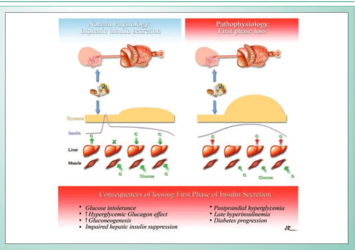
The synchronization between food intake and insulin secretion is a precise and intriguing mechanism. This synchronism depends on a cephalic phase of insulin secretion that precedes the food intake, being small in absolute terms, but directly related to the total amount of insulin secreted right after the beginning of food intake16. In response to an
intravenous or oral glucose overload, there is, therefore, a biphasic insulin secretion, characterized by an initial peak between 5 and 7 minutes. This first secretion phase lasts up to 10 or 15 minutes and is followed by a prolonged secretion at lower levels, usually for up to 4 hours, until the glucose levels return to normal basal levels. In absolute terms, approximately 1% of the beta cell content is secreted in the first phase and 10% more are secreted in the second phase.
After the oral glucose overload, there is an inverse relation between the 30-min insulin concentration (a marker of the first phase of the secretion) and the 2-hour glycemia17. On the
other hand, there is a direct relation between the insulinemia level and the 2-hour glycemia, and the loss of the first phase of insulin secretion can lead to hyperglycemia and late hyperinsulinemia. Without the initial insulin peak, there is no suppression of glucagon production and, consequently, a blockage of the hepatic glucose production18. The suppression
of the hepatic production in diabetic individuals is 50% lower when compared to normal individuals. The effects of the loss of the first phase are summarized in Figure 119,20.
Patients with type 2 diabetes, at the time of the diagnosis, demonstrate a virtually lost first phase of secretion, despite presenting an increase in the second phase and fasting hyperinsulinemia. A prospective study with the Pima Indians of Arizona, who evolved from normality to diabetes in a period of 5 years of follow-up, showed that the progression from normal to glucose intolerance, and subsequently, to diabetes, was associated to weight gain, to the worsening of insulin sensitivity and the deterioration of the first phase of insulin secretion21. In summary, the loss of the first phase is
a crucial event in the secretory defect related to the genesis of type 2 diabetes, being associated with glucose intolerance and postprandial hyperglycemia20.
Postprandial hyperglycemia and
cardiovascular morbi-mortality
glucose tolerance test (OGTT). Although the OGTT does not represent a stimulus identical to a complex meal, with fibers and lipids, studies have shown a good correlation between the glycemic peaks observed after the test and after a complex meal, being the OGTT then considered a surrogate marker for postprandial glycemia.
Studies such as the Hoorn study32, the Honolulu
Heart Study33 and the DECODE (Diabetes Epidemiology:
Collaborative Analysis of Diagnostic Criteria in Europe) study34
showed that the hyperglycemia 2 hrs after the glucose overload was an important predictor of cardiovascular risk, which has also been confirmed by Coutinho and cols. in a meta-analysis involving more than 90,000 individuals35. Similarly,
prospective cohort studies such as the Whitehall Study, the Paris Prospective Study and the Helsinki policemen study showed that the risk of CV mortality had a 2-fold increase in those with a hyperglycemic response to the stimulation test when compared to those with normal response to the glucose overload36-38. Thus, prospective clinical assays with correction
of postprandial hyperglycemia will be able to characterize the actual role of this control in the prevention of cardiovascular events. Notably, the presence of multiple associated risk factors in diabetic patients presupposes an intensive therapeutic intervention in these patients39.
The hypothesis of atherogenesis as a postprandial phenomenon was raised as early as the 70’s40.
(CVD) in patients with type 2 diabetes has been recognized for some time22,23. Diabetic patients present a 2 to 4-fold
increased risk of CVD, when compared to non-diabetic individuals. The mortality risk due to CVD is 2 to 10-fold increased in diabetic patients. A case-control study, which assessed risk factors for acute myocardial infarction (AMI) in the metropolitan region of the city of São Paulo, Brazil, showed that the presence of diabetes antecedents had an independent association, which was not observed in relation to the glycemic measurement alone, evaluated in the study. It is noteworthy that, although the study was not explicit about it, one can assume that the measurement was carried out in the fasting state, as the same blood sample was used for the measurement of lipid parameters24. Other studies carried out
with the Brazilian population showed conflicting results25,26.
CVD is the main cause of mortality among diabetic patients, being responsible for up to 50% of the deaths27-29. Despite
the fact that hyperglycemia was related to a higher risk of cardiovascular events in the United Kingdom Prospective Diabetes Study (UKPDS), the treatment of hyperglycemia was not able to significantly reduce the risk of cardiovascular events in this important study30.
Observational epidemiological studies, however, have shown that postprandial hyperglycemia is an independent risk factor for CVD31. The majority of these studies were
carried out through the measurement of glycemia after an oral
Effect of the postprandial state on lipids
(Postprandial lipemia)
The proposal of the characterization of atherogenesis as a postprandial phenomenon more than 20 years ago led to an increasing interest in the postprandial lipid metabolism. The progression of atherosclerosis is associated to postprandial hyperlipidemia in epidemiological and case-control studies41,42. This process depends on the direct deposition of
lipoprotein residues on the arterial wall43, or, indirectly, due
to its contribution in the generation of small and dense LDL particles and HDL3 cholesterol44. Thus, the interventions in
postprandial dyslipidemia can be beneficial.
Postprandial hyperlipemia is a common atherogenic situation in patients with type 2 diabetes45,46. In normal
conditions, the postprandial levels of triglycerides and the conversion of VLDL particles into LDL are controlled by a dynamic metabolic process that involves the lipoprotein lipase (LPL) and hepatic lipase (HPL) enzymes47(. LPL is responsible
for the conversion of lipoproteins rich in triglycerides into free fatty acids, allowing the uptake of the latter by the peripheral tissues. The HPL removes the triglycerides and phospholipids from the chylomicron residues and VLDL, increasing the uptake of chylomicrons by the liver46. In the normal lipoprotein
transportation, there is the maintenance of low triglyceride and VLDL cholesterol levels, with predominance of LDL and HDL lipoproteins. After food intake, there is an expected increase in the levels of lipoproteins and increased lipase action, resulting in a return to basal lipid levels 4 to 6 hours later. In patients with type 2 diabetes, the presence of insulin resistance is associated to LPL reduction with an increased production of VLDL. Elevated levels of VLDL compete with the chylomicrons for LPL action, resulting in marked postprandial hyperlipidemia in these patients. The basal levels of triglycerides can be predictive of the intensity of the postprandial lipid excursion48.
The association of atherogenicity with the elevated levels of lipoproteins rich in triglycerides in the postprandial state has been demonstrated in several studies49,50. The main lipoprotein
abnormalities observed in the postprandial state in diabetes51
are shown in Figure 2.
Some authors mention the presence of insulin resistance, abdominal obesity and postprandial dyslipidemia as interrelated factors for the risk of coronary disease, calling this condition “the deadly triad”52.
The postprandial state and the non-traditional cardiovascular risks
The mechanisms by which the postprandial increases of glucose and lipids are related to the increase of macrovascular disease will discussed in this session.
The extracellular concentrations of a nutrient can alter the functions of a cell, modifying the cell membrane structure and function from the increase of the concentration of this nutrient in the intracellular environment. The hyperglycemia exemplifies this process. When the plasma concentrations of glucose are high, there is an increase of nonenzymatic glycosilation of the cell membrane and the circulating proteins. The glucose-permeable cells (nerve, retina, and glomerular cells) activate protein kinase C (PKC) through the hyperglycemia effect, causing cellular stress. The mechanisms proposed
for tissue injury caused by hyperglycemia include: PKC activation, activation of the polyol pathway that correspond to the intracellular effects, in addition to the increase of the formation of advanced glycosilated end-products (AGEs), which correspond to the extracellular effects of hyperglycemia. The increased oxidative stress in the postprandial state is represented by the increase of glucose-oxidation and lipid-oxidation products in plasma, the AGEs, which are highly reactive. The intracellular oxidative stress leads to an activation of redox-sensitive transcription, nuclear factor κB (NFκB) and expression of the tissue growth factors53.
Oxidative stress of the arterial wall and
postprandial endothelial dysfunction
Several phenomena that contribute to arterial wall stress occur by influence of postprandial hyperglycemia, culminating in an increase of endothelial dysfunction. The oxidative stress assessed by the serum increase of nitrotyrosine, in association to the presence of AGEs, activation of polyol pathway and PKC increase, leads to arterial wall injury53,54.
The endothelial function is essential for the elasticity of the vessel wall and its dysfunction contributes to the process of atherosclerosis. The preserved endothelium is responsible for the regulation of muscle tonus through the production of nitric oxide (NO), which promotes vasodilation, and of endothelin, which is vasoconstrictor. Other functions refer to the control of the protein matrix synthesis, cell migration and growth stimulation, regulation of thrombogenesis (modulation of PAI-1 and platelet aggregation) and the inflammatory response through the production of cytokines and adhesion molecules. The endothelium-dependent NO is synthesized from the amino acid L-arginine through the NOS (nitric oxide synthase) enzyme, and the availability of this amino acid can be a limiting factor for the availability of endothelial NO.
The endothelial function is altered in patients with type 2 diabetes and is manifested by the PKC activation, increased presence of adhesion molecules, increase of endothelin and type IV collagen levels, in addition to the characteristic reduction of endothelial NO production. In diabetic individuals, the vasodilating response to the stimulation is altered and this alteration is related to the glycemic control. The hyperglycemia induces endothelial dysfunction in diabetic
Fig. 2 - Lipoprotein alterations in the postprandial period in type 2 diabetes. DIaBeteS MeLLItUS tYPe 2: lipoprotein alterations in the postprandial period
Increase of
-Triglycerides -VLDL -HDL3 -Free fatty acids -LDL (small-dense)
Postprandial dyslipidemia
reduction of
- HDL 2 - Apo-A1
as well as non-diabetic individuals, and increase of systolic as well as diastolic arterial pressure, increase of cardiac frequency and decreased blood flow to the limbs have been demonstrated, suggesting vasoconstriction. These alterations might be related to the lower availability of NO, as they are reverted after L-arginine infusion55. These processes are
altered mainly during the postprandial state, being equally influenced by the presence of postprandial hyperglycemia and hyperlipidemia56,57. Studies have shown that some of
the effects induced by hyperglycemia were also reverted with the administration of antioxidants such as glutathione58,
ascorbic acid59 and sinvastatin60, supporting the probable
etiological role of oxidative stress in the physiopathology of these alterations, such as the endothelial dysfunction.
Postprandial hypercoagulability
In the postprandial state, diabetic patients exhibit a series of alterations that culminate with the characterization of a pro-coagulant state. There is an increase of coagulation factors and serum markers of hypercoagulation and hypofibrinolysis such as factor VII61, prothrombin62, PAI-1, D-dimer and increase
in platelet aggregation63. The alteration is also characterized
by fibrinogen half-life decreas64. The result of these events is
a prothrombotic condition in these patients, which manifests at a special degree in the postprandial state.
Postprandial hyperlipemia and hyperglycemia are also related to the increase in the levels of cell adhesion molecules, which regulate the adhesion of leucocytes to the endothelium65,66, as well as nitrotyrosine increase, a marker
of oxidative stress.
Intravascular inflammation in the
postprandial state
The recognition of the presence of a subclinical, chronic inflammatory state in obesity and type 2 diabetes has been increasingly accepted in the scientific community, being held responsible for part of the progression of the atherosclerotic disease in diabetes. Recent studies have shown that, during the postprandial period, there is an increased recruiting of neutrophils due to the independent influence of hyperlipidemia and hyperglycemia. Additionally, other inflammation markers such as the increased presence of hydroperoxides and elevated levels of interleukins (IL6 and
IL8) are characteristic of the postprandial state67.
All these phenomena, acting dynamically and jointly in diabetic patients in the postprandial state probably correspond to the physiopathology of atherogenesis, attributed to postprandial hyperglycemia.
The abnormal glycemic excursions can contribute to oxidative stress, endothelial dysfunction, prolonged QT interval at the electrocardiogram56 (marker for sudden death),
atherosclerotic plaque formation and instability, culminating with the process of accelerated atherogenesis associated to coronary artery disease responsible for the premature death of type 2 diabetes patients.
Thus, in addition to being a therapeutic objective of the glycemic control itself, the postprandial hyperglycemia can be
considered a marker of underlying atherosclerotic processes. Long-term studies on the treatment of the postprandial state, with special focus on postprandial hyperglycemia, will better characterize the effect of the reduction of the cardiovascular risk and associated mortality.
Control of postprandial hyperglycemia
The increase in postprandial glycemia can be present even under conditions of normal fasting glycemia, constituting one of the initial stages of type 2 diabetes. This stage is known to contribute for the development of early micro and macrovascular complications, in addition to accelerating the process of progression to symptomatic diabetes through peripheral glucotoxicity and that inside the beta cell68. The
early identification of postprandial hyperglycemia and its effective control constitute a potential therapeutic objective for the prevention of chronic diabetes complications. According to the most recent consensus, the postprandial glycemia must be kept below 140 mg/dL for the prevention of macrovascular complications of diabetes such as the coronary artery disease69,70.
A randomized prospective clinical intervention study was carried out aiming at comparing the treatment of diabetes based on the preprandial glycemic control with the postprandial state in women with gestational diabetes. De Venciana and cols. showed a reduction in the overall glycemic control, evaluated by the glycated hemoglobin (A1C), which was more intense in the postprandial control group when compared to the preprandial one (reduction of 3.2 ± 2.2 % vs. 0.6 ± 1.6 %). The children born to women with control of the postprandial period had lower indices of neonatal hypoglycemia and macrosomia, as well as lower indices of Caesarean section indication due to cephalopelvic disproportion71.
The treatment focused on the reduction of postprandial glycemia and not on the fasting glycemia seems more effective in promoting a more intense reduction of the glycated hemoglobin levels, supporting the hypothesis of postprandial hyperglycemia reduction as the main objective to improve the overall control and possible reduction of macrovascular complications72. The relative contribution of the postprandial
or fasting glycemia for the overall glycemic control seems to vary according to the stage of the disease, or with the degree of control. In patients with HbA1c levels that are close to the desirable ones, for instance, those in earlier stages, the postprandial glycemia can respond for up to 70% of the overall glycemic control73.
Physical exercises
The physical activity is considered one of the mainstays of type 2 diabetes mellitus treatment. The effect of physical exercise on the glycemic control is attained mainly through the sensitization effect of the exercises to insulin, as well as the glucose uptake stimulation regardless of the insulin action.
In the post-absorptive period, the moderate physical activity decreases glycemia and increases insulin sensitivity74.
postprandial period is more potent in reducing glycemia after a meal than the effect of exercise performed in the fasting state on fasting glycemia75. The effect of exercising on postprandial
glycemia is present at the moment it is being performed, but it does not extend until the next meal.
The beneficial effects of exercise on postprandial glucose homeostasis are more related to the total energy consumed than with the peak of exercise intensity74. The effects of
postprandial exercises however, do not seem so relevant in preventing postprandial lipid excursion. The comparison between the effect of exercises and dietary restrictions on the postprandial lipemia shows the remarkable advantage of exercises performed after meals. In a comparative study, the exercises were numerically three-fold more potent in reducing the postprandial lipid excursion when compared to calorie intake reduction76.
Diet
Carbohydrates are the most determinant dietary component in postprandial glycemic excursion. Other macronutrients can also influence the glycemic excursion, such as dietary fiber content and fats, which decrease glucose absorption. The postprandial glycemia depends on the amount of carbohydrates ingested in grams as well as on the type of the carbohydrates in the diet (glycemic index)77. Although
carbohydrates are the mostly responsible for the glycemic elevation after meals, carbohydrate-poor diets are not recommended for diabetic patients, who must ingest 45-65% of their total caloric intake in the form of carbohydrates, with a minimum of 130 g/day for adults. However, the monitoring of the amount of carbohydrates ingested - carbohydrate counting - can be a useful tool to help diabetes treatment. Soluble fibers, such as guar gum and pectin, improve the postprandial glycemia, due to their viscosity and capacity to delay carbohydrate absorption78.
For the diet planning, one must take into account the carbohydrate counting, the lifestyle (physical activity) and the pharmacological treatment79. Additionally, the substitution
of saturated fat for unsaturated fat (for instance, cold water fish, olive oil, etc), increase of soluble and insoluble fiber consumption, and consumption of fruit and vegetables instead of refined carbohydrates, all improve the profile of postprandial glycemic absorption and are beneficial from the cardiovascular point of view80. The consumption of small
and moderate amounts of alcohol is also related to a lower prevalence of metabolic syndrome and insulin sensitivity improvement81.
Antidiabetic agents and their effect on
postprandial hyperglycemia
The behavior therapy through diet and exercises can prevent the progression of carbohydrate intolerance to diabetes82. However, in established diabetes, the evolution of
the pathology requires the use of pharmacological agents to maintain the postprandial state within the normal range.
Many times a combination of antidiabetic agents is necessary, as there can be several physiopathological
mechanisms acting in a same patient, resulting in metabolic disorder (Table 1).
Insulin secretagogues
Sulphonylureas
These substances have been broadly used in the treatment of type 2 diabetes. Its action mechanism depends on the binding and activation of a receptor in the pancreatic beta cell membrane, in the KATP-dependent calcium channel. They act by promoting the depolarization of the membrane with calcium inflow and subsequent insulin release. Thus, the sulphonylureas such as glibenclamide, glimepiride and glicazide, among others, act by increasing the circulating insulin levels, but do not correct the eventual deficiency of the first phase of the secretion, as its binding to the receptor is slow and prolonged. The sulfas correct the late postprandial hyperglycemia, but not the early one83-85.
Meglitinides
Repaglinide – It was the first class agent to be launched in the market. Although it has a mechanism of action similar to that of sulphonylureas, repaglinide binds to a different binding site at the potassium channel of the beta cell and interacts, little or not at all, with the potassium-channel of the cardiovascular tissue (in opposition to what is demonstrated by some sulphonylureas)86. Repaglinide allows the mimicking
of the physiology of insulin secretion by stimulating a fast and short-duration insulin secretion trying to reproduce the first phase of secretion, usually abolished in established diabetes. This intense and short-duration action allows a therapy that encompasses postprandial hyperglycemia, minimizing the risk of hypoglycemia between meals. Regarding the reduction of HbA1C, repaglinide presents an efficacy that is similar to that of sulphonylureas. The primary site of Repaglinide metabolism is the liver and, therefore, it can be used in patients with renal insufficiency87,88. The treatment with repaglinide showed to
improve the oxidative stress parameters in an open study89.
Nateglinide – It is a derivate from D-phenylalanine (DPA) that stimulates fast insulin secretion, allowing the reduction of postprandial hyperglycemia when taken before meals. Similarly to repaglinide, it is rapidly absorbed and eliminated, supplying an insulin secretion that is close to the physiological one; however, it seems to have a lesser power of reduction over HbA1C and fasting glycemia, when compared to other secretagogues90,91.
In clinical practice, the glinides are usually more often used in association to a sensitization agent, such as metformin or thiazolidinediones.
Insulin action sensitization agents
Biguanides – The most frequently used drug in this therapeutic class is metformin, which is the number one oral anti-diabetic agent prescribed in the world. This substance acts mainly by reducing the glucose production by the liver and also by slightly improving the peripheral sensitivity to insulin92.Its
is described mainly on fasting glycemia and overall glycemic control93. There is evidence, however, that metformin also
acts on the postprandial glycemic excursion, especially by increasing glucose availability for the tissues94. In addition,
metformin also acts on non-classical cardiovascular markers, such as PAI-I reduction and fibrinolysis increase89. At the
UKPDS, the treatment with metformin provided a reduction in the myocardial infarction risk of 39% in overweight patients. Additionally, there was a reduction of the risk of death related to diabetes of 42%95.
Thiazolidinediones (TZD) – TZDs act by reducing insulin resistance in skeletal muscle, liver and fat tissue. When they interact with the activated peroxisome proliferator-activated receptor gamma (PPARγ), these agents modulate the transcription of some genes, related to insulin sensitivity96,97.
Several studies have shown the efficacy of TZDs in promoting a good glycemic control in type 2 diabetes as monotherapy or in combination with other agents, with significant reduction in postprandial and fasting glycemia98. The association with
glinides can potentialize this benefit regarding postprandial glycemia99. TZDs also have an important effect on the
metabolic syndrome markers, such as increase of fibrinolysis and improvement in endothelial function91.
Alpha-glucosidase inhibitors (AGIs) – The AGI available in Brazil is acarbose, the first agent directed specifically to postprandial hyperglycemia. This drug inhibits the action of intestinal glucosidases, limiting the breakdown of oligosaccharides into monosaccharides, delaying the absorption of glucose and promoting a lower postprandial glycemic excursion100. It is effective in combination with
other agents, improving glycemic control, although patients’ compliance might be low due to the gastrointestinal side effects101.
Insulin – From the clinical point of view, insulin replacement
consists in prandial (bolus) and basal insulin. Prandial insulin is administered to restore the rapid physiological insulin response to feeding. The availability of new insulin analogs such as insulin-aspart and insulin Lispro allowed a better reproduction of rapid insulin secretion – first phase – in patients with insulin deficiency. These agents present an earlier and short-lasting peak of action, when compared to regular human insulin, which has a later and longer-lasting action. Thus, they are closer to the normal physiology, allowing a more adequate suppression of the hepatic glucose production and better glucose uptake by the peripheral tissues. The fast-acting insulin analogs provide a better control of postprandial glycemia and a lower risk of hypoglycemia between meals102. Additionally, an
improvement in the oxidative stress and endothelial function parameters has also been described with the administration of these fast-acting analogs103,104. One option in the face of
low compliance to the multiple-dose regimen of prandial and basal insulin doses is the use of premixed insulin analogs such as AspartMix 30, in which 30% of the dose consists of ultra fast acting insulin analog Aspart, and the remainder of N-protaminated insulin, or LisproMix25, with insulin Lispro. The use of this insulin schedule, despite being predetermined, can be useful in the postprandial glycemic control and also as the start of insulin therapy in patients with oral drug failure, but with a minimum insulin reservoir105,106. In January 2006,
the first inhaled insulin was approved for clinical use in the USA by the FDA (Food and Drug Administration), which, due to its fast action, is indicated for the correction of the prandial peak107.
Combined therapy – considering the physiopathology of postprandial hyperglycemia, there is a rationale in the use of insulin action sensitization agents together with insulin secretagogues, or even insulin itself. This is consistent with the progressive character of diabetes mellitus, which usually implicates in a time-phased treatment of different associated agents108.
Active substance product
reference Available dose usual posology
Reduction hbA1c (%)
Se
cr
etago
gu
es
Sulphas
Chlorpropamide Diabinese® 250mg 100-500mg;1x day
1.5 to 2.0
Glibenclamide Daonil® 5mg 5-20mg; 1-2 x day
Glimepiride Amaryl® 1, 2 and 4mg 1-4mg; 1x day
Glipizide Minidiab® 5mg 5 - 20mg; 1-2x day
Gliclazide Diamicron MR® 30mg 30-60mg ; 1x day
Glynides Repaglinide NovoNorm® 0.5, 1 and 2 mg 3 - 6 mg/day before meals 1.0 to 2.0
Nateglinide Starlix® 120 mg 1 tablet before meals 1
Se
n
sib
iliz
er
s
Biguanide Metformin Glifage®
500, 850 and 1,000 mg
500 to 2550 mg /day after
meals 1.5 to 2.0
Glifage XR®* 500 mg 500 to 1,000 mg /day;1x day
Glitazones Rosiglitazone Avandia® 4 and 8mg 4-8mg ; 1-2x day 0.7 to 1.8
Pioglitazone Actos® 15, 30, 45mg 15-45mg ; 1x day
α-glucosidase
inhibitor Acarbose Glucobay® 50 and 100mg 50-300mg ; 1-3x day(<60Kg) 0.5 to 1.0
* will be launched in the Brazilian market in 2006.
1. De Fronzo RA. Lilly lecture 1987. The triumvirate: beta cell, muscle, liver. A collusion responsible for IDDM. Diabetes 1988; 37: 667-87.
2. Stratton IM, Adler AI, Neil HA, et al. Association of glycaemia with macrovascular and microvascular complications of type 2 diabetes (UKPDS 35): prospective observational study. BMJ 2000; 321: 405-12.
3. American Diabetes Association. Standards of care for patients with diabetes mellitus (Position Statement). Diabetes Care 2004 (Suppl 1): S15-35.
4. DECODE Study Group. Glucose tolerance and cardiovascular mortality. Arch Intern Med 2001; 161: 397-405.
5. Dinneen S, Gerich J, Rizza R. Carbohydrate metabolism in non-insulin dependent diabetes mellitus. N Engl J Med 1992; 327: 707-13.
6. Gavin III JR. Pathophysiologic mechanisms of postprandial hyperglycemia. Am J Cardiol 2001; 88(suppl 1): 4H-8H.
7. Lebovitz HE. A Symposium: Managing the atherogenic potential of the postprandial state. Am J Cardiol 2001; 88(Suppl 1): 1H-3H.
8. Meyer U, Gressner A M. Endocrine regulation of energy metabolism: review of pathobiochemical and clinical chemical aspects of leptin, ghrelin, adiponectin, and resistin. Clinical Chemistry 2004; 50(9): 1511-25.
9. Polonsky KS, Given BD, Hirsch LJ, et al. Abnormal patterns of insulin secretion in non-insulin-dependent diabetes mellitus. N Engl J Med 1988; 318: 1231-39.
10. Service FJ, Hall LD, Westland RE, et al. Effects of size, time of day and sequence of meal ingestion on carbohydrate tolerance in normal subjects. Diabetologia 1983; 25: 316-21.
11. Yaney GC, Corkey BE. Fatty acid metabolism and insulin secretion in pancreatic beta cells. Diabetologia 2003; 46(10): 1297-12.
12. Kahn SE. The importance of beta-cell in the pathogenesis of type 2 diabetes melitus. Am J Med 2000; 108: 2S-8S.
References
Other treatments – perspectives
Ginseng – The use of herbal agents for the treatment of diabetes is broadly used by patients worldwide, although there is no medical evidence of their therapeutic efficacy. The American Ginseng (Panax quinquefolius) showed to be effective in reducing the glycemic excursion after glucose overload in patients with and without type 2 diabetes109. The results are
preliminary ones and do not allow recommendations regarding its clinical use.
Pramlintide – This agent is an of amylin analog, a second glucoregulatory hormone co-secreted with insulin by the beta cell. The addition of this agent to clinical assays has shown efficacy in promoting better glycemic control when in association with insulin, with no higher risk of hypoglycemia. Pramlintide was recently approved for clinical use in the USA by the FDAa10,111 .
GLP-1 - Exenatide, or Exendin-4, a GLP-1 receptor agonist isolated from the Gila monster saliva, showed to have an effect on reducing both fasting and postprandial glycemia, in addition to delaying gastric emptying and promoting lower calorie intake in healthy individuals112. Similarly, the administration
of the long-term analog of GLP-1 analog, liraglutide, in clinical assays has shown to be efficient in improving glycemic control in type 2 diabetes patients, being effective in reducing fasting hyperglycemia as well as the postprandial one113. In individuals
with type 2 diabetes, exenatide has shown to be also effective
in improving both glycemic measurements114. Physiologically,
GLP-1 is rapidly metabolized by an enzyme called Dipeptidyl Peptidase IV (DPP-IV) and an inhibitor of this enzyme is a perspective agent for the postprandial control of patients with type 2 diabetes, which has been demonstrated in the first studies carried out with it115. Exenatide was approved
by the FDA for launching in the USA market in May 2005 as an adjunct therapy in the treatment of patients with type 2 diabetes using metformin, sulphonylurea or both.
Final considerations on available treatment
Physicians who treat patients with type 2 diabetes agree that one of the treatment aims is to maintain glycemic control close to the normal range, in fasting as well as in the postprandial period. Postprandial hyperglycemia has a central role in the development of micro and macrovascular complications in type 2 diabetes. The success in maintaining the glycemic and perhaps, the lipid excursion must be acknowledged as the therapy target.Most available treatments reduce fasting glycemia, but have a lesser effect on the postprandial glucose excursions. The availability of new agents directed at the postprandial state as the aforementioned ones, leads to the possibility of better long-term management of patients with type 2 diabetes, aiding in the prevention of the high morbi-mortality associated to diabetes, especially cardiovascular disease.
13. Poitout V, Robertson RP. Minireview: secondary beta-cell failure in type 2 diabetes: a convergence of glucotoxicity and lipotoxicity. Endocrinology 2002; 143: 339-42.
14. McKillop AM, Abdel-Wahab YH, Mooney MH, et al. Secretion of glycated insulin from pancreatic beta-cells in diabetes represents a novel aspect of beta-cell dysfunction and glucose toxicity. Diabetes Metab 2002; 28: 3S61-69.
15. Visboll T, Holst JJ. Incretins, insulin secretion and Type 2 diabetes mellitus. Diabetologia 2004; 47(3): 357-66.
16. Bruce DG, Storlien LH, Furler SM, Chisholm DJ. Cephalic phase metabolic responses in normal weight adults. Metabolism 1987; 36: 721-5.
17. Mitrakou A, Kelley D, Mokan M, et al. Role of reduced suppression of glucose production and diminished early insulin release in impaired glucose tolerance. N Engl J Med 1992; 326: 22-9.
18. Luzi L, De Fronzo RA. Effect of loss of first-phase insulin secretion on hepatic glucose production and tissue glucose disposal in humans. Am J Physiol 1989; 257: E241-E246.
19. Consoli A, Nurjhan N, Capani F, Gerich J. Predominant role of gluconeogenesis in increased hepatic glucose production in NIDDM. Diabetes 1989; 38: 550-7.
20. Del Prato S, Marchetti P, Bonadonna RC. Phasic insulin release and metabolic regulation in type 2 diabetes. Diabetes 2002; 51(Suppl 1): S109-16.
21. Weyer C, Borgadus C, Mott DM, Pratley RE. The natural history of insulin secretory dysfunction and insulin resistance in the pathogenesis of type 2 diabetes mellitus. J Clin Invest 1999; 104: 787-94.
22. Freskens EJ, Kromhout D. Glucose tolerance and the risk of cardiovascular disease: the Zutphen Study. J Clin Epidemiol 2001; 54: 869-76.
47. Pastch J. Influence of lipolysis on chylomicron clearance and HDL cholesterol levels. Eur Heart J 1998; 19(suppl H): H2-H6.
48. Ginsberg HN, Illingworth DR. Postprandial dyslipidemia: an atherogenic disorder common on patients with diabetes mellitus.Am J Cardiol 2001; 11(suppl.): 4H-8H.
49. Daughert YA, Lange LG, Sobel BE, Schonfeld G. Aortic accumulation and plasma clearance of beta-VLDL and HDL: effects of diet-induced hypercholesterolemia in rabbits. J lipid Res 1985; 16: 955-63.
50. Rapp JH, Lespine A, Hamilton RL, et al. Triglyceride-rich lipoproteins isolated by selected-affinity anti-apolipoprotein B immunosortion from human atherosclerotic plaque. Artherioscler Thromb 1994; 14: 1767-74.
51. Cohn, J S. Oxidized fat in the diet, postprandial lipaemia and cardiovascular disease. Current Opinion in Lipidology 2002; 13(1): 19-24.
52. Frayn KN. Insulin resistance, impaired postprandial lipid metabolism and abdominal obesity. A deadly triad. Med Princ Pract 2002; 11(suppl 2): 31-40.
53. Lebovitz HE. Effect of the postprandial state on nontraditional risk factors.Am J Cardiol 2001; 88(suppl): 20H-25H.
54. Haller H. Postprandial glucose and vascular disease. Diabetic Medicine 1997; 14: S50-S56.
55. Giuliano D, Marfella R, Coppola L, et al. Vascular effects of acute hyperglycemia in humans are reversed by L-arginine: evidence for reduced availability of nitric oxide during hyperglycemia. Circulation 1997; 95: 1783-90.
56. Heine RJ, Dekker JM. Beyond postprandial hyperglycaemia: metabolic factors associated with cardiovascular disease. Diabetologia 2002; 45: 461-75.
57. Lee IK, Kim HS, Bae JH. Endothelial dysfunction: its relantionship with acute hyperglycemia and hyperlipidemia. Int J Clin Pract 2002; 129(suppl): 59-64.
58. Marfella R, Verrazzo G, Acampora R, et al. Glutathione reverses systemic hemodynamic changes by acute hyperglycemia in healthy subjects. Am J Physiol 1995; 268: E1167-73.
59. Beckman JA, Goldfine AB, Gordon MB, Creager MA. Ascorbate restores endothelium-dependent vasodilation impaired by acute hyperglycemia in humans. Circulation 2001; 103: 1618-23.
60. Ceriello A, Quagliaro L, Piconi L et al. Effect of postprandial hypertrigliceridemia and hyperglycemia on circulating adhesion molecules and oxidative stress generation and the possible role of simvastatin treatment. Diabetes 2004; 53: 701-10.
61. Ceriello A, Giugliano D, Quatraro A, et al. Blood glucose may condition factor VII levels in diabetic and normal subjects. Diabetologia 1988; 31: 889-91.
62. Ceriello A, Giacomello R, Stel G, et al. Hyperglycemia-induced thrombin formation in diabetes: the possible role of the oxidative stress. Diabetes 1995; 44: 924-8.
63. Sakamoto T, Ogawa H, Kawano H, et al. Rapid change of platelet aggregability in acute hyperglycemia: detection by a novel laser-light scattering method. Thromb Haemost 2000; 83: 475-9.
64. Jones RL, Peterson CM. Reduced fibrinogen survival in diabetes mellitus a reversible phenomenon. J Clin Invest 1979; 63: 485-93.
65. Ceriello A, Falleti E, Motz E, et al. Hyperglycemia-induced circulating ICAM-1 increase in diabetes mellitus: the possible role of oxidative stress. Horm Metab Res 1998; 30: 146-9.
66. Marfella R, Esposito K, Giunta R, et al. Circulating adhesion molecules in humans: role of hyperglycemia and hyperinsulinemia. Circulation 2000; 101: 2247-51.
67. Van Osostrom AJHHM, Sijmonsma TP, Verseyden C. Postprandial recruitment of neutrophils may contribuye to endotelial dysfunction. J Lipid Res 2003; 44: 576-83.
68. Woerle HJ, Pimenta WP, Meyer C, et al. Diagnostic and therapeutic implications of relationships between fasting , 2-hour postchallenge plasma glucose and hemoglobin A1c values. Arch Intern Med 2004; 164: 1627-32.
69. The American Association of Clinical Endocrinologists Medical Guidelines for the Management of Diabetes Mellitus: The AACE System of Intensive Diabetes Self-Management. Endocr Pract 2002; 8 (Suppl 1): 40-82. 24. Avezum A, Piegas LS, Pereira JCR. Risk factors associated with acute
miocardial infarction in the São Paulo metropolitan region. A developed region in a developing country. Arq Bras Cardiol 2005; 84(3): 206-13.
25. Moraes SA, de Souza JM. Diabetes Mellitus and ischemic heart disease. Comparison by gender. Arq Bras Cardiol 1996; 66(2): 59-63.
26. Matsubara BB, Okoshi K, Okoshi MP, et al. Arterial dysfunction as a possible mechanism of cardiac injury in type. A Doppler-echocardiographic study. Arq Bras Cardiol 1997; 69(3): 155-9.
27. Kannel WB, McGee DL. Diabetes and cardivascular disease. The Framingham study. JAMA 1979; 241: 2035-8.
28. Stamler J, Vaccaro O, Neaton JD, Wentworth D. Diabetes, other risk factors, and 12-yr cardiovascular mortality for men screened in the Multiple Risk Factor Interventional Trial. Diabetes Care 1993; 16: 434-44.
29. Lee WL, Cheung AM, Cape D, Zinman B. Impact of diabetes on coronary artery disease in women and men: a meta-analysis of prospective studies. Diabetes Care 2000; 23: 962-68.
30. UK Prospective Diabetes Study Group. Intensive blood-glucose control with sulphonylurea or insulin compared with conventional treatment and risk of complications in patients with type 2 diabetes (UKPDS 33). Lancet 1998; 352: 837-53.
31. Laakso M. Hyperglycemia and cardiovascular disease in type 2 diabetes. Diabetes 1999; 48: 937-42.
32. De Vegt F, Dekker JM, Ruhè HG, et al. Hyperglycaemia is associated with all-cause and cardiovascular mortality in the Hoorn population: the Hoorn Study. Diabetologia 1999; 42: 926-31.
33. Donahue RP, Abbott RD, Reed DM, Yano K. Postchallenge glucose concentration and coronary heart disease in men of Japanese ancestry: Honolulu Heart Program. Diabetes 1987; 36: 689-92.
34. The DECODE Study Group, the European Diabetes Epidemiology Group. Glucose tolerance and mortality: comparison of WHO and American Diabetes Association diagnostic criteria. Lancet 1999; 354: 617-21.
35. Coutinho M, Gerstein HC, Wang Y, Yusuf S. The relationship between glucose and incident cardiovascular events: a metaregression analysis of published data from 20 studies of 95,783 individuals followed for 12.4 years. Diabetes Care 1999; 22: 233-40.
36. Fuller JH, Shipley MJ, Rose G et al. Coronary heart disease risk and impaired glucose tolerance: the Whitehall Study. Lancet 1980; 8183: 1373-76.
37. Fontbonne A, Eschwege E, Cambien F, et al. Hypertriglyceridaemia as a risk factor of coronary heart disease mortality in subjects with impaired glucose tolerance or diabetes. Results from the 11-year follow-up of the Paris Prospective Study. Diabetologia 1989; 32(5): 300-4.
38. Pyorala K. Relationship of glucose tolerance and plasma insulin to the incidence of coronary heart disease: results from two population studies in Finland. Diabetes Care 1979; 2(2): 131-41.
39. Bahia L, Gomes MB, da Cruz P di M, Gonçalves M de F. Coronary artery disease, microalbuminuria and lipid profile in patients with non-insulin dependent diabetes mellitus. Arq Bras Cardiol 1999; 73(1): 11-22.
40. Zilversmidt DB. Atherogenesis: a postprandial phenomenon. Circulation 1979; 60: 473-85.
41. Groot PHE, Van Stiphout WAHJ, Krauss XH, et al. Postprandial lipoprotein metabolism in normolipidemic men with and without coronary artery disease. Arterioscler Thromb 1991; 11: 653-62.
42. Patsch JR, Miesenböck G, Hopferwieser T, et al. Relation of triglyceride metabolism and coronary artery disease. Studies in the postprandial state. Arterioscler Thromb 1992; 12: 1336-45.
43. Mamo JCL, Proctor SD, Smith D. Retention of chylomicron remnants by arterial tissue; importance of an efficient clearance mechanism from plasma. Atherosclerosis 1998; 141(suppl): S63-69.
44. Miesenböck G, Patsch JR. Postprandial hyperlipidemia: the search for the atherogenic lipoprotein. Curr Opin Lipidol 1992; 3: 196-201.
45. Ginsberg HN. Lipoprotein physiology in nondiabetic and diabetic states: relationship to atherogenesis. Diabetes Care 1991; 14: 839-55.
70. Consenso Brasileiro sobre Diabetes 2002. Diagnóstico e classificação do diabetes melito e tratamento do diabetes melito do tipo 2. Rio de Janeiro: Diagraphic Editora; 2003.
71. De Venciana, Major CA, Morgan MA, Tamerou A, Toohey JS, Lien JM, EVANS AT.Postprandial versus preprandial blood glucose monitoring in women with gestacional diabetes mellitus requiring insulin therapy. N Engl J Med 1995; 333: 1237-41.
72. Bastyr EJ, Stuart CA, Brodows RG, et al. Therapy Focused on Lowering Postprandial Glucose, Not Fasting Glucose, May be Superior for Lowering HbA1c. Diabetes Care 2000; 23: 1236-41.
73. Monnier L, Lapinski H, Colette C. Contributions of fasting and postprandial plasma glucose increments to overall diurnal hyperglycemia of type 2 diabetic patients. Diabetes Care 2003; 26: 881-5.
74. Larsen JJ, Dela F, Kjaer M, Galvo H. The effect of intense exercise on postprandial glucose homeostasis in type 2 diabetic patients. Diabetologia 1999; 42: 1282-92.
75. Poirier P, Mawhinney S, Grondin L, et al. Prior meals enhances the plasma glucose lowering effect exercise in type 2 diabetes. Med Sci Sports Exerc 2001; 33: 1259-64.
76. Gill JMR, Hardman AE. Postprandial lipemia: effects of exercise and restriction of energy intake compared. Am J Clin Nutr 2000; 71: 465-71.
77. Wolever TM, Bolgnesi C. Source and amount of carbohydrate affect postprandial glucose and insulin in normal subjects. J Nutr 1996; 126: 2798-806.
78. Brenelli SL, Campos SDS, Saad MJA. Viscosity of gums in vitro and their ability to reduce postprandial hyperglycemia in normal subjects. Brazilian J Med and Biol Res 1997; 30: 1437-40.
79. Sheard NF, Clark NG, Brand-Miller JC et al. Dietary carbohydrate (amount and type) in the prevention and management of diabetes. A Statement by the American Diabetes Association. Diabetes Care 2004; 27(9): 2266-71.
80. Hu FB, Willet WC. Optimal diets for prevention of coronary heart disease. JAMA 2002; 288: 2569-78.
81. Freiberg MS, Cabral HJ, Heeren TC et al. Alcohol Consumption and the prevalence of the Metabolic Syndrome in the U.S. Diabetes Care 2004; 27: 2954-59.
82. Diabetes Prevention Program Research Group. Reduction in the incidence of type 2 diabetes with lifestyle intervention or metformin. N Engl J Med 2002; 346: 393-403.
83. Clark HE, Mattews DR. The effect of glimepiride on pancreatic beta-cell function under hyperglycaemic clamp and hyperinsulinaemic, euglycaemic clamp conditions in non-insulin-dependent diabetes mellitus. Horm Metab Res 1996; 28: 445-50.
84. Ashcroft FM. Mechanisms of the glycaemic effercts of sulfonylureas. Horm Metab Res 1996; 28: 456-63.
85. Shapiro ET, Van Cauter E, Tillil H, et al. Glyburide enhances the responsiveness of the beta cell to glucose but does not correct the abnormal patterns of insulin secretion in non-insulin-dependent diabetes mellitus. J Clin Endocrinol Metab 1989; 69: 571-6.
86. Dabrowski M, Wahl P, Holmes WE, Ashcroft FM. Effect of repaglinide on cloned beta cell, cardiac and smooth muscle types of ATP-sensitive potassium channels. Diabetologia 2001; 44: 747-56.
87. Dornhorst A. Insulinotropic meglitinide analogues. Lancet 2001; 358: 1709-16.
88. Nattrass M, Lauritzen T. Review of prandial glucose regulation with repaglinide: a solution to the problem of hypoglycaemia in the treatment of Type 2 diabetes? International Journal of Obesity 2000; 24(Suppl 3): S21-31.
89. Tankova T, Dragomir K, Dakovska L, KiriloVG. The effect of repaglinide on insulin secretion and oxidative stress in type 2 diabetic patients. Diab Res Clin Pract 2003; 59: 43-9.
90. Hanefeld M, Bouter KP, Dickinson S, Guitard C. Rapid and short-acting mealtime insulin secretion with nateglinide controls both prandial and mean glycemia. Diabetes Care 2000; 23: 202-7.
91. Inzucchi SE. Oral Antihyperglycemic Therapy for Type 2 Diabetes. Scientific Review. JAMA 2002; 287: 360-72.
92. Bailey C. Biguanides and NIDD. Diabetes Care 1992; 15(2): 755-72.
93. Bailey CJ, Turner RC. Metformin. N Engl J Med 1996; 334: 574-79.
94. Fery F, Plat L, Balasse EO. Effects of metformin on the pathways of glucose utilization after oral glucose in non-insulin-dependent diabetes mellitus patients. Metabolism 1997; 46(2): 227-33.
95. UK Prospective Diabetes Study Group. Effect of intensive glucose control with metformin on complications in overweight patients with type 2 diabetes (UKPDS 34). Lancet 1998; 352: 854-65.
96. Day C. Thiazolidinediones: a new class of antidiabetic drugs. Diabetic Med 1999; 16: 1-14.
97. Rosen ED, Spiegelman BM. PPAR-γ: a nuclear regulator of metabolism, differentiation, and cell growth. J Biol Chem 2001; 276: 37731-34.
98. Raskin P, Rappaport EB, Cole ST, et al. Rosiglitazone short-term monotherapy lowers fasting and post-prandial glucose in patients with Type II diabetes. Diabetologia 2000; 43: 278-84.
99. Fonseca V, Grunberger G, Gupta S, et al. Addition of nateglinide to rosiglitazone monotherapy suppresses mealtime hyperglycemia and improves overall glycemic control. Diabetes Care 2003; 26(6): 1685-90.
100. Lebovitz HE. Alfa-Glucosidase inhibitors as agents in the treatment of diabetes. Diabetes Revs 1998; 6: 132-45.
101. Holman RR, Cull CA, Turner RC on Behalf of The UKPDS Study GROUP. A randomized double-blind trial of acarbose in type 2 diabetes shows improved glycemic control over 3 years (U.K. Prospective Diabetes Study 44). Diabetes Care 1999; 22(6): 960-4.
102. Hirsh IB. Insulin Analogues. N Engl J Med 2005; 352: 174-83.
103. Evans M, Anderson RA, Smith JC, Khan N, et al. Effects of insulin lispro and chronic vitamin C therapy on postprandial lipaemia, oxidative stress and endothelial function in patients with type 2 diabetes mellitus. Europ J Clin Invest 2003; 33: 231-8.
104. Ceriello A, Cavarape A, Martinelli L, et al. The post-prandial state in Type 2 diabetes and endothelial dysfunction: effects of insulin aspart. Diabet Med 2004; 21: 171-5.
105. Hermansen K, Colombo M, Storgaard H, et al. Improved postprandial glycemic control with biphasic insulin aspart relative to biphasic human insulin in patients with type 2 diabetes. Diabetes Care 2002; 25(5): 883-8.
106. Raskin P, Allen E, Hollander P, et al. Initiating insulin therapy in type 2 diabetes: a comparison of biphasic and basal insulin analogs. Diabetes Care 2005; 28(2): 260-5.
107. Odegard PS, Capoccia KL. Inhaled Insulin: Exubera. Ann Pharmacother 2005; 39: 843-53.
108. Turner RC, Cull CA, Frighi V, et al. Glycemic control with diet, sulfonylurea, metformin, or insulin in patients with type 2 diabetes mellitus: progressive requirements for multiple therapies. JAMA 1999; 281: 2005-12.
109. Vuskan V, Sievenpiper JL, Koo VYY, et al. American ginseng (Panax quinquefolius L) reduces postprandial glycemia in nondiabetic subjects and subjects with type 2 diabetes mellitus. Arch Intern Med 2000; 160: 1009-13.
110. Levetan C, Want LL, Weyer C, et al. Impact of pramlintide on glucose fluctuations and postprandial glucose, glucagons, and triglyceride excursions among patients with type 1 diabetes intensively treated with insulin pumps. Diabetes Care 2003; 26: 1-8.
111. Weyer C, Gottleb A, Kim DD, et al. Pramlintide reduces postprandial glucose excursions when added to regular insulin or insulin lispro in subjects with type 1 diabetes. Diabetes Care 2003; 26: 3074-9.
112. Edwards CM, Stanley SA, DaVis R, et al. Exendin-4 reduces fasting and postprandial glucose and decreases energy intake in healthy volunteers. Am J Physiol Endocrinol Metab 2001; 281: E155-61.
113. Juhl CB, Hollingdal M, Sturis J et al. Bedtime administration of NN2211, a long-acting GLP-1 derivative, substantially reduces fasting and postprandial glycemia in type 2 diabetes. Diabetes 2002; 51: 424-9.
114. Kolterman OG, Buse JB., Fineman MS, et al. Synthetic Exendin-4 (Exenatide) significantly reduces postprandial and fasting plasma glucose in subjects with type 2 diabetes. J Clin Endocrinol Metab 2003; 88: 3082-89.