MYASTHENIA GRAVIS AND THYMOMA
EVALUATION OF 41 PATIENTS
JOSÉ LAMARTINE DE ASSIS*, ANTONIO ALBERTO ZAMBON **, PATRICIA S. SOUZA***, PAULO EURIPEDES MARCHIORI*
ABSTRACT – We evaluated the epidemiological, clinical, laboratory and therapeutical aspects of 41 patients with thymomatous myasthenia gravis. Thirty five patients (85.36%) were submitted to thymectomy. Follow-up ranged from two to 18 years. Diagnosis of thymoma was based upon clinical investigations and CT scan of the anterior mediastinum and in 11 patients supported by immunological tests of anti-striated muscle antibodies with a positive result in more than 80% of cases. Histopathologic examination of all thymomectomized patients confirmed the diagnosis of thymoma. There was a significant predominance of benign over malignant thymoma. Occurred higher prevalence of male patients and of patients over 40 years of age. The therapeutical strategy to control myasthenic clinical findings was the same as that for non-thymomatous myasthenia gravis. The corticosteroids associated to cytotoxic drugs were less often used. Radiotherapy of the anterior mediastinum was more often used in patients having invasive tumors submitted to surgery or not. With regard to survival and control of myasthenia gravis, especially in younger patients and in those submitted to early surgery, results of treatment were surprisingly favorable.
KEY WORDS: myasthenia gravis, thymoma, thymectomy.
Miastenia grave e timoma: avaliação de 41 pacientes
RESUMO – Avaliamos 41 pacientes com miastenia grave timomatosa sob os aspectos epidemiológico, clínico e terapêutico. Trinta e cinco pacientes (85,36%) foram timectomizados. O seguimento clínico variou de dois meses até 18 anos. O diagnóstico do timoma foi fundamentado no estudo de imagem do mediastino (tomografia axial computadorizada) e, em 11 pacientes, complementado com a determinação sérica de anticorpos para músculo estriado com resultado positivo em mais de 80% dos casos e confirmado pelo exame anátomo-patológico do timo realizado em todos os pacientes operados. Ocorreu predomínio significante de timomas benignos sobre timomas malignos, forma clínica generalizada severa, frequente envolvimento do sexo masculino e, em pacientes com mais de 40 anos de idade. A estratégia terapêutica para o controle dos sintomas miastênicos foi a mesma que para os pacientes não timomatosos. O emprego de imunossupressão medicamentosa esteróide associada a drogas citotóxicas foi menos frequente. A radioterapia foi usada com mais frequência nos pacientes portadores de tumores invasivos operados ou não.
PALAVRAS-CHAVE: miastenia grave, timoma, timectomia.
Acquired myasthenia gravis (MG) is an immunological disease with antibody activity against the acetylcholine nicotine receptor (antiAChR) of the neuromuscular junction, with fluctuant weakness of the skeletal muscle which improves with the administration of cholinergic drugs (CD). These antibodies are produced by B lymphocytes activated by helper T lymphocytes antigen-specific (CD4)1,2 in the thymus and peripheral blood of patients with MG3,4.
Neurologic Clinic of São Paulo University School of Medicine, São Paulo: *Associated Professor Neurologic Clinic; **Assistant; ***Post-graduate Student. Aceite: 9-novembro-1998.
At the Hospital das Clinicas (General Hospital) of the Faculdade de Medicina da Universidade de São Paulo (School of Medicine of the São Paulo University - HCFMUSP) where until now about 1100 patients with MG are registered, 110 cases of thymoma submitted to surgery or not, between 1950-1995, were reviewed. In this series the association of MG and thymoma (TMG) is of 11% which agrees with references found in literature2,4-7.
The current purpose of this work is to evaluate the epidemiological, clinical, laboratory and therapeutic survey of a 41 patients series, since 1960.
METHODS
Forty-one out of 110 TMG patients from the HCFMUSP (Table 1), were evaluated. Thirty-five patients (85.36%) underwent surgery; six (14.63%) non-thymectomized myasthenic patients received only symptomatic management with antiacetylcholinesterase drugs, and immunosuppressive agents to which, in two cases, was added X-irradiation. The evaluation of serum antiacetylcholine by radioimmunoassay and anti-striated muscle antibodies by enzymeimmunoassay (ELISA) and determination of antigen fraction of striated muscle by Western-blot were made.
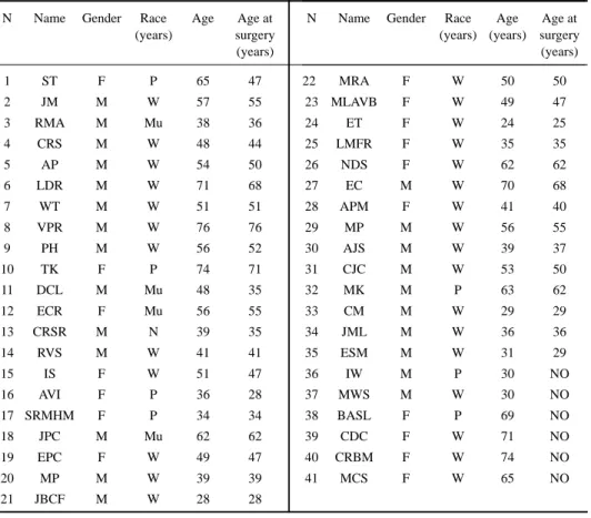
Management strategy of the myasthenic symptoms in TMG was the same as that of thymoma-free MG. There were 24 (58.53%) male and 17 (41.46%) female patients (Table 1). Upon surgery and at first examination of the nonsurgical ones, ages of patients ranged from 25 to 76 years; four (11.42%) were under 30 years old at the time of surgery, the youngest being 25 years old and with MG onset at 24. Twenty-one (60.0%) patients were over 40 years of age and one was 40 at the time of surgery (Table 1).
Table 1. Identification.
N Name Gender Race Age Age at
(years) surgery
(years)
1 ST F P 65 47
2 JM M W 57 55
3 RMA M Mu 38 36
4 CRS M W 48 44
5 AP M W 54 50
6 LDR M W 71 68
7 WT M W 51 51
8 VPR M W 76 76
9 PH M W 56 52
10 TK F P 74 71
11 DCL M Mu 48 35
12 ECR F Mu 56 55
13 CRSR M N 39 35
14 RVS M W 41 41
15 IS F W 51 47
16 AVI F P 36 28
17 SRMHM F P 34 34
18 JPC M Mu 62 62
19 EPC F W 49 47
20 MP M W 39 39
21 JBCF M W 28 28
F, female; M, male; W, white; Mu, mulatto; P, pale; N, negro; NO, non operated.
N Name Gender Race Age Age at
(years) (years) surgery (years)
22 MRA F W 50 50
23 MLAVB F W 49 47
24 ET F W 24 25
25 LMFR F W 35 35
26 NDS F W 62 62
27 EC M W 70 68
28 APM F W 41 40
29 MP M W 56 55
30 AJS M W 39 37
31 CJC M W 53 50
32 MK M P 63 62
33 CM M W 29 29
34 JML M W 36 36
35 ESM M W 31 29
36 IW M P 30 NO
37 MWS M W 30 NO
38 BASL F P 69 NO
39 CDC F W 71 NO
40 CRBM F W 74 NO
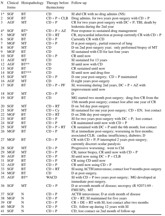
Table 2. Clinical forms, histopathology, therapy, follow-up.
N Clinical Histopathology Therapy before Follow-up
forms thymectomy
1* SGF BT CD SI abd CR with no drug admins (NS)
2 SGF BT CD + P + CLB Drug admins. for two years post-surgery with CD + P
3 AGF MT CD + P CR for two years post-surgery with DC + P; TBI, death by
leukemia during the 2nd year
4* SGF BT* CD + P + AZ Poor response to sustained drug management
5 MGF MT CD + RT CR, myocardial infarction at postop currently CR with CD + P
6 SGF BT CD + P Currently CR with CD
7 MGF MT CD + P D at post-surgery., partial removal of lung
8 SGF MT CD D on 2nd post-surgery year; only performed biopsy of MT
9 MGF BT CD SI sustained with CD for last four years
10 SGF BT CD + P CR until now
11 AGF MT CD SI sustained for 13 years
12 AGF BT** CD SI until now with CD
13 SGF BT CD CR sustained until now
14 SGF BT*** CD SI until now and drug-free
15 SGF MT CD + P D one year post-surgery; CD + P maintained
16 AGF MT CD D eight years post-surgery
17 SGF BT CD + P + PF SI, worsening during 2nd years, DC + P + AZ with improvement until now
18 SGF MT CD + P D
19 SGF BT CD MG started two month post-surgery; drug-free CR from the
15th month post-surgery; contact lost after one year of CR
20 SGF MT CD + RT D on 3rd day post-surgery
21 SGF MT CD + Cy SI sustained for one year post-surgery; CD + EN; lost contact
22 MGF BT CD + RT D on 20th day post-surgery
23 SGF BT CD + P SI for two years post-surgery with DC + P; lost contact
24 SGF BT CD + P CR maintained until now with CD + P
25 SGF MT CD + P + RT CR sustained with DC + P until the 6th month; lost contact 26 MGF BT CD + P SI at immediate post-surgery; worsening in first months;
associated CLB; cardiac insufficiency, diabetes; D
27 MGF BT CD + P CR with CD + P; P interrupted 2 years post-surgery;
currently discreet ocular paralysis
28 SGF MT CD + P Progressive worsening; went to Chl
29 MGF MT CD + P CR; tumor biopsy, CR until now with CD + P
30 AGF BT CD + P SI until now using DC + P + CLB
31 SGF BT CD + P CR using CD until now
32 AGF BT CD + P CR until now using CD + P
33 SGF BT CD + RT SI using ACTH intravenous; contact lost 9 months post-surgery
34 MGF BT CD D at post-surgery.
35 AGF BT* WACD SI with CD + P two years post-surgery; MG developed at
immediate post-surgery.
36 SGF MT CD + P D at seventh month of disease; necropsy (R 92071/69
-FMUSP); MT
37 SGF N CD + P ACTH intravenous; D at sixth month of disease
38 MGF N CD + P CD + RT; SI maintained for five years
39 OF N CD + P CR + DE + RT with SI; lost contact after two months
40 MGF N CD + P CD; follow-up during 12 years with SI
41 SGF N CD + P CD; lost contact on 2nd month of follow-up
Among those submitted and not submitted to thymectomy, 23 (56.09%) developed severe generalized clinical form (SGF - Osserman-IV), six (14.63%) had the generalized accentuated form (AGF - Osserman-IIb), 10 (24.39%) progressed with mild generalized form (MGF - Osserman-IIa), one (2.43%) presented with the ocular form (OF - Osserman-I) and thus remained during the short term (two months) follow-up; one patient of the surgical group developed MG after thymomectomy (Case 35) (Table 2). The clinical forms that of Osserman7 modified were adopted.
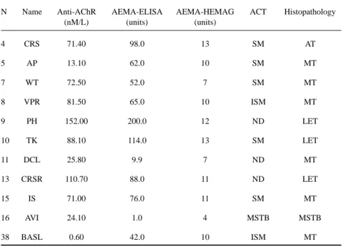
Diagnosis was mainly based on clinical examination, on the CT scans of the thorax and anatomopathologic tests of the surgical specimen of the thymus in all the thymectomized patients. A patient of the nonsurgical group (Case 36) and another of the surgical group (Case 34) were submitted to necropsy. For diagnostic support in 10 of the patients submitted to surgery and in one of the nonsurgical group (Case 38) immunologic investigations were performed: tests for the titering of antibody against human muscular antigens (AEMA)8 and dosage of antibody against acetylcholine receptor (anti-AChR)9. A comparative study of the tests with the findings of the axial computerized tomography (ACT) and thymus histopathology, were undertaken (Table 3) 9. Thymectomy was performed following the partial or total sternum-splitting approach. In one case of a very large thymoma (case 35) which invaded the right hemithorax, the chosen procedure was a posterior lateral thoracotomy.
Pathological examination of the thymus - 22 (62.85%) of patients had benign (BT) and 13 (37.14%)
malignant thymoma (MT). In one patient (Case 35) tumor was large and invaded the right hemithorax. Malignancy criterion was based upon the thymoma invasiveness. In one female patient (Case 12) adjacent to the tumor ectopic thyroid tissue and hyperplasic thymic tissue were found. In one patient (Case 14) adjacent to a lymph epithelium thymoma and hyperplasic thymic tissue was found and in another patient (Case 4), besides thymoma an atrophic thymus was found.Thymomas were lymphocytic in 43%, lymphoepithelial in 29%, epithelial in 22% and spindle cells in 6%. The comparative morphological and histologic evaluation between TMG and thymomas without MG (TWMG) did not show differences.
Tabela 3. Immunological, morphologic and histopathological data in TMG
N Name Anti-AChR AEMA-ELISA AEMA-HEMAG ACT Histopathology
(nM/L) (units) (units)
4 CRS 71.40 98.0 13 SM AT
5 AP 13.10 62.0 10 SM MT
7 WT 72.50 52.0 7 SM MT
8 VPR 81.50 65.0 10 ISM MT
9 PH 152.00 200.0 12 ND LET
10 TK 88.10 114.0 13 SM LET
11 DCL 25.80 9.9 7 ND MT
13 CRSR 110.70 88.0 11 ND LET
15 IS 71.00 76.0 11 SM MT
16 AVI 24.10 1.0 4 MSTB MSTB
38 BASL 0.60 42.0 10 ISM MT
RESULTS
In the group of 41 patients with TMG, there was a significant prevalence of males (58.5%) over females (41.4%). Prevailing age was over 30 years at MG onset, with a predominance of those over 40 years.
Severe clinical forms (Osserman-IV) prevailed in 70.73% of patients, against 24.39% of the mild forms Osserman-IIa); only one patient (Case 39) had OF (Osserman-I)and another (Case 35) presented with MG at postoperative stage.
Myasthenic crises (MC) occurred in 20% of patients submitted to surgery, in their majority (12.50%) in the immediate postoperative. In the group of patients not submitted to surgery, two (cases 36 and 37) evolved in a relatively short term to MC six and seven months after the start of myasthenic symptoms.
In the group of nonsurgical patients X-ray scans strongly suggested thymoma, except in one (Case 36) in which diagnosis was reached at by anatomopathologic results. Another patient of this group (Case 38) presented with positive immunologic assays (AEMA-ELISA and AEMA-HEMAG) for TMG, in agreement with the ACT, exhibiting a solid invasive mass.
In the 10 patients with TMG, submitted to surgery and with titers of anti-AChR, all had higher antibody levels ranging from 13.0 nM/L a 152.00 nM/L. Antibody serum levels against human skeletal muscle (AEMA-ELISA e AEMA-HEMAG) were increased in all of the patients, except in two (cases 11 and 16), whose titers were low. A variation from 1.0 to 200 units on the AEMA-ELISA and from 4 to 13 units in AEMA-HEMAG was found (Table 3).
Follow-up from two months to 18 years of 68.29% of surgical and nonsurgical patients and of 71.42% of those in the first group from two to 18 years discloses the following results for those submitted to thymectomy: 23 patients (65.70%) evolved with complete remission (CR) or significant improvement (SI); 62.85% of patients were 40 years old or more at the time of surgery and, in this subset, 60.86% evolved with SI or CR; 28.57% were not yet 40 years old and 91.66% had SI or CR; in this subset, six (60%) had MT, two (Cases 16 and 21) were under 30 years of age and one of them (Case 16) survived with short periods of SI for eight years and had pulmonary tuberculosis; in the group of those submitted to surgery with 40 years of age or more, seven (20%) had MT and only two survived: one (Case 5) has for the last five years been in CR with CD and prednisone (P); one (Case 29), solely submitted to biopsy of the tumor, has for two years been in CR with CD + P; this patient has been examined 2.5 years after surgery and is still under CR, keeping up the same therapeutic protocol.
In the group of patients not submitted to surgery two (Cases 38 and 40) had a lengthy evolution with the MGF, follow-up from five to 12 years respectively; these patients were on CD and in only one radiotherapy (RT) was used (Case 38). The female patient with OF (Case 39) had a favorable response to dexamethasone at the onset of treatment, however after a two months follow-up she did not return.
DISCUSSION
Although thymectomy is a very important approach to management of MG, it remains quite controversial10,11. However, its indication in presence of thymoma is consensual2,6,11-14. In an effort to lessen risks of local infiltration and to maintain CR of MG, complete excision of the tumor, of the thymic tissue and of other non vital adjacent structures is recommended in invasive thymoma, but such objectives are not always met6,13,15-17.
In the series under study there was a significant prevalence of thymomas in the male gender and in patients with over 40 years of age. This result corresponds to the age group of patients with TMG from 50 to 60 years of age 4. Age influenced results in the group submitted to surgery, as a higher number of patients (91.66%), under 40 years of age had favorable responses. Nevertheless, for both age groups, early surgery is of vital importance, especially in cases of invasive thymoma.
In almost all cases, thymomas come with MG, nevertheless in some there are no evident clinical findings and MG will only develop after a variable time lapse after thymectomy2,7,18,19.
In the current series only one patient (Case 35) developed post-surgery MG. As a rule TMG develops with a severe or pronounced symptomatology and with more frequent MC, although it may evolve with mild symptoms, and seldom exhibit the ocular form2,5. In this series there was a significant prevalence of the severe forms (SGF and AGF) over the mild ones and only one of the OF (Case 39).
Presence of MT in the group submitted to surgery did not always shorten the survival rate or cause a worsening of MG, as 14.28% of the patients had an adequate survival with SI or CR of MG for two to five years or more.
In the current series a significant prevalence of BT over MT was found, which agrees with other authors5,20-22, although some13,15 have reported a high incidence of MT.
In the current paper, for the identification of thymoma malignancy, its invasiveness was adopted as the most significant factor22. From a histologic point of view criteria for the identification of thymoma malignancy are poor, the spreading of metastases is rare and in general takes place in the pleura and lungs13,23,24. In most cases thymomas start in the thymic site and exceptionally develop in an ectopic site such as the lung23.
Diagnosis of thymomas is reached at by clinical examination and ACT supported by immunologic tests3,5,8,9,25-33. Using such procedures most thymomas are detected prior to surgery21. Immunologic assays may lead to suspect thymoma even prior to X-rays detection3,4,8. This is possible because of the high incidence of both antibodies anti-receptors such as anti-AChR and anti-ryanodine as well as antibodies against proteins (antigens) of skeletal muscle, in TMG3-5,8,25-28.
Almost all of the TMG patients had autoantibodies against skeletal muscle class IgG 5,8,26; they may be found in only 10%-15% of MG and atrophic thymus is rarely detected in patients with MG and thymic hyperplasia 5.
Antibodies against myofibrils (myosin, actin and actimyosin) are found in 85% of TMG patients 4. In the present series, AEMA enabled to detect antibodies against skeletal muscle antigen in 11 patients and especially, when to this test was added the enzymeimmunoassay (AEMA-ELISA). A positive result was reached at in over 80% of patients. This test is highly sensitive and specific for TMG diagnosis.
Numerous efforts have been made to correlate prognosis to histopathology or with associated diseases that would be more frequent in TMG35,36.
In conclusion, TMG has a relatively favorable prognosis with regard to the survival rate. Such prognosis depends mainly on the thymoma characteristics; if invasive or not, on its total and early excision, on MG response to treatment and to an eventual associated pathology.
In the series under study the number of patients requiring long term management with steroid immunosuppressants was significantly high. The utilization of P associated to CD led to SI or CR of the myasthenic symptomatology in most patients. Some authors37 recommend RT in all patients with invasive thymomas and, also in those with a non excisable tumor. Others15 use this therapeutic approach almost exclusively when the tumor has not been totally excised. Bernatz et al.35 and Wilkins et al.36 report a survival rates of five years in 50% and 63% respectively, with the combination of surgery-radiotherapy. Such association is considered the best treatment strategy for invasive thymomas24.
How we did not show any differences about morphologic and histologic changes in TMG and TWMG, is possible that the development of MG can depend on the presence of protein p-153 (153 Kd) antigen in epithelial cells of cortical and well-differentiated thymomas associated myasthenia gravis37.
REFERENCES
1. Lisak RP, Barchi RL. Myasthenia gravis. Philadelphia: Saunders, 1982: 121-125. 2. Oosterhuis HJGH. Myasthenia gravis. Edinburgh: Churchill-Livingstone, 1984.
3. Mygland Å, Kuwajima G, Mitoshi K, Tysnes O, Aarli JA, Gilhus NE. Thymomas express epitopes shared by the ryanodine receptor. J Neuroimmunol 1995;62:79-83.
4. Penn AS, Rowland LP. Disorders of the neuromuscular junction. In Rowland LP. Merritt’s textbook of neurology. 9.Ed. Philadelphia: Williams & Wilkins, 1995:754-762.
5. Aarli JÁ. Myasthenia gravis and thymoma. In Lisak RP. Handbook of myasthenia gravis and myasthenic syndromes. New York, 1994:207.
6. Newsom-Davis J. Myasthenia gravis. In Matthews WB, Glaser GH. Recent advances in clinical neurology 4. Edinburgh: Churchill-Livingstone, 1984:1-18.
7. Osserman KE. Myasthenia gravis. London: Grune, 1958.
8. Marchiori PE. Identificação de elementos imunológicos e morfológicos na miastenia grave: valor diagnóstico para o timoma. Tese, Faculdade de Medicina da Universidade de São Paulo, São Paulo, 1987.
9. Marchiori PE. Anticorpo anti-receptor de acetilcolina em miastenia grave. Tese, Faculdade de Medicina da Universidade de São Paulo, São Paulo, 1985.
10. Rowland LP. Symposium on therapeutic controversies: myasthenia gravis. Trans Am Neurol Assoc 1978;103:277-298. 11. Rowland LP. Controversies about the treatment of myasthenia gravis. J Neurol Neurosurg Psychiatry 1980;43:644-649. 12. Assis JL. Miastenia grave. São Paulo: Sarvier, 1990.
13. Braitman H, Li W, Herrman C Jr., Mulder GD. Surgery for thymic tumors. Arch Surg 1971;103:14-16.
14. Genkins G, Sivak M, Tartter PL. Treatment strategies in myasthenia gravis and related disorders. Ann NY Acad Sci 1993;681:603-608.
15. Slater G, Papatestas AE, Genkins G, Kornfield P, Horowitz H, Bender A. Thymomas in patients with myasthenia gravis. Ann Surg 1978;188:171-174.
16. Scadding GK, Harvard CWH, Lange MJ, Domeb I. The long-term experience of thymectomy for myasthenia gravis. J Neurol Neurosurg Psychiatry 1985;48:401-406.
17. Simpson JA. An evaluation of thymectomy in myasthenia gravis. Brain 1958;81:112-144. 18. Kimura J, van Allen MW. Pos-thymectomy myasthenia gravis. Neurology 1967;17:413-420.
19. Rowland LP. Myasthenia gravis. In Goldensohn ES, Appel SH. Scientific approaches to clinical neurology. Philadelphia: Lea, 1977: 1518-1554.
20. Castleman B. The pathology of the thymus gland in myasthenia gravis. Ann NY Acad Sci 1966;135:495-505. 21. Keesey J, Bein ME, Mink J, et al. Detection of thymoma in myasthenia gravis. Neurology 1980;30:233-239. 22. Zambon AA. Timectomia em miastenia grave: avaliação de 150 pacientes com estudo histopatológico do timo. Tese,
Faculdade de Medicina da Universidade de São Paulo, São Paulo, 1991.
23. Deron HA, Schneider MJ, Persky L. Myasthenia gravis: a clinical and pathological study of a case associated with a primary mediastinal thymoma and solitary intrapulmonary thymoma. N Engl J Med 1950;243:478-486.
24. Ohmi M, Ohuchi M. Recurrent thymoma in patients with myasthenia gravis. Ann Thorac Surg 1990;50:243-247. 25. Aarli JA, Thunold S. Serological detection of thymoma in myasthenia gravis. Eur Neurol 1981;20:38-387.
27. Baron RL, Lee JKT, Sagel SS, Levitt RG. Computed tomography of the abnormal thymus. Radiology 1982;141:127-134. 28. Gautel M, Lakey A, Barlow DP, et al. Titin antibodies in myasthenia gravis: identification of a major immunogenic region
of titin. Neurology 1993;43:1581-1585.
29. Janssen RS, Kaye AD, Lisak RP, Schatz NJ, Argel PA, Savino PJ. Radiologic evaluation of the mediastinum in myasthenia gravis. Neurology 1983;33:534-539.
30. Mink JH, Bein ME, Sukov R, et al. Computed tomography of the anterior mediastinum in patients with myasthenia gravis and suspected thymoma. Am J Roentgenol 1978;130:239-246.
31. Mygland Å, Tysnes OB, Matre R, Volpe P, Aarli JA, Gilhus WE. Ryanodine receptor autoantibodies in myasthenia gravis patients with a thymoma. Ann Neurol 1992;32:589-591.
32. Mygland Å, Aarli JA, Matre R, Gilhus NE. Ryanodine receptor antibodies related to severity of thymoma-associated myasthenia gravis. J Neurol Neurosurg Psychiatry 1994;57:843-846.
33. Stump XMGRC. Tomografia axial computadorizada do mediastino anterior. In Assis JL. Miastenia Grave. São Paulo: Sarvier, 1990:47-52.
34. Pescarmona E, Rendina ER, Venuta F, Ricci C, Ruco LP, Barone CD. The prognostic implication of thymoma histologic subtyping. Am J Clin Pathol 1990;93:190-195.
35. Bernatz PE, Harrison EG, Clagett OT. Thymoma: clinicalpathologic study. J Thorac Cardiovasc Surg 1961;42:424-444. 36. Wilkins EW Jr, Edmunds LH Jr, Castleman B. Cases of thymoma at the Massachusetts General Hospital. J Thorac Cardiovasc
Surg 1966;52:322-330.