Surgical Mitral Valve Repair in Children with Rheumatic Fever
Andréa Rocha e Silva
1, Gesmar Volga Haddad Herdy
1, Alan Araujo Vieira
1, Luiz Carlos Simões
2Universidade Federal Fluminense (UFF)1, Niterói, Rio de Janeiro; Instituto Nacional de Cardiologia (INC)2, Rio de Janeiro, RJ - Brasil
Summary
Background: Mitral repair is well accepted in children with rheumatic fever.
Objective: To analyze the outcomes of surgical mitral repair in children with rheumatic lesions after four years of follow-up.
Methods: Retrospective study of 40 patients younger than 18 years, who underwent surgery in the National Institute of Cardiology (Rio de Janeiro) between January 1998 and January 2003. The echocardiographic degree of mitral regurgitation; surgical technique used; pre and postoperative functional class; patient outcome; need for valve replacement; and deaths were analyzed.
Results: Twenty one patients (52.5%) were females. Severe mitral regurgitation was observed in 32 patients (80%) and moderate in eight (20%). Three immediate deaths occurred (7.5%). Three months after surgery, echocardiography showed no valve regurgitation or mild regurgitation in 35 of 37 cases (94.6%) patients, and severe regurgitation in two (5.2%). Thirty three cases (82.5%) were in functional class III or IV in the preoperative period, and three months after surgery all the 37 cases (100%) were in functional class I or II. The differences between the degree of mitral regurgitation and functional class in pre and postoperative periods were statistically significant (p<0.01). Seven (19%) patients underwent heart valve replacement before four years of follow-up.
Conclusion: Mitral valve repair showed favorable results in most of the cases as regards the degree of mitral regurgitation and the pre and postoperative functional class. Only 19% of the patients required surgical valve replacement before four years of follow-up. (Arq Bras Cardiol 2009; 92(6) : 400-404)
Key words: Mitral valve/surgery; mitral valve insufficiency; rheumatic fever.
Mailing address: Gesmar Volga Haddad Herdy •
Travessa Antonio Pedro, 10/301, 24.230-030, Niteroi, RJ - Brasil E-mail: [email protected]
Manuscript received July 29, 2008; revised manuscript received October 16, 2008; accepted October 24, 2008
Introduction
The incidence of rheumatic fever among children, adolescents and young adults is of 0.3% to 3% of the individuals1. There are few Brazilian studies available on the
prevalence of this disease. Among children from public schools of the city of Belo Horizonte this rate was 3.6/1,0002. In Brazil,
rheumatic fever accounts for 30% of the cardiac surgeries in general, especially for mitral valve regurgitation3.
Mitral regurgitation results from annulus dilatation and changes in the papillary muscles, chordae tendineae and leaflets4-7. The available prostheses lead to complications
which are difficult to be controlled, especially calcification and rupture in bioprostheses, thromboembolism in mechanical prostheses, and the need for continuous use of anticoagulation and its monitoring with periodic laboratory tests8. In lower age
ranges, prosthesis replacement is necessary because of height and weight growth. Based on these facts, surgeons have used reconstructive techniques in the treatment of mitral valve regurgitation with increasing frequency9,10.
The objectives of the present study were to analyze the results of mitral valve repair in patients with rheumatic fever, to evaluate the patients’ outcome, and to determine the factors that could influence the need for valve replacement before four years of follow-up.
Methods
aorta and venae cavae were cannulated, and extracorporeal circulation (ECC) was established. All patients underwent moderate hypothermia (28o C), continuous total aortic clamp,
and myocardial protection with blood cardioplegia at 4o C. The
mitral valve was approached via the left atriotomy, and both mitral valve cusps were preserved in all patients.
Echocardiographic studies for the analysis of mitral regurgitation were performed in a SONUS instrument (Pharmaceutical-Abbott Laboratories, USA), and regurgitation was rated as mild, moderate or severe, according to the characteristics of the regurgitant jet. The main surgical techniques used for valve repair were: mitral ring placement, quadrangular resection of the posterior leaflet, chordal shortening, chordal transfer, and commissurotomy. The patients were followed-up for at least four years postoperatively.
Data were described as relative frequencies (percentage), mean, median, minimum and maximum values, and the groups were compared using the Chi square test. Fisher’s correction was used when indicated. The possible risk factors for heart valve replacement before four years of clinical follow-up were analyzed using simple logistic regression. Statistical significance was set at 95%.
The cases studied are filed in the NIC database for the long-term follow-up and further description of the outcomes.
This study was approved by the Research Ethics Committee of NIC under number 0181/26.11.07.
Results
Twenty one patients (52.5%) were females. The age at bout of rheumatic fever and at surgery ranged from three to 14 years (median of eight years), and from four to 17 years (median of 12 years), respectively. The difference between the age at the first bout and at surgery ranged from zero to 10 years (median of three years) (Table 1).
In the first visit prior to surgery, 12 patients (30%) were not taking any medication, 16 (40%) were on four or more medications, and the remaining twelve cases were on continuous use of one to three medications. The drugs more frequently used were: furosemide, spironolactone, captopril, hydralazine, and penicillin G benzathine.
The degree of mitral regurgitation was analyzed by echocardiography in the preoperative period and was classified as mild, moderate or severe, according to the regurgitant jet. Mitral regurgitation was severe in 32 patients (80.0%) and moderate in eight (20.0%). Immediate echocardiographic analysis was performed in all patients, except for two, due to death. The analyses showed that 20 of the 38 patients
Table 1 – Demographic characteristics of the patients undergoing mitral valve repair (n=40)
Variables Minimum value Maximum value Median
Age at bout 3 14 8
Age at surgery 4 17 14
Time between bout and surgery 0 10 3
(52.6%) had mild or no valve regurgitation, and 18 (47.4%) had moderate regurgitation. Another death occurred three days after surgery.
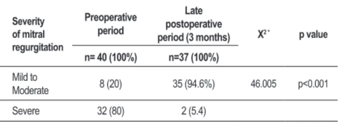
The echocardiographic studies performed three months after surgery in the 37 patients that survived showed that 19 (51.3%) had mild or no valve regurgitation, 16 (43.3%) had moderate regurgitation, and two (5.4%) had severe regurgitation (the latter presented a new bout of rheumatic fever). Therefore, 35 patients (94.6%) presented mild or moderate mitral regurgitation in the postoperative period. Statistically significant difference was observed (p<0.001) (Table 2).
The length of hospital stay ranged from four to 103 days (median of 12). ECC time ranged from 50 to 220 minutes, with mean of 120±36 minutes, and time of anoxia from 35 to 170 minutes, with mean of 93±32 minutes. The mean length of stay in the children’s postoperative care unit ranged from one to 60 days (median of four) (Table 3).
The main surgical techniques used were: mitral ring placement (n= 28), quadrangular resection of the posterior leaflet (n=17), chordal shortening (n=23), chordal transfer (n=5), and commissurotomy (n=11). Eleven patients (27.5%) required only one surgical technique, whereas 29 patients (72.5%) required two or more techniques (Table 4).
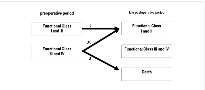
The preoperative NYHA functional class ranged from II to IV: FC II (n=7) 17.5%, FC III (n=17) 42.5%, and FC IV (n=16) 40.0%, that is, 33 patients (80.5%) were in functional class III and IV in the preoperative period. In the outpatient follow-up three months after surgery, all the 37 patients (100%) became FC I (n=28) and II (n=29). Statistically significant difference was observed (p<0.001) (Table 5; Figure 1)
Table 2 – Preoperative and late postoperative severity of mitral regurgitation as assessed by echocardiography in the patients
Severity of mitral regurgitation
Preoperative period
Late postoperative
period (3 months) X2 * p value
n= 40 (100%) n=37 (100%) Mild to
Moderate 8 (20) 35 (94.6%) 46.005 p<0.001
Severe 32 (80) 2 (5.4)
* Cochrane’s method and Fisher’s correction.
Table 3 – Length of hospital stay and operative time
Maximum time Mean±SD Median Minimum
value Maximum value Days of hospital
stay 20±20 dias 12 4 103
Time of ECC * 120±36 min 120 50 220
Time of anoxia * 93±32 min 97 35 170
Days of IPO 6±9 dias 4 1 60
Table 4 – Most frequently used surgical techniques (n=40)
Technique n %
Mitral ring placement (Carpentier’s) 28 70
Chordal shortening 23 57.5
Quadrangular resection 17 42.5
Commissurotomy 11 27.5
Chordal transfer 5 12.5
More than one technique was used in several cases.
Table 5 – Pre (n=40) and postoperative (n=37) variation of NYHA functional class
Assessment of the Functional Class
Preoperative Postoperative
X2* p value
n = 40 n = 37
Functional class
I and II 7 (17.5%) 37 (100%)
Functional class
III and IV 33 (80.5%) 0 54.340 p<0.001
* Cochrane’s method and Fisher’s correction.
Table 6 – Possible risk factors for surgical valve replacement before four years of mitral valve repair (n=7)
Variable analyzed OR* IC 95% p
Functional class** 2.61 0.25 – 24.38 0.397
Medications*** 1.08 0.25 – 4.63 0.914
Perfusion∞ 1.16 0.20 – 6.80 0.864
Surgery▪ 2.44 0.56 – 10.55 0.231
Anoxia∞ 2.18 0.47 – 10.05 0.314
Amines◊ 1.47 0.32 – 6.83 0.620
OR - odds-ratio; 95% CI, 95% conidence interval; * Odds-ratio - adjusted for
gender, age, functional class, medications, time of perfusion, operative time, time of anoxia, use of vasoactive amines; ** - New York Heart Association functional class heart failure; *** - Number of medications used per patient at
the moment of surgery; ∞ - Time in minutes; ▪ - Number of surgical techniques used by the surgeon; ◊ - Number of patients using vasoactive amines.
Figure 1 - Progression of NYHA functional class in patients undergoing mitral repair
Transesophageal echocardiography was performed in 28 patients (70.0%) during surgery. Blood products were used in 21 cases (52.5%). Vasoactive amines were necessary in 26 patients (65.0%) in the postoperative period. Three immediate deaths occurred (7.5%): one in the operation room, one 24 hours after surgery, and one three days after surgery (two patients of cardiogenic shock and one of coagulation disorders).
Heart valve replacement was required in eight patients (20.0%), seven of whom with less than four years of mitral repair (Table 6).
Risk factors that could affect the outcome of patients in relation to the mitral valve replacement before four years, and whose analysis is shown in Table 6, were not statistically significant and included functional class and degree of pre and postoperative mitral regurgitation; number of medications used by the patients at the moment of surgery; time of perfusion and anoxia; number of surgical techniques used; and need for vasoactive amines in the immediate postoperative period.
Discussion
Mitral valve repair is a technique universally accepted as superior to valve replacement, especially in children, in whom further replacements may be necessary as they grow6,7,11. The
risk of endocarditis, and the poorer preservation of the ventricular function, mitral valve repair is superior to mitral valve replacement12. An important advantage of the surgical
reconstruction technique is the low thrombogenicity of the repaired valve, which eliminates the need for anticoagulation therapy. Absence of thromboembolism after mitral repair is reported in 87% to 99% of the cases after five years. Most of the embolic manifestations are cerebrovascular events10-12.
The following valve repair techniques were used: mitral ring placement, quadrangular resection, chordal shortening, commissurotomy and chordal transfer, with the aim of preserving both the mitral apparatus and the ventricular function. In rheumatic fever, valve reconstruction is technically more difficult and the mid and long-term outcomes may be influenced by new bouts of the disease6,9. Initial studies
presented techniques which addressed only the valve annulus treatment13. One of the largest personal experiences
in conservative procedures in the mitral valve is that of Carpentier’s14, with 1,421 patients with mitral regurgitation.
The techniques used in his series were partial resections, chordal shortening or transfer, reinsertion of the papillary muscles, and annuloplasty with rigid ring.
In the present study, 39% of the patients underwent surgery with significant impairment of the whole valvular structure and myocardial dysfunction. These situations, which are not uncommon in our midst (surgical treatment performed when the patient already presents ventricular dysfunction and/or significant clinical symptoms), mainly result from social factors and difficulties found in our health system. However, mitral repair should be indicated early, on the basis of the severity of regurgitation and on the degree of valve impairment; thus, favorable results can be achieved with the conservative treatment, even when symptoms are scarce or absent15. For
valve reconstruction, perfect knowledge of the anatomy and multiplicity of techniques available is required of the surgical team. Additionally, excellent visualization of the valve is important, especially of the subvalvular plane, to enable good assessment of the chordae tendineae and papillary muscles. If necessary, these structures are shortened, in an attempt to achieve good cusp coaptation. In the Department of Pediatric Cardiovascular Surgery of NIC, surgeons perform papillary muscle shortening by means of a cross-sectional wedge resection because they believe this technique permits more homogeneous shortening of the chordae tendineae. In addition to this technique, they also use mitral ring placement, quadrangular resection, commissurotomy, and chordal transfer.
Annuloplasty was the single procedure used in only eight of the patients studied (20%), thus demonstrating that in most of the cases (80%) the association with more than one technique was necessary, and in many occasions with modifications of the initial planning, a situation that has also been described by other authors6,10,16. Mitral annulus dilatation is present
in almost all patients with mitral regurgitation. Thus, ring implantation offers more stability in valve reconstruction, preventing further dilatations of the posterior cusp as suggested by Carpentier14, according to whom remodeling the valve
with repair is always important for the long-term outcomes. Deloche et al demonstrated excellent durability of mitral repair
using Carpentier’s technique, and only 6.2% of their series required further mitral valve replacement9. In our study, 19%
underwent early mitral valve replacement.
Mitral regurgitation may be secondary to multiple lesions in the commissures, in the leaflets, and in the subvalvular apparatus. As previously mentioned, mitral annulus dilatation is observed in all patients. This is due to the displacement of its posterior portion, since the anterior portion is limited by the right and left fibrous trigones of heart. According to the literature, repair is considered a good option, because it permits the conservation of the native valve cusps and of the subvalvular apparatus, thus preserving the left ventricular geometry and ventricular function10.
In-hospital mortality in the present study was 7.5%. The three patients who died were in functional class IV, and two of them were in the acute phase of the disease. This mortality rate may be considered high if compared with those of most of the authors, which range from 0 to 4.8%17-20. Carpentier reported his 10-year experience with
repair, with an in-hospital mortality of 4.2% in adults and children14. Surgical mortality is higher in the acute phase of
rheumatic feverwhen operation is indicated in patients with severe valve dysfunction and ineffective clinical treatment. Even in this group, repair may provide better results than valve replacement, as it preserves the whole subvalvular apparatus, which results in better postoperative ventricular function12,19.
In our case series, reoperation with indication of valve replacement before four years was necessary in seven (19%) of the 37 cases, a very high rate in comparison with that of other authors. Pomerantzeff et al21 reported
reoperation-free survival in 70% after 17 years21. Patients with rheumatic
valve disease have had a high incidence of reoperation, as demonstrated by Deloche et al9. Reoperation rates range
from 8% to 10%7,18. Additionally, the outcomes also depend
on the status of the valve at the moment of reconstruction, that is, the more severely it is damaged, the worse the outcome16. Gillinov et al reported reoperation-free survival
in 93% after 10 years22; the reason for the need of a new
surgery was new bouts of rheumatic fever in most of the patients. In Machado et al18 case series, 72.6% of the cases
were free from reoperation after 188 months. The major causes of failure in rheumatic mitral repair are wrong indications, use of inadequate techniques, and progression of the rheumatic valve disease7,16.
Transesophageal echocardiography has been established as the most adequate method for the assessment of immediate outcomes, guiding the need for valve replacement in the cases of failure in mitral repair or significant residual regurgitation10. Murad et al6 reported
the use of transesophageal echocardiography in only 4% of their surgeries. In the present study, transesophageal echocardiography was used in most of the patients (68.3%) during the surgical procedure. Also, we observed improved regurgitation in 94.6%, a result similar to that of other Brazilian studies16,18.
References
1. World Health Organization. Rheumatic fever and rheumatic heart disease. Report of a WHO Expert Consultation .Geneve, 29 October-1 November. Geneve:WHO, 2004.
2. Meira ZM, Castilho SR, Barros MV. Prevalência de febre reumática em crianças de escola da rede pública de Belo Horizonte. Arq Bras Cardiol. 1995; 65: 331-4.
3. Ministério da Saúde (Homepage na Internet). Secretaria Executiva. DATASUS. Informes hospitalares (SIH–Sus) Consulta eletrônica. [Acesso em 2006 jan 09]. Disponível em http://www.datasus.gov.br.
4. Herdy GVH, Pinto CAM, Carrinho M, Olivaes MC, Medeiros CC, Souza D. Estudo clínico e ecocardiográfico das alterações do aparelho mitral em crianças com cardite reumática grave: aspecto de prolapso ou ruptura. Arq Bras Cardiol.1996; 66 (3): 125-8.
5. Herdy GVH, Pinto CAM, Olivaes MC, Carvalho EA, Tchou Y, Herdy AH. Rheumatic carditis treated with high dosis of methylprednisolone (pulsetherapy): results in 70 children over 12 years. Arq Bras Cardiol. 1999; 72 ( 5): 604-6.
6. Murad H, Goes Ec, Pinheiro AA, Azevedo JA, Noronha AP. Surgical treatment of mitral valve insufficiency by valve repair. Rev Bras Cir Cardiovasc. 2002; 17 (4): 299-306.
7. Talwar S, Rajesh MR, Subramanian A, Saxena A, Kumar AS. Mitral valve repair in children with rheumatic heart disease. J Thorac Cardiovasc Surg. 2005; 129 (4): 875-9.
8. Ren JF, Aksut S, Lighty GW. Mitral valve repair is superior to valve replacement for the early preservation of cardiac function. Am Heart J. 1996; 131 (5): 974-81.
9. Deloche A, Carpentier A, Jebara VA, Chavaud S, Fabiani JN, Dreyfus G. Mitral valve repair with Carpentier’s techniques :a third decade. In 81st Annual meeting of the American Association for Thoracic Surgery. 2001; San Diego.
10. Wood AE, Healy DG, Nolke L, Duff D, Walsh K. Mitral valve reconstruction in a pediatric population: late clinical results and predictors of long-term outcome. J Thorac Cardiovasc Surg. 2005; 130 (1): 66-73.
11. Hillman ND, Tani LY, Veasy G, Lambert LL, Russo GB, Doty DB. Current
status of surgery for rheumatic carditis in children. Ann Thorac Surg. 2004; 78 (4): 1403-8.
12. Skoularigis J, Sinovich V, Joubert G, Sareli P. Evaluation of the long-term results of mitral valve repair in 254 young patients with rheumatic mitral regurgitation. Circulation. 1994; 90 (5): 167-74.
13. Lillehei CW, Levy MJ. Mitral valve replacement with preservation of the papillary muscles and the chordae tendinea. J Thorac Cardiovasc Surg. 1964; 47 (3): 532-43.
14. Carpentier A. Cardiac valve surgery: the “French correction”. J Thorac Cardiovasc Surg. 1983; 86 (2): 323-37.
15. Sousa Uva M, Dreyfus G, Rescigno G, al Aile N, Mascagni R, La Marra M, et al. Surgical treatment of asymptomatic and mildly symptomatic mitral regurgitation. J Thorac Cardiovasc Surg. 1996; 112 (5): 1240-9.
16. Volpe MA, Braile DM, Vieira RW, Rossi D, Souza DRS. Mitral valve repair with a malleable bovine pericardium ring. Arq Bras Cardiol. 2000; 75 (5): 389-96.
17. Provenzano Jr SC, Sá MPL, Bastos ES, Murad H, Gomes EC. Plastia valvar mitral na doença cardíaca reumática e degeneração mixomatosa: estudo comparativo. Rev Bras Cir Cardiovasc. 2002; 17 (1): 24-34.
18. Machado VHS, Gregori Jr F. Avaliação cardiológica tardia de crianças com insuficiência mitral reumática submetidas a cirurgia reconstrutora com anel de Gregori. Arq Bras Cardiol. 2005; 85 (6): 403-11.
19. Kumar AS, Rao PN, Saxena A. Results of mitral valve reconstruction in children with rheumatic heart disease. Ann Thorac Surg. 1995; 60 (4): 1044-7.
20. Gunther T, Mazzitelli D, Schreiber C. Mitral valve replacement in children under 6 years of age. Eur J Cardio-Thorac Surg. 2000; 17 ( 4): 426-30.
21. Pomerantzeff PMA, Brandão CMA, Faber CM, Fonseca MH, Puig LB, Grinberg M, et al. Plástica da valva mitral: resultado aos 17 anos de experiência. Rev Bras Cir Cardiovasc. 1999; 14 (3): 185-90.
22. Gillinov AM, Cosgrove DM, Lytle BW, Taylor PC, Stewart R, McCarthy PM, et al. Reoperation for failure of mitral valve repair. J Thorac Cardiovasc Surg. 1997; 113 (3): 467-75.
verified in the assessment at the third postoperative month, however with a high need for reoperation before four years of follow-up.
Potential Conflict of Interest
No potential conflict of interest relevant to this article was reported.
Sources of Funding
There were no external funding sources for this study.
Study Association