Case Report
Key words
Fistula; hemoptysis; aorta, thoracic / surgery.
Background and objectives: In patients with hemoptysis and a history of aortic surgery, the possibility of aortobronchopulmonary fistula must always be considered. The objective of this study was to report a rare case of hemoptysis due to aortobronchopulmonary fistula in the late postoperative period of aortic surgery.
Case report: Female patient, 34 years, surgical correction of aortic coarctation, presenting massive hemoptysis. The echocardiogram disclosed a pseudoaneurysm. The surgical correction was performed and a Dacron tube graft was implanted in the affected aortic segment successfully.
Conclusions: Aortobronchopulmonary fistulas must be considered in patients with previous aortic surgery, due to the elevated morbimortality if they are not promptly diagnosed and treated.
Aortobronchopulmonary Fistula in the Postoperative Period of Aortic
Coarctation
Antônio Fernando Coelho Júnior
1,2; Antônio Amorim de Araújo Filho
2; João Paulo de Vasconcelos Leitão
2; Francisco
Cabral Júnior
2Hospital do Coração de Natal1; Hospital Central Coronel Pedro Germano2 - Natal - RN - Brazil
Mailing address: Antônio Fernando Coelho Júnior•
Rua Raimundo Chaves, 1652 C-12, Candelária, 59.064-390, Natal - RN - Brazil.
E-mail: afcoelho@cardiol.br
Manuscript received October 18, 2007; revised manuscript received January 24, 2008; accepted January 29, 2008.
Introduction
Aortobronchopulmonary fistulas are rare and potentially fatal complications of thoracic aorta reconstruction surgeries1.
Among the most common causes are the aneurysm and the infection in the thoracic aorta graft2,3. Currently, the
surgeons offer good chances for cure, either through the less invasive technique (endovascular treatment) or open-chest surgery. Therefore, it is important to consider the diagnosis of aortobronchopulmonary fistula in patients that present hemoptysis and were submitted to thoracic aorta surgery in the past. The delay in the diagnosis is potentially catastrophic, despite being a common problem. The following case report describes the case of an aortobronchopulmonary fistula that occurred 26 years after the aortic coarctation surgery.
Case Report
JFMS, a 34-year-old female patient with systemic arterial hypertension and previous surgical correction of aortic coarctation – isthmoplasty – at eight years of age, presented
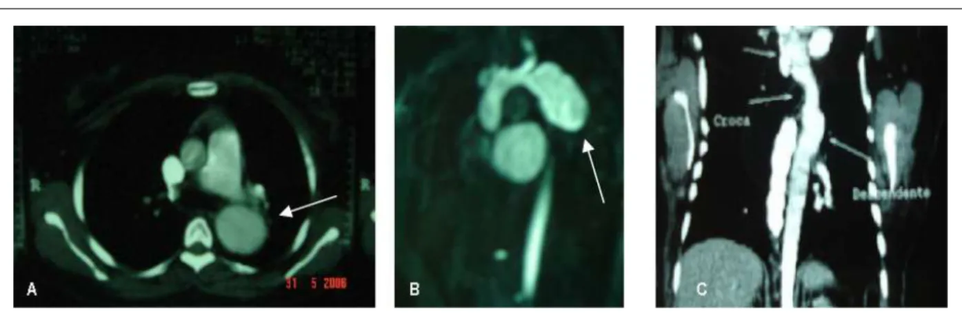
hemoptysis and respiratory failure, requiring intubation and ventilatory support. She was admitted at the Intensive Care Unit (ICU) with tachydyspnea, normal skin color, large pulses, good perfusion, regular cardiac rhythm, normal heart sounds without murmurs, clicks or rubs, presence of bilateral diffuse pulmonary rhonchi and rales, heart rate of 132 bpm and systemic arterial pressure of 135 X 90 mmHg. Hemoglobin: 9.3g/dL, hematocrit: 29.7%, platelets: 232,000, INR (International Normalized Ratio): 1.2, activated partial thromboplastin time (PTT): 30 seconds, fibrinogen: 400mg/ dL. The chest x-ray showed an enlarged mediastinum and normal pulmonary fields. The electrocardiogram showed sinus tachycardia. After two days, a computed tomography (CT) of the chest showed aneurysmatic dilatation of the descending aorta after the emergence of the left subclavian artery up to the left atrium, measuring 5.8 x 4.5 cm at the largest diameters, causing a mass effect and compressing the bronchus of the apicoposterior segment (Figure 1). A transesophageal echocardiogram showed the presence of a pseudoaneurysm (Figure2). The percutaneous treatment with endoprosthesis was programmed. On the 8th day of hospital
admission she presented two episodes of massive hemoptysis associated to hemodynamic instability and needed emergency surgery. During the intraoperative period, a large hematoma, which involved the thoracic aorta, was identified, extending to and in contact with the segmental bronchus (apicoposterior segment of the upper lobe of the left lung). A Dacron tube was implanted in the affected aortic segment, with extracorporeal circulation and the affected bronchus was repaired; the patient presented good evolution and was discharged on the 15th
postoperative day.
Discussion
The aortobronchopulmonary fistula is a rare entity that can be fatal if not adequately treated4. It was first described in the literature
by Keefer and Malory in 19345. Most aortobronchopulmonary
fistulas originate from a thoracic aortic aneurysm, which causes erosion of the pulmonary parenchyma or of the bronchial tree5. It can have different etiologies, such as tuberculosis,
syphilis, fungal infections, trauma and atherosclerosis, mainly in elderly individuals. Particularly in young individuals, as in the case reported here, an aortobronchopulmonary fistula can be secondary to the history of surgical correction of congenital heart defects4. In cases that are secondary to aortic surgical
interventions, where prosthesis implants are involved, such as in the coarctation correction, aneurysms or pseudoaneurysms can occur in the prosthesis suture lines, either proximally or distally.
Case Report
Coelho et al Fistula after correction of aortic coarctation
Arq Bras Cardiol 2009; 92(4) : e23-e25
Figure 1 -Computed tomography; A - Transversal view showing the dilated descending aorta; B - Dilation in descending thoracic aorta after left subclavian artery; C
- Control - 3 months after surgery.
The surgical correction of the aortic coarctation with the use of prosthesis or synthetic repair has a high risk of postoperative complications and reoperations6-8. Knyshov et al7 studied
the long-term outcomes (1 to 24 years) of the postoperative period of surgical correction of the aortic coarctation in 891 patients, showing that 48 patients (5.4%), with a mean age of 30 years, developed aneurysms in the repair site. Among these, 43 patients (89.6%) were submitted to aortoplasty with prosthesis implant or synthetic repair7. This case becomes
rarer as it occurred 26 years after the procedure, which did not use synthetic repair.
Once the aneurysm is formed, the weakened vascular wall is continuously submitted to distension due to the arterial pressure, damaging the aorta and the subjacent pulmonary parenchyma, creating a communication4.
In the case described here, the initial clinical manifestation was hemoptysis, which was repeated 8 days later, with a massive episode, leading to hemorrhagic shock that required immediate intervention. The initially proposed procedure was not carried out due to the immediate non-availability of the auto-expandable endoprosthesis with the adequate dimensions for the case at that moment. Classically, the
aortobronchopulmonary fistula is associated to a history of hemoptysis, with the latter being the main clinical manifestation5,9. The existence of an aortobronchopulmonary
fistula must always be considered in patients presenting hemoptysis and a history of thoracic aortic surgery1. It can
occur within three weeks to 25 years after the surgery4. The
formation of thrombi can result in an interval of days or weeks between the episodes of hemoptysis. It tends to be self-limited and recurrent and increasingly worsens with time, until a massive bleeding episode occurs. Precordial pain may be another symptom, found in approximately 45% of the cases4,
althoughit did not occur in the case described here. The diagnosis of an aortobronchopulmonary fistula is difficult to attain, as the chest x-ray tends to be normal or present alveolar infiltrate caused by the bloody material, disclosing the aneurysm in only 16% of the cases6. The CT
and the angiography generally identify the aneurysm and, to a lesser extension, the fistula4. The visual identification of
the fistula is possible in approximately 17% of the cases by CT5. The CT can disclose, in addition to the aneurysm, the
periaortic hematoma, mural thrombus and contrast leakage into the pulmonary parenchyma5.
Figure 2 -Transesophageal echocardiogram; A - Descending thoracic aorta showing the true and the false lumen; B - Doppler showing inlow and outlow in the
pseudoaneurysm.
Case Report
Coelho et al
Fistula after correction of aortic coarctation
Arq Bras Cardiol 2009; 92(4) : e23-e25
References
1. Quintana AL, Aguilar EM, Heredero AF, Riambau V, Paul L, Acín F. Aortobronchial fistula after aortic coarctation. J Thorac Cardiovasc Surg. 2006; 131: 240-3.
2. Favre JP, Gournier JP, Adham M, Rosset E, Barral X. Aortobronchial fistula: report of three cases and review of the literature. Surgery. 1994; 115: 264-70.
3. Ishizaki Y, Tada Y, Takagi A, Sato O, Takayama Y, Shirekawa M, et al. Aortobronchial fistula after an aortic operation. Ann Thorac Surg. 1990; 50: 975-7.
4. Calderón AA, Chinarro BJ, Fernández AA, Navarrete OI, Martos AR, Moragues MAJ. Recurrent hemoptysis secondary to an aortobronchial fistula. Arch Bronconeumol. 2005; 41 (6): 352-4.
5. Posacioglu H, Apaydin AZ. Pseudoaneurysm and aortobronchial fistula after aortic coarctation repair by patch aortoplasty. Tex Heart Inst J. 2004; 31:
319-21.
6. Hamilton MCK, Holemans JA, Entwisle J. Aortobronchopulmonary fistula after repair of aortic coarctation. Clin Radiol. 2004; 59: 1044-7.
7. Knyshov GV, Sitar LL, Glagola MD, Atamanyuk MY. Aortic aneurysms at the site of the repair of coarctation of the aorta: a review of 48 patients. Ann Thorac Surg. 1996; 61 (3): 935-9.
8. Bhuyan RR, Rajendran S, Unnikrishnan M, Jayakumar K. Successful repair of aortobronchial fistula using circulatory arrest. ANZ J Surg. 2007; 77(5): 398-9.
9. Kalkat MS, Bonser RS. Management of aortobronchial fistula following coarctation repair. Eur J Cardiothorac Surg. 2003; 23: 116-8.
10. Heikkinen LO, Järvinen AA. Aortopulmonary fistula after coarctation repair with dacron patch aortoplasty. Ann Thorac Surg. 2002; 73: 1634-6. The magnetic resonance imaging (MRI) is another
examination recommended by the literature for the diagnosis4. Heikkinen et al10 demonstrated the trajectory of
the fistula through MRI, although its visual identification is rarely possible10. The bronchoscopy must be indicated with
caution when an aortobronchopulmonary fistula is suspected, mainly when there is abundant hemoptysis, as there can be mobilization of thrombi formed in the trajectory of the fistula, which can lead to a massive hemorrhage6,10.
Another important risk factor for aneurysm formation and aortic dissection is systemic arterial hypertension (SAH), as it causes additional stress and turbulent flow at the site of the coarctation repair7. Our patient reported here had a
history of uncontrolled SAH due to poor adherence to the drug treatment and this fact might have contributed to the development of the clinical picture.
Conclusion
The recognition of this severe clinical entity is of great importance, as it is crucial to attain a rapid diagnosis, considering that the aortobronchopulmonary fistula can be fatal and must be considered an emergency situation. The
surgical treatment must be promptly carried out, even before the diagnosis is fully confirmed. The prognosis is usually good, resulting in a survival rate of 76%4-6.
The endovascular treatment with auto-expandable prosthesis has also been used; however, randomized prospective studies are still necessary to establish the procedure outcomes and to allow a comparative analysis with the surgical treatment outcomes1.
Potential Conflict of Interest
No potential conflict of interest relevant to this article was reported.
Sources of Funding
There were no external funding sources for this study.
Study Association
This study is not associated with any post-graduation program.