Genomic imbalances detected
through array CGH in fetuses
with holoprosencephaly
Isabela Nelly Machado, Juliana Karina Heinrich, Ricardo Barini
ABSTRACT
Objective: Holoprosencephaly (HPE) is heterogeneous in pathogenesis, integrating genetic susceptibility with the influence of environmental factors. Submicroscopic aberrations may contribute to the etiology of HPE. Our aim was to report the molecular analysis of 4 fetuses with HPE and normal metaphase karyotype. Method: A whole genome BAC-array based Comparative Genomic Hybridization (array CGH) was carried out in fetal blood samples. All potential cytogenetic alterations detected on the arrays were matched against the known copy number variations databases. Results: The array CGH analysis showed copy number gains and losses in all cases. We found a recurrent deletion in 15q14 (clone RP11-23J11) and in 15q22 (clone RP11-537k8) in 2 out 4 cases analyzed. We also observed submicroscopic gain in 6p21 in 3 out of 4 fetuses in nearby clones. All these regions were tested in known databases and no copy number variations have been described for them. Conclusion: This is the first report of molecular characterization through a whole genome microarray CGH of fetuses with HPE. Our results may contribute to verify the effectiveness and applicability of the molecular technique of array CGH for prenatal diagnosis purposes, and contributing to the knowledge of the submicroscopic genomic instability characterization of HPE fetuses.
Key words: holoprosencephaly, comparative genomic hybridization, prenatal diagnosis, genetic testing, genomic instability.
Instabilidades genômicas detectadas através de array CGH em fetos com holopro sencefalia
RESUMO
Objetivo: Holoprosencefalia (HPE) é uma malformação heterogênea na patogênese, integrando a suscetibilidade genética com a influência de fatores ambientais. Aberrações submicroscópicas podem contribuir para a etiologia da HPE. Nosso objetivo foi relatar a análise molecular de 4 fetos com HPE e cariótipo normal. Método: Foi realizado um estudo descritivo prospectivo dos achados da técnica de hibridação genômica comparativa baseada em microarranjos utilizando BAC clones de ampla cobertura genômica (BAC-array CGH) em amostras sanguíneas de fetos portadores de holoprosencefalia e com cromossomos numericamente normais ao bandamento G. Todas as potenciais alterações citogenéticas detectadas foram comparadas com bancos de dados com variações do número de cópias conhecidas. Resultados: A análise de array CGH evidenciou ganhos e perdas do número de cópias em todos os 4 casos. Foram encontradas deleções recorrentes em 15q14 (clone RP11-23J11) e em 15q22 (clone RP11-537k8) em 2 dos 4 casos analisados. Observou-se em 3 fetos ganho genômico na região 6p21 em clones próximos. Todas estas regiões não apresentaram variações do número de cópias descritas em bancos de dados conhecidos. Conclusão: Este é o primeiro relato de caracterização molecular através de um microarray CGH de fetos com HPE. Nossos resultados podem contribuir para verificar a eficácia e aplicabilidade da técnica molecular de array CGH para fins de diagnóstico pré-natal, contribuindo para o conhecimento da caracterização de instabilidades genômicas submicroscópicas de fetos com HPE. Palavras-chave: holoprosencefalia, hibridização genômica comparativa, diagnóstico pré-natal, análise genética, instabilidade genômica.
Correspondence
Isabela Nelly Machado
Department of Obstetrics and Gynecology Rua Alexander Fleming 101
13083-970 Campinas SP - Brasil E-mail: [email protected]
Support
This study was sponsored by São Paulo State Research Foundation FAPESP (Grant Nº 2007/04684-0)
Received 25 November 2009 Received in final form 7 July 2010 Accepted 14 July 2010
Holoprosencephaly (HPE) OMIM 2361000, is the most common developmental defect of midline cleavage in human embryonic forebrain, with a variable pheno-typic expression. Its estimated prevalence is of 1:16,000 live-births1 and 1:250 conceptuses2, but it should be
high-er considhigh-ering the current advances in neuroimaging that allow the diagnosis of less severe forms of this malfor-mation. he association of holoprosencephaly with oth-er fetal structural and chromosomal abnormalities justi-ies a detailed investigation of the fetal morphology and karyotype.
Our current understanding of the pathogenesis of HPE attempts to integrate genetic susceptibility with the epigenetic inluence of environmental factors. Identiiable genetic causes in humans account for about 15-20% of all cases3. he most common chromosomal abnormality
as-sociated to holoprosencephaly is the trisomy 13. To date, known human mutations in at least 12 diferent genetic loci have been associated with HPE4, but a very small
per-centage of all cases has a molecularly deined HPE. here-fore, submicroscopic aberrations may contribute to the etiology of HPE.
The aim of this study was to report the molecular indings through a whole genome array comparative ge-nomic hybridization (array CGH) of 4 fetuses with pre-natal ultra-sound diagnosis of HPE in an attempt to im-prove the knowledge of the submicroscopic abnormali-ties presented in these malformed fetuses.
METHOD
Patients and samples
For this study, fetuses with holoprosencephaly as an isolated brain malformation, no recognized genetic syn-drome, and normal metaphase karyotype delivered be-tween January 2008 and July 2009 were prospectively in-cluded. his study was carried out after the protocol ap-proval by the institution’s ethical committee.
The fetal and parental karyotype analysis was per-formed using G-banded metaphase chromosomes at ap-proximately the 500 band level. All the parents gave in-formed consent. he presence and morphologic classii-cation of HPE was conirmed by postnatal MRI (magnetic resonance imaging) or autopsy reports. Maternal diabe-tes mellitus, drug ingestion, exposure to alcohol and in-fections were excluded.
Fetal blood samples were collected by cordocentesis at diferent week’s gestation for karyotype, according to the guidelines of the Fetal Medicine Unit of the Women’s Hospital of the State University of Campinas (Unicamp).
Molecular study
Genomic DNA was extracted and puriied from fetal blood by means of the Wizard® Genomic DNA
Puriica-tion Kit (Promega Corp., Madison, WI, USA), according to manufacturer’s protocol for whole blood.
Array Comparative Genomic Hybridization was car-ried out using the Constitutional Chip® 4.0 (PerkinElmer Inc., Turku, Finland), comprised of approximately 5000 BAC (Bacterial Artiicial Chromosome) clones, covering the whole human genome with an average resolution of <650 kb and spotted in duplicate.
For each experiment, a sex-mismatched normal ref-erence DNA (Promega Corp., Madison, WI, USA) was used. he DNA concentrations used were 40 ng/µl. All experiments included dye reversal and two array hybrid-izations to obtain an accurate ratio. After post-hybrid-ization washes, slides were scanned, and captured imag-es were analyzed by either the GenePix® Pro 6.0 (Molec-ular Devices Corp.) or the ScanArray Express® (Microar-ray Analysis System 4.0.0.4) software.
After quantiication, the cyanine 5 and cyanine 3 av-erage ratio luorescence intensities for each BAC clone on each of the duplicate arrays (gpr iles) were uploaded into the web-based SpectralWare® v2.3.3 software (PerkinEl-mer Inc.), normalized with linear regression algorithms (on a log2 scale) and plotted according to the BAC chro-mosomal location. he raw data from dye-reversed pairs were combined, and threshold values were ascertained to make inferences according to a clone-by-clone clas-siication procedure to determine the gain, loss and no change status of each clone for each subject, relative to the diploid reference DNA. he threshold values were determined by the software using the “Iterative 2.5X Sig-mas” algorithm. Subsequent normalization of the data with “Block Lowess” method was performed for verii-cation of copy number changes. he P values for each probe were also calculated, furnishing additional objec-tive statistical criteria to determine whether deviation of each probe from zero is a signiicant change5. he quality
criteria adopted included standard deviation of the inten-sity ratios among the duplicates less than 10% and more than 97.5% of spots with adequate intensity ratio values for analysis6. For each analysis, all quality control metrics
were noted to be optimal. Clone-by-clone changes were reviewed and only those aberrations detected in both hy-bridizations were studied further.
All potential cytogenetic alterations detected on the arrays were matched against the known online databases to determine whether they encompassed described copy number variations (CNV) regions.
RESULTS
chromosomes at G-band analysis of peripheral blood. he maternal age ranged from 14 to 36 years old, with a par-ity varying from zero to two. One woman had previous spontaneous abortions, with no investigation for cytoge-netics aberrations. he fetuses’ hearts were morpholog-ically normal at the fetal echocardiography. All the four babies were born alive with adequate weight for the ges-tational age and two of them, the semi-lobar and the alo-bar cases, died in the same day. An autopsy conirmed the prenatal indings and classiication of HPE in all cas-es. he array CGH analysis showed copy number gains and losses in all cases.
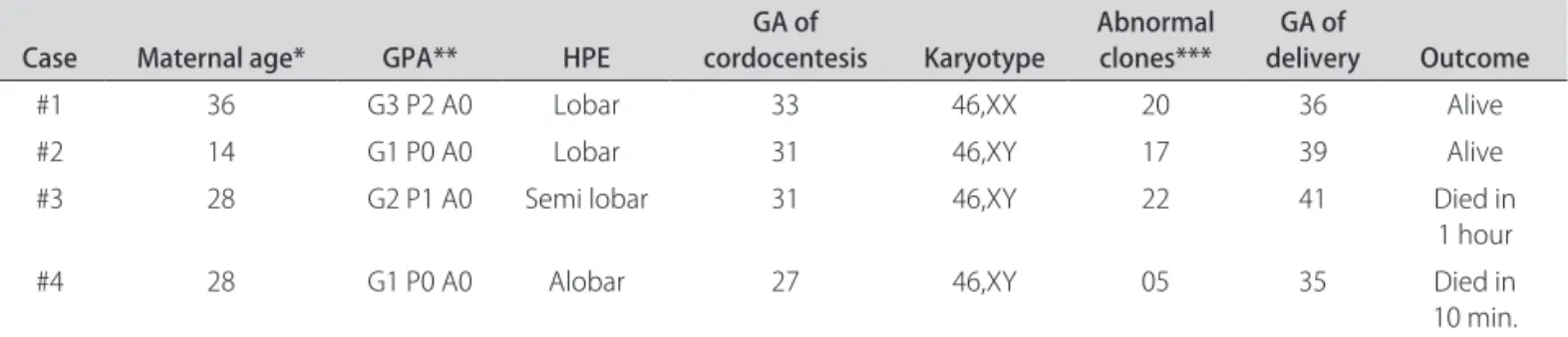
Clinical aspects, the gestation outcome and the total number of clones with genomic instability observed in each case are summarized in Table 1.
We identified recurrent deletion in 15q14 and in
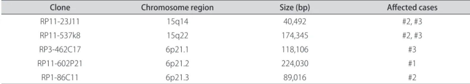
15q22 in 2 out 4 cases analyzed. Based on the physi-cal mapping positions as obtained from the March 2006 and February 2009 Assembly of the UCSC Genome Browser (http://genome.ucsc.edu/), the size of the delet-ed regions were determindelet-ed to be 40,492 bp (37,806,124-37,846,615) and 174,345 bp (62,082,000-62,256,344), re-spectively. We also observed a recurrent copy number gain in 6p21 region in 3 out of 4 evaluated fetuses in-volving diferent but close clones (Figure). he complete list of abnormal clones found in the four fetuses is listed in Table 2. Details about the recurrent abnormal clones are listed in Table 3.
All the identified common regions were tested in known databases and four copy number variations (CNV) were found and excluded. hey were gain at 1p36, 2q37.3 and 9q34, and loss at 5q13.
Table 1. Clinical aspects, gestation outcome and the total number of clones with genomic instability in 4 fetuses with holoprosencephaly.
Case Maternal age* GPA** HPE cordocentesisGA of Karyotype Abnormal clones*** deliveryGA of Outcome
#1 36 G3 P2 A0 Lobar 33 46,XX 20 36 Alive
#2 14 G1 P0 A0 Lobar 31 46,XY 17 39 Alive
#3 28 G2 P1 A0 Semi lobar 31 46,XY 22 41 Died in
1 hour
#4 28 G1 P0 A0 Alobar 27 46,XY 05 35 Died in
10 min. GA: gestational age (weeks); *Maternal age (completed years); **G: gravidity, P: parity, A: number of abortions; ***Total number of abnormal clones.
Table 2. Abnormal clones detected in 4 fetuses with holoprosencephaly using whole genome array CGH.
#1 #2 #3 #4
G L G L G L G L
RP1-283E3 RP11-328L16 RP4-628J24 RP11-23J11 RP11-625N16 RP11-91G12 RP11-353K11 RP1-160H23 RP3-375M21 RP1-77N19 RP11-537K8 RP11-118M12 RP11-352A18 RP5-1011O17 RP11-602P21 RP11-173D3 RP5-856G1 RP11-300G13 RP3-462C17 RP11-551B22 RP11-15M20
RP11-48C7 RP1-86C11 RP11-483F11 RP11-3G21 RP1-103M22
RP1-44H16 RP3-428L16 GS-261-B16 RP11-107O19 RP11-80F22
GS-908-H22 RP3-366N23 RP11-416K5 RP11-23J11
RP5-908H22 RP11-173G21 RP11-160E2 RP11-537K8
RP11-598F7 RP11-738I14 RP11-46E14
RP11-277E18 RP11-91H5 GS-325-I23
RP11-256C2 RP11-79M19 RP5-860F19
RP11-26M6 RP11-142I8 RP11-379J5
CTD-2184G2 RP11-103B5 RP4-745C22
RP11-64L12 RP11-173D3 RP1-141I3
RP11-67A5 RP1-81F12
DISCUSSION
HPE seems to be a multiple hit pathology, requiring two or more events involving several genes and/or envi-ronmental factors7. he pathology of HPE can be caused
by environmental (drugs, infections) and metabolic (dia-betes mellitus, alcohol, smoking) factors. Among genet-ic causes, it can be part of deined malformations syn-dromes with normal karyotype, chromosomal abnormal-ities as trisomy 13, trisomy 18 and triploidy, or it can be due to known sequence mutations on described chro-mosome regions.
he currently identiied HPE genes only account for a small portion of all sporadic HPE cases (15-20%)3, and
mutations in the currently recognized HPE genes explain only a very small proportion of all sporadic HPE cases8.
In a cohort of 424 unrelated postnatal cases with severe central nervous system indings, normal karyotype and negative for the for four main HPE genes, micro deletions were found in 4.7% and no micro deletions were found in 85 individuals with HPE microsigns9. he same group
of researchers found a percentage of 8.5% of microdele-tions in the prenatal period in 97 fetuses10. he remaining
cases are assumed to be a non-known etiology manifes-tation: neither environmental, nor syndromic, nor chro-mosomal4. he diiculties in identifying these genes may
relate to the multigenic nature of HPE. Loss of function in a single HPE gene may not lead to the disease. In hu-man, there will be other modiier genes acting in addi-tion11. A recent study involving twenty members of an
af-fected family showed the importance of perseverance de-spite initially negative tests, including applying new tech-nology and testing newly discovered genes and highlight-ed the fact that genetic disorders may manifest in ways not exactly as traditionally described12.
herefore, we hypothesized that there are still uniden-tiied genes causing underlying submicroscopic aberra-tions that could contribute to the etiology of HPE. In a irst attempt to identify novel candidate regions involved in the pathology of this heterogeneous disease, and to evaluate the feasibility of BAC arrays in the analysis of prenatal samples, we used an array CGH pangenomic ap-proach to report the molecular characterization of group of 4 fetuses with normal karyotype and diagnosis of HPE visible by ultra-sound prenatal care.
Table 3. Recurrent abnormal clones in 4 fetuses with holoprosencephaly.
Clone Chromosome region Size (bp) Afected cases
RP11-23J11 15q14 40,492 #2, #3
RP11-537k8 15q22 174,345 #2, #3
RP3-462C17 6p21.1 118,106 #3
RP11-602P21 6p21.2 224,030 #1
RP1-86C11 6p21.3 89,016 #2
bp: base pairs.
he array CGH analysis showed copy number gains and losses in all cases. Interestingly, the alobar case, the more severely afected fetus, presented the smallest num-ber of genomic abnormalities. Indeed, our results did not allow an inkling of correlation between the number and size of these imbalances and the severity of the phenotype.
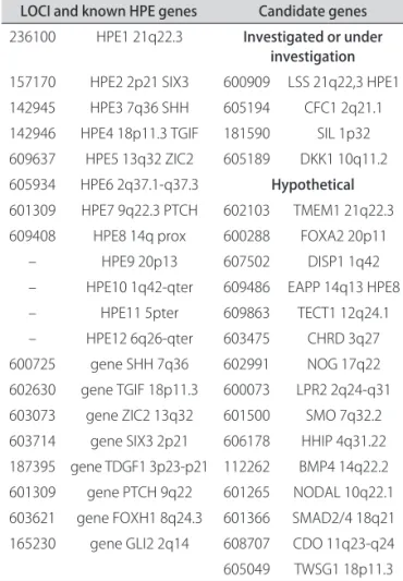
he current described genes and candidate genes for HPE, with its respective chromosome regions are pre-sented in Table 44. It was not a goal of this study to search
for mutations in the four main HPE genes (SHH, ZIC2, SIX3, TGIF), but we could observe that there were no ab-normal clones in their known loci.
A Medline search using the keywords 15q14 and ho-loprosencephaly could not ind any citation. he same occurred using the association between 15q22 or 6p21 and holoprosencephaly. One patient was related with a de novo reciprocal translocation afecting the breakpoints 6p21.1 and 7q36, presenting premaxillary agenesis (part of the HPE spectrum) as well as skeletal abnormalities and impacted teeth reminiscent of cleidocranial dyspla-sia (CCD). But, in this patient, the HPE phenotype could be explained by the 7q36 breakpoint that maps to the sonic hedgehog gene (SHH), the HPE3 described locus13.
As the mutations in genes mapping the 6p21 region can
cause CCD, the breakpoint in this region in this case ap-pears to explain the CCD phenotype14.
A possible association between the 15q22 region and holoprosencephaly could be postulated considering that, in some instances, the agenesis of corpus callosum can be part of the holoprosencephaly spectrum3. Of the
clin-ical manifestations reported cases of individuals with deletions encompassing 15q15-q22 region, one patient showed partial agenesis of corpus callosum15 and other
showed hypoplastic corpus callosum16.
The inheritance background of our findings is un-known as blood samples from the parents were not avail-able for array CGH analysis to determine if the copy num-ber gains were inherited or de novo.
he greatest care must be taken for molecular prena-tal diagnosis in HPE. Even if a mutation has been identi-ied and seems to be transmitted with clinical manifesta-tions in the family, another event, like a mutation in an-other gene (not yet identiied) or an environmental fac-tor, may be necessary to generate the holoprosencepha-ly phenotype7. In this case, molecular biology performed
prenatally provides only an additional criterion with re-gard to prenatal ultrasound or MRI, which still takes pre-cedence over molecular analysis.
Microarray-based CGH is a powerful method to de-tect and analyze genomic imbalances that are well below the level of detection on high resolution banded karyo-type analysis providing a better opportunity for genokaryo-type/ phenotype correlations in other similarly afected indi-viduals. Array CGH is relatively widely used in genetic testing of children. Recently, a case with the middle in-terhemispheric variant, a milder variant of HPE, was de-scribed carrying a deletion of ~10.4 Mb at 6q22.31-q23.2, suggesting a novel candidate gene of HPE17. But its true
potential is still under-explored in prenatal diagnosis. Some advantages of this molecular method is that it does not involve cell culture, does not require prior knowledge of the genomic region involved and the ability to study cases where only DNA is available and no chro-mosomes can be obtained. he method was reproduc-ible in a clinical standpoint, with reliable results within 48 hours. hus, we demonstrate that the technique of array CGH can become an excellent tool for prenatal diagnosis. But, it is important to emphasize that array CGH may not replace conventional G-banded karyotype analysis, but it can complement and expand current methods for a pre-cise prenatal diagnosis and syndromes’ characterization. One advantage of G banding analysis is that it allows the detection of somatic chromosomal mosaicism, which has been described in some patients with PHE.
Based on our results, array CGH results are promising in prenatal genetic testing and a study for submicroscopic deletions in fetuses with non-syndromic HPE should be
Table 4. Known genes and candidate genes for HPE.
LOCI and known HPE genes Candidate genes
236100 HPE1 21q22.3 Investigated or under investigation
157170 HPE2 2p21 SIX3 600909 LSS 21q22,3 HPE1 142945 HPE3 7q36 SHH 605194 CFC1 2q21.1 142946 HPE4 18p11.3 TGIF 181590 SIL 1p32 609637 HPE5 13q32 ZIC2 605189 DKK1 10q11.2 605934 HPE6 2q37.1-q37.3 Hypothetical
601309 HPE7 9q22.3 PTCH 602103 TMEM1 21q22.3 609408 HPE8 14q prox 600288 FOXA2 20p11
– HPE9 20p13 607502 DISP1 1q42
considered as part of the routine laboratory evaluation, in addition to high resolution chromosomal and mutation analysis. Positive results in any of these studies will help to better understand the etiology of HPE and aid the es-tablishment of the recurrence risk for family counseling. Moreover, additional research is needed to further estab-lish the role of genes from related chromosome regions in brain development and to determine the prevalence of copy number gain in the 15q and 6p regions among HPE patients. Also, in accordance with others authors, epide-miologic investigations should be conducted to check of environmental factors that could act in coordination with genetic events to give rise to holoprosencephaly.
ACKNOWLEDGMENTS – The authors have no conflict of interest with any of the information presented in this article. We are grateful to the families who participate in this research. We thank members of the Cell Culture and Cytogenetics Laboratory of the Women’s Hospital (CAISM – State University of Campinas – UNICAMP) for their con-tribution throughout the course of this project and Christopher Wil-liams (PerkinElmer Inc., Waltham, MA) for skilled technical assistance. Eletronic-Database and online software: he URL for data presented herein is as follows: SpectralWare® v2.3.3 software (PerkinElmer Inc.), http://service.spectralgenomics.com (for the array analysis). Database of Genomics Variants, http://projects.tcag.ca/variation/ (for CNV search). Copy Number Variation, http://cnv.chop.edu/ (for CNV search). UCSC Genome Browser, http://genome.ucsc.edu/. (for physical mapping posi-tions and size determination of chromosomal regions). Medline, http:// www.ncbi.nlm.nih.gov/pubmed/. (for literature search).
REFERENCES
1. Rasmussen S, Moore C, Khoury M, Cordero J. Descriptive epidemiology of holoprosencephaly and arhinencephaly in metropolitan Atlanta, 1968-1992. Am J Med Genet 1996;66:320-333.
2. Matsunaga E, Shiota K. Holoprosencephaly in human embryos: epidemio-logic studies of 150 cases. Teratology 1977;16:261-272.
3. Cohen MJ. Holoprosencephaly: clinical, anatomic, and molecular dimensions. Birth Defects Res A Clin Mol Teratol 2006;76:658-673.
4. Dubourg C, Bendavid C, Pasquier L, Henry C, Odent S, David V. Holoprosen-cephaly. Orphanet J Rare Dis 2007;2:8.
5. Ng G, Huang J, Roberts I, Coleman N. Deining ploidy-speciic thresholds in array comparative genomic hybridization to improve the sensitivity of de-tection of single copy alterations in cell lines. J Mol Diagn 2006;8:449-458. 6. Vermeesch J, Melotte C, Froyen G, et al. Molecular karyotyping: array CGH
quality criteria for constitutional genetic diagnosis. J Histochem Cytochem 2005;53:413-422.
7. Ming J, Muenke M. Multiple hits during early embryonic development: di-genic diseases and holoprosencephaly. Am J Hum Genet 2002;71:1017-1032. 8. Nanni L, Croen L, Lammer E, Muenke M. Holoprosencephaly: molecular study
of a California population. Am J Med Genet 2000;90:315-319.
9. Bendavid C, Haddad B, Griin A, et al. Multicolour FISH and quantitative PCR can detect submicroscopic deletions in holoprosencephaly patients with a normal karyotype. J Med Genet 2006;43:496-500.
10. Bendavid C, Dubourg C, Gicquel I, et al. Molecular evaluation of foetuses with holoprosencephaly shows high incidence of microdeletions in the HPE genes. Hum Genet 2006;119:1-8.
11. Shen J, Walsh C. Targeted disruption of Tgif, the mouse ortholog of a human holoprosencephaly gene, does not result in holoprosencephaly in mice. Mol Cell Biol 2005;25:3639-3647.
12. Solomon BD, Lacbawan F, Jain M, et al.A novel SIX3 mutation segregates with holoprosencephaly in a large family. Am J Med Genet Part A 2009;149: 919-925.
13. Huang J, Hofman JD, Zhang Y, et al. Identiication of a submicroscopic dele-tion of SHH associated with the holoprosencephaly spectrum by array-based CGH. Clin Genet 2006;69:367-369.
14. Fernandez B, Siegel-Bartelt J, Herbrick J, Teshima I, Scherer S. Holoprosen-cephaly and cleidocranial dysplasia in a patient due to two position-efect mutations: case report and review of the literature. Clin Genet 2005; 68:349-359.
15. Lalani S, Sahoo T, Sanders M, Peters S, Bejjani B. Coarctation of the aorta and mild to moderate developmental delay in a child with a de novo deletion of chromosome 15(q21.1q22.2). BMC Med Genet 2006;7:8.
16. Koivisto P, Koivisto H, Haapala K, Simola K. A de novo deletion of chromo-some 15(q15.2q21.2) in a dysmorphic, mentally retarded child with congen-ital scalp defect. Clin Dysmorphol 1999;8:139-141.