DOI: 10.1590/0004-282X20160050
VIEW AND REVIEW
Collateral blood vessels in acute ischemic
stroke: a physiological window to predict
future outcomes
Circulação colateral no acidente vascular cerebral isquêmico: uma janela fisiológica para
prever resultados futuros
Heitor Castelo Branco Rodrigues Alves
1,2, Felipe Torres Pacheco
1,2, Antonio J. Rocha
1,2Stroke is the second most common cause of death and was
responsible for approximately 6.7 million deaths worldwide
in 2012
1. Ischemia, or restricted blood low, is the main cause
of stroke and is typically due to abrupt occlusion of a cerebral
artery as a result of progressive atherosclerosis or embolism
2.
Acute ischemic stroke (AIS) can result in severe neurologic
disability or death
3.
Since the late 1990s intravenous thrombolysis has been
a recommended treatment for AIS
4,5. Additionally, in the last
decade, several clinical trials have investigated the efects of
endovascular treatment (EVT) in the setting of an intracranial
or extracranial large artery occlusion. Several studies,
includ-ing MR CLEAN
6, ESCAPE
3, EXTEND-IA
7and SWIFT-PRIME
8,
recently proved EVT to be more efective than standard med
-ical care, with or without intravenous thrombolysis, using
stentrievers in the majority of the patients in the EVT arms.
However, although EVT has been shown to be generally
efective, the trials have documented erratic individual and
overall patient outcomes. hese diferences may not be solely
the result of the various methods used but may also be
relat-ed to patient-speciic characteristics
9. Among these
patient-speciic characteristics, collateral status has emerged as an
independent factor that is associated with angiographic and
clinical outcomes in AIS patients
10,11.
he collateral circulation is a physiologic pathway of spe
-cialized endogenous bypass vessels that is present in most
1Santa Casa de São Paulo, Faculdade de Ciências Médicas, Divisao de Neuroradiologia, São Paulo SP, Brasil;
2Fleury Medicina e Saúde, Divisao de Neuroradiologia, São Paulo SP, Brasil.
Correspondence: Antonio Jose da Rocha; Santa Casa de Misericordia de Sao Paulo, Serviço de Diagnostico por Imagem; Rua Dr. Cesario Motta Jr. 112; 01221-020 São Paulo SP, Brasil; E-mail: a.rocha@uol.com.br
Conflict of interest: There are no conlicts of interest to declare. Received 31 December 2015; Accepted 08 February 2016.
ABSTRACT
Collateral circulation is a physiologic pathway that protects the brain against ischemic injury and can potentially bypass the effect of a
blocked artery, thereby inluencing ischemic lesion size and growth. Several recent stroke trials have provided information about the role
of collaterals in stroke pathophysiology, and collateral perfusion has been recognized to inluence arterial recanalization, reperfusion,
hemorrhagic transformation, and neurological outcomes after stroke. Our current aim is to summarize the anatomy and physiology of the
collateral circulation and to present and discuss a comprehensible review of the related knowledge, particularly the effects of collateral
circulation on the time course of ischemic injury and stroke severity, as well as imaging indings and therapeutic implications.
Keywords: stroke; brain schemia; collateral circulation; tomography; angiography; perfusion.
RESUMO
A circulação colateral é um circuito isiológico de proteção contra alterações isquêmicas que, potencialmente, evita os efeitos de
uma oclusão arterial e com isso pode inluenciar nas dimensões e no crescimento de uma lesão isquêmica. Vários estudos recentes
forneceram informações a respeito do papel das colaterais na isiopatologia do acidente vascular encefálico isquêmico e demonstraram
a capacidade da circulação colateral de inluenciar as taxas de reperfusão, recanalização, transformação hemorrágica e com isso
desfecho clínico dos pacientes. O objetivo desta revisão é sintetizar a anatomia e a isiologia da circulação colateral encefálica,
apresentando e discutindo, o que se conhece atualmente acerca do seu efeito na cronologia e gravidade da lesão isquêmica, além dos
achados de imagens e implicações terapêuticas.
tissues and protects against ischemic injury during initial
oligemic status
12. In the setting of AIS, the extent of
collat-eral circulation inluences the size of the inal infarct and the
growth of the penumbra. Hence, the relationship between
the collateralization grade and the predictability of infarct
evolution has been a primary focus in recent years
11,13,14.
he reinement of diagnostic techniques for evaluation
of collateral circulation may contribute to improved
ana-tomical and pathophysiological characterization of this
vas-cular network and its potential therapeutic and prognostic
implications
15. he ability to deine physiologic parameters
through diagnostic techniques is particularly useful when
the time of the stroke is unknown or a wider
nonconvention-al window for treatment is being considered.
In this review, we summarize the basic anatomy and
physiology of the collateral circulation and its potential
as an endogenous therapeutic target in AIS. The relevance
of multidetector computed tomography (MDCT) in
clini-cal settings to support mediclini-cal decisions regarding AIS is
highlighted, and recent evidence indicating that good
col-lateral circulation can prevent or delay permanent neural
damage is presented.
THE ANATOMY OF COLLATERAL CIRCULATION
Two main routes underlie collateral perfusion of the
brain parenchyma. he anatomy of this arterial circulation
includes extracranial sources of blood low that can supply
intracranial vessels, as well as intracranial routes that can
supplement other intracranial areas when pathophysiologic
mechanisms become actived
15.
he extracranial sources consist of large connections be
-tween the extracranial and intracranial arteries. he external
carotid artery gives rise to many branches in the neck that
are potential sources of collateral blood low, particularly
when chronic stenosis or occlusion has developed in the
in-ternal carotid artery
16. he facial, maxillary, middle menin
-geal, and occipital arteries are the main branches that can
shunt low via anastomoses to the intracranial arteries. Apart
from these branches, common anastomotic routes include
the ophthalmic artery, which may ill in a retrograde direc
-tion, as well as smaller and unnamed dural arteries
17.
Intracranial collateral routes can be further subdivided
into primary and secondary routes. he primary pathways
include the permanently active components of the circle
of Willis, and the secondary pathways include less direct
routes that develop over time. he blood supply to the brain
is unique because four major arteries coalesce to form an
equalizing distributor, i.e., the circle of Willis, which, despite
its variability and asymmetry, can redistribute blood low in
the event of sudden occlusion of a parent vessel.
he secondary pathway comprises leptomeningeal
anastomoses that link distal sections of the major cerebral
arteries. It has been reported that some small arteriolar
con-nections (~50–400
μ
m) allow retrograde perfusion of
adja-cent territories
18. It is assumed that these connections are
important routes for collateral blood low, especially when
an acute arterial occlusion occurs. hese arteriolar anasto
-moses mimic the circle of arteries but connect a much larger
extension of the microvasculature, joining territories of the
middle cerebral artery (MCA) with both the anterior cerebral
artery (ACA) and the posterior cerebral artery (PCA)
19.
he development of native pial collateral circulation (col
-laterogenesis), which begins in the embryo, has been shown
to determine the extent of the collaterals in adulthood
20.
Acute obstructions induce blood low across the collateral
network (recruitment) followed by remodeling and, poten
-tially, formation of additional collaterals in chronic
obstruc-tive disease (neocollateral formation)
17.
here is wide variation in collateral status among healthy
adults, and recent animal studies indicate that genetic
back-ground may be a major factor
21. {Zhang, 2010 #110} A single
polymorphic locus on chromosome 7 in mice, i.e., the
deter-minant of collateral extent 1 (Dce1), has been shown to
in-luence the extent of collateralization, blood low and infarct
volume after middle cerebral artery occlusion
22. Whether
hu-man Dce1 or related loci are responsible for the wide
varia-tion in collateral status in humans is still under investigavaria-tion.
A number of other factors, including environmental and
clinical features, have also been shown to afect the quality
and quantity of collaterals (rarefaction) at the time of presen
-tation in AIS. Of these, the strongest predictor by far is age
23.
Other clinical features include elevated glucose at the time of
presentation, uric acid level, history of hypertension, and
his-tory of smoking
24,25.
PHYSIOLOGY OF COLLATERAL BLOOD FLOW
REGULATION
he importance of the collateral circulation in brain phys
-iology may be demonstrated with the concept of the
collat-erome, which is an extension of the connectome concept
26.
he collaterome provides a physiologically relevant approach
to the management of stroke and the inluential balance of
collateral perfusion that determines both stroke evolution
and related clinical sequelae
27.
Beyond structural assessments of collateral
circula-tion, advances in perfusion-based imaging have allowed
for functional evaluations of the quality of collateral blood
low (efective parenchymal perfusion). Cerebral blood low
Normal CBF ranges between 50 and 60 mL/100 g/min and
is tightly controlled by cerebral autoregulation
29. he pace
of cellular death in the brain after an arterial occlusion is
closely linked to the severity of the decrease in blood low
within the local environment. When blood low is less than
10 mL/100 g/min, damage is rapid, and most cells die
with-in mwith-inutes of the with-insult
30,31. When CBF is between 10 and
20 mL/100 g/min (hypoperfusion), neurons cease to function
but remain structurally intact and are potentially revivable if
normal blood low is restored
31. herefore, neuronal damage
is not uniform when an intracranial artery is occluded,
espe-cially in the irst few hours after an insult. Depending on the
extent of collateral perfusion, infarction may not be complete
for hours or even days
32.
In thrombotic and embolic strokes, the intravascular
pres-sure distal to the occlusion falls immediately. Concurrently, the
pressure within the pial vessels is relatively well preserved,
re-sulting in a gradient that is able to promote low through anas
-tomoses
18. he efectiveness of collateral vessel low can be
assessed only with measurements of tissue perfusion, which
relect the statuses of both the microcirculation and the mac
-rocirculation. Computed tomography (CT) and magnetic
resonance imaging (MRI) perfusion techniques and other
methods, such as positron emission tomography (PET) and
single-emission computed tomography (SPECT), can provide
insight into the collateral low in patients with cerebrovascu
-lar disease
17,33,34. Physiologically efective collateral perfusion is
evident when CBF and cerebral blood volume (CBV) are
main-tained within the territory of an occluded artery.
IMAGING OF COLLATERAL VESSELS
Digital subtraction angiography (DSA) remains the gold
standard for the anatomic evaluation of the collateral
circu-lation. his technique allows for the dynamic visualization of
blood low through pial collaterals or other secondary collat
-erals
17,35. he main limitations of DSA are its invasive nature,
its reliance on iodinated contrast and ionizing radiation, and
its inability to evaluate brain parenchyma. Furthermore,
per-forming DSA in AIS when intra-arterial therapy is not consid
-ered may generate an additional delay to treatment
36.
Several noninvasive approaches have been proposed to
evaluate intracranial collateral blood low and the network,
but none of these techniques have been shown to be as efec
-tive as a reference standard for quantifying collateral low
37.
Computed tomography angiography (CTA) is fast,
reproduc-ible, and widely available, and its reasonable cost-to-efectiveness
ratio makes this technique one of the most widely used methods
of evaluating the locations of vascular occlusions and the
collat-eral system. Analysis of CTA source images (CTA-SI) has a higher
sensitivity for demonstrating the infarct core than non-contrast
computed tomography (NCCT)
38. Post-processed CTA data
in-volving maximal intensity projections (MIP) and multiplanar
reconstruction (MPR) allow for better visualization of the
oc-cluded vessel and the extent of leptomeningeal low
39. he main
limitation of CTA is that it is a snapshot of arterial contrast
en-hancement, providing limited information about low dynam
-ics. Some studies have attempted to circumvent this limitation
with dual-phase CTA
40. Such variations in acquisition protocols
and diferences in classiication have led to low levels of agree
-ment among the relevant results even among experienced
ob-servers (K-alpha 0.3-0.6)
41.
Dynamic CTA has become available for clinical practice
in recent years and merges the noninvasive nature of CTA
and the dynamic acquisition of DSA. his technique, also re
-ferred to as 4D-CTA, enables the noninvasive evaluation of
the low dynamics of the intracranial vasculature by multiple
subsequent CT acquisitions or continuous volume CT acqui
-sition over a period of time
42.
Several protocols of acquisition have been proposed, in
-cluding a toggling-table technique, shuttle mode scanning,
and volume mode. he volume mode is considered the most
versatile option and allows for complete or partial coverage of
the whole brain during 1 rotation of the scanner
42. Dynamic
acquisitions in volume mode can be performed discontinu
-ously or continu-ously, depending on the required temporal
resolution. When collateral low, as in the case of an arterial
occlusion, needs to be evaluated, a lower temporal resolution
is necessary. In the setting of AIS, 4D-CTA better estimates
thrombus burden and the presence of collateral vessels than
conventional CTA
42. he challenge of the radiation dose level
remains, although recently available noise-reduction ilters
have dramatically reduced radiation exposure
43.
Magnetic resonance angiography (MRA) using
time-of-light technique (TOF) is one of the most used MR tech
-niques for accessing collateral circulation, but it remains
controversial. MRA provides structural information based on
low-sensitive images but is less efective for collateral evalu
-ations than CTA, especially when the objective is to estimate
the occlusion of distal branches
36,44. Contrast-enhanced MRA
(CE-MRA) allows better delineation of slow-moving blood in
the distal branches and is a better predictor of infarct
out-come, but it provides lower spatial resolution
44.
Transcranial Doppler (TCD) also provides information
regarding cerebral autoregulation and cerebral circulation.
Flow direction changes, such as those found in the
ophthal-mic artery, and increased velocity in vessels ipsilateral to a
stenosis are correlated with the presence of
leptomenin-geal collaterals
45. However considerable variability has been
found in TCD performance and interpretation
35,45.
IMAGING STUDIES OF COLLATERAL CIRCULATION IN
THE CLINICAL SETTING OF ACUTE ISCHEMIA
he concept that a vascular network can potentially by
ischemic lesion size and growth
37,46has recently had an
in-creased impact on the management of stroke patients
4.
Assessments of collaterals in angiographic studies
have proven be widely variable. A recent meta-analysis by
Leng et al.
9found 12 studies that used the American Society
of Interventional and herapeutic Neuroradiology/Society of
Interventional Radiology (ASITN/SIR) collateral low grading
system by DSA and primarily deined grades 3-4 and 0-2 as good
and poor collateral statuses, respectively. Eleven studies have
used other grading methods for DSA, 9 studies have used
dif-ferent grading methods for CTA, and others have used CTP or
combined grading methods with diferent imaging modalities.
Two DSA classiications stand out: Higashida
47used the
ASITN/SIR assessment, which was based on the extent and
delay of retrograde illing, whereas Christoforidis
48proposed
a distinct model that is based solely on the extent of the
feedback.
he ASITN/SIR classiication
47seems to be the most
appropriate because it has higher reproducibility and
ac-counts for both the parameters of the extent and the delay
of the feedback via collaterals. he ASITN/SIR classiication
involves a ive-point system (Table 1) that has been used in
several endovascular trials
9,10,49,50,51,52. Grades 0 and 1 indicate
only marginal low, grade 2 indicates only partial illing of the
ischemic territory, and grades 3 and 4 indicate varying rates
of complete illing of the occluded arterial territory.
Additional non-invasive grading systems for assessing
collateral blood low circulation have also been described
53.
Several strategies have been used to describe CTA-visualized
vessels by either comparing them with the contralateral brain
hemisphere or estimating the percentage of MCA branches
that become illed by contrast media during examination
54,55.
Souza et al.
56proposed a simple and reliable grading system
that correlates collateral scores and difusion-weighted im
-aging (DWI) lesion volumes on admission.
he best method of evaluating and grading collateral low
remains controversial. he currently proposed methods of as
-sessing collaterals are largely qualitative or semiquantitative,
and there are no clear indications of the superiority of any of
the available techniques
57. his lack of consensus may con
-tribute to an overall under appreciation of the fundamental
role of collateral circulation in outcomes following AIS.
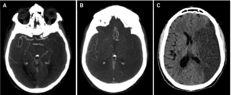
Nevertheless, Shet and Liebeskind
58, when revising the
sta-tus of collateral circulation in endovascular therapy for stroke,
concluded that collateral blood supply is pivotal in determining
clinical outcomes. In the setting of vascular occlusion, patients
with more robust collaterals have smaller infarcts (Figure 1)
17,59.
Regardless of the method used to determine the collateral score,
Table 1.
American Society of Interventional and Therapeutic
Neuroradiology / Society of interventional Radiology
(ASITN/SIR) collateral grade scale.
Grade
Angiographic collaterals
(Digital subtraction angiography)
0
No collaterals visible to the ischemic site
1
Slow collaterals to the periphery of the ischemic site
with persistence of some of the defect
2
Rapid collaterals to the periphery of ischemic site with
persistence of some of the defect and to only a portion
of the ischemic territory
3
Collaterals with slow but complete angiographic blood
low of the ischemic bed by the late venous phase
4
Complete and rapid collateral blood low to the vascular
bed in the entire ischemic territory by retrograde
perfusion
A
B
C
extremely poor outcomes are predicted when collateral blood
low is reduced or absent (Figures 2 and 3)
60,61,62,63.
Physiology is now considered to be more relevant than time
in AIS because it is the degree of collateral low and not simply
the time elapsed since the stroke that is the main factor
deter-mining core infarct volume within the irst 6 hours of stroke on
-set
46. Collateral status is recognized as having an inluence on
stroke prognosis, particularly in terms of recanalization,
reper-fusion, hemorrhagic transformation and subsequent neurologi
-cal outcomes
59. Collateral grading (Table 2) may represent a
cur-rently available opportunity to predict possible future outcomes,
whereas elapsed time solely relects the past and does not have a
causal connection with the future of infracted brain tissue.
RELATIONSHIP WITH PARENCHYMAL PERFUSION
Collaterals are usually evaluated by examining arterial low
with angiography techniques, and parenchymal perfusion is
profoundly inluenced by downstream microcirculation
59.
he microcirculation is crucial to the restoration of the blood
supply to the brain, and collateral circulation may increase
ischemic tolerance by enhancing microvascular perfusion
35.
A relatively simple method of evaluating brain perfusion
and viable tissue is the assessment of capillary blush using DSA.
he capillary index score (CIS) can deine an ischemic area in
two ways: either by a lack of anterograde low (in an area that
receives blood supply in a retrograde fashion through pial
collat-erals) or by a signiicant delay in anterograde low due to a prox
-imal partially recanalized clot
64. his angiographic index has
been shown to be a good predictor of outcomes and a powerful
strategy for improving outcomes in endovascular treatment
65.
Beyond structural assessments, perfusion-based images
have allowed functional evaluations to diferentiate critically
hypoperfused areas (infarct cores), penumbral areas
(poten-tially savable areas) and benign oligemic tissues. Current
evi-dence indicates that the goals of acute stroke treatment should
be to determine tissue viability by noninvasive techniques, use
this information to individualize thrombolytic therapy, extend
the therapeutic time window and rescue penumbral tissue
46.
herefore, knowing the factors that inluence the loss of pen
-umbral tissue is crucial, and collaterals have emerged as a
ma-jor feature that is relevant to this knowledge.
he presence of robust collaterals both markedly reduces and
slows down penumbra loss
14. his can be seen in patients without
signiicant reperfusion after treatment, reinforcing the fact that
poor collaterals alone are able to predict larger infarcts. In
con-trast, smaller infarcts, at least in part, result from good collaterals.
Reperfusion can occur not only via successful recanalization of
the primary occlusion and restoration of downstream low but
also via viable collateral blood low
66. However, it is important to
note that mismatch (the diference between the penumbra and
the infarct core) is also an independent prognostic factor with a
strong association with better outcomes in target mismatch
pa-tients, adding information to the study of collateral proiles
67.
Some studies have demonstrated the use of
ischemia-induced vascular damage estimates in AIS in
combina-tion with collateral scores and brain perfusion analysis
to predict hemorrhagic complications
17,68,69. In the setting
of poor collaterals, a finding of hyperperfusion may
indi-cate a higher risk of hemorrhagic transformation. When
revascularization is achieved, symptomatic
hemorrhag-ic transformation may occur more frequently in patients
who have presented with poor collaterals
49. Higher
fre-quencies of infarct growth and symptomatic hemorrhagic
A
B
C
transformation in patients with poor collaterals in whom
therapeutic recanalization has been achieved may
sup-port the concept of reperfusion injury
70.
TEACHING POINTS FOR CLINICAL USE
Larger recent trials have established the use of
intra-cranial vascular studies in the setting of AIS to detect
proximal obstructions, define the treatment subtype and
select an intraarterial approach
3,6,7. Therefore, the
collat-eral profile should be determined in all of these patients.
Some critical findings may be established with the
knowl-edge of collateral status.
Abundant native (preexisting) collateral circulation is
di-rectly correlated with better clinical status and smaller
vol-umes of infarcted brain
10. Improved collateral circulation also
predicts higher rates of recanalization, favorable outcomes
and lower rates of mortality
9,10,50.
he absence or relative paucity of a collateral network
is a major predictor of extensive infarct on admission
56, and
a proximal thrombus associated with such an absence has
been termed a “malignant proile”
71. Recent studies have also
demonstrated that worse collaterals are associated with
in-creased hemorrhagic complications, reinforcing the
rele-vance of speciic approaches for these patients
49,70.
FINAL REMARKS
his literature review supports the view that noninva
-sive vascular studies should be used to identify proximal
ar-terial occlusions and to estimate collateral grading in the
setting of AIS. A personalized approach that is not solely
re-stricted by time should be provided to maximize the efect
of therapy, including appropriate patient selection for EVT.
he currently available knowledge has increased the
pathophysiologic understanding of intrinsic compensatory
vascular mechanisms, supports the use of MDCT techniques
to rapidly evaluate hyperacute AIS, and provides evidence for
therapeutic decisions. Modern techniques for reducing radi
-ation exposure should be employed to ensure that diagnostic
tests preserve patient safety.
Table 2.
Collateral grade by Miteff system.
Grade
Computed tomography angiography collaterals
1
The contrast opaciication is merely seen in the distal
supericial branches
2
Vessels can be seen at the Sylvian issure
3
If the vessels are reconstituted distal to the occlusion
A
B
C
References
1. World Health Organization. Global burden of Stroke. Geneva: World Health Organization; 2012 [cited 2015 May 4]. Available from: http://www.who.int/mediacentre/factsheets/fs310/en/
2. Truelsen T, Piechowski-Jóźwiak B, Bonita R, Mathers C, Bogousslavsky J, Boysen G. Stroke incidence and prevalence in Europe: a review of available data. Eur J Neurol. 2006;13(6):581-98. doi:10.1111/j.1468-1331.2006.01138.x
3. Goyal M, Demchuk AM, Menon BK, Eesa M, Rempel JL, Thornton J et al. Randomized assessment of rapid endovascular treatment of ischemic stroke. N Engl J Med. 2015;372(11):1019-30. doi:10.1056/NEJMoa1414905
4. Jauch EC, Saver JL, Adams HP Jr, Bruno A, Connors JJ, Demaerschalk BM et al. Guidelines for the early management of patients with acute ischemic stroke: a guideline for healthcare professionals from the American Heart Association/American Stroke Association. Stroke. 2013;44(3):870-947. doi:10.1161/STR.0b013e318284056a
5. Martins SC, Freitas GR, Pontes-Neto OM, Pieri A, Moro CH, Jesus PA et al. Guidelines for acute ischemic stroke treatment: part II: stroke treatment. Arq Neuropsiquiatr. 2012;70(11):885-93. doi:10.1590/S0004-282X2012001100012
6. Berkhemer OA, Fransen PS, Beumer D, Berg LA, Lingsma HF, Yoo AJ et al. A randomized trial of intraarterial treatment for acute ischemic stroke. N Engl J Med. 2015;372(1):11-20. doi:10.1056/NEJMoa1411587
7. Campbell BC, Mitchell PJ, Kleinig TJ, Dewey HM, Churilov L, Yassi N et al. Endovascular therapy for ischemic stroke with perfusion-imaging selection. N Engl J Med. 2015;372(11):1009-18. doi:10.1056/NEJMoa1414792
8. Saver JL, Goyal M, Bonafe A, Diener HC, Levy EI, Pereira VM et al. Solitaire with the Intention for Thrombectomy as Primary Endovascular Treatment for Acute Ischemic Stroke (SWIFT PRIME) trial: protocol for a randomized, controlled, multicenter study comparing the Solitaire revascularization device with IV tPA with IV tPA alone in acute ischemic stroke. Int J Stroke. 2015;10(3):439-48. doi:10.1111/ijs.12459
9. Leng X, Fang H, Leung TW, Mao C, Miao Z, Liu L et al. Impact of collaterals on the eficacy and safety of endovascular treatment in acute ischaemic stroke: a systematic review and meta-analysis. J Neurol Neurosurg Psychiatry. 2015;pii: jnnp-2015-310965. doi:10.1136/jnnp-2015-310965
10. Liebeskind DS, Tomsick TA, Foster LD, Yeatts SD, Carrozzella J, Demchuk AM et al. Collaterals at angiography and outcomes in the Interventional Management of Stroke (IMS) III trial. Stroke. 2014;45(3):759-64. doi:10.1161/STROKEAHA.113.004072
11. Liebeskind DS, Jahan R, Nogueira RG, Zaidat OO, Saver JL,
Investigators S. Impact of collaterals on successful revascularization in Solitaire FR with the intention for thrombectomy. Stroke. 2014;45(7):2036-40. doi:10.1161/STROKEAHA.114.00478
12. Faber JE, Chilian WM, Deindl E, Royen N, Simons M. A brief etymology of the collateral circulation. Arterioscler Thromb Vasc Biol. 2014;34(9):1854-9. doi:10.1161/ATVBAHA.114.303929
13. Verma RK, Gralla J, Klinger-Gratz PP, Schankath A, Jung S, Mordasini P et al. Infarction Distribution pattern in acute stroke may predict the extent of leptomeningeal collaterals. PLoS One. 2015;10(9):e0137292. doi:10.1371/journal.pone.0137292
14. Jung S, Gilgen M, Slotboom J, El-Koussy M, Zubler C, Kiefer C et al. Factors that determine penumbral tissue loss in acute ischaemic stroke. Brain. 2013;136(12):3554-60. doi:10.1093/brain/awt246
15. Liebeskind DS. Collateral circulation. Stroke. 2003;34(9):2279-84. doi:10.1161/01.STR.0000086465.41263.06
16. Krishnaswamy A, Klein JP, Kapadia SR. Clinical cerebrovascular anatomy. Catheter Cardiovasc Interv 2010;75(4):530-9. doi:10.1002/ccd.22299
17. Sheth SA, Liebeskind DS. Imaging evaluation of collaterals in the brain: physiology and clinical translation. Curr Radiol Rep. 2014;2(1):29. doi:10.1007/s40134-013-0029-5
18. Brozici M, Zwan A, Hillen B. Anatomy and functionality of
leptomeningeal anastomoses: a review. Stroke. 2003;34(11):2750-62. doi:10.1161/01.STR.0000095791.85737.65
19. Shuaib A, Butcher K, Mohammad AA, Saqqur M, Liebeskind DS. Collateral blood vessels in acute ischaemic stroke: a potential therapeutic target. Lancet Neurol. 2011;10(10):909-21. doi:10.1016/S1474-4422(11)70195-8
20. Lucitti JL, Mackey JK, Morrison JC, Haigh JJ, Adams RH, Faber JE. Formation of the collateral circulation is regulated by vascular endothelial growth factor-A and a disintegrin and metalloprotease family members 10 and 17. Circ Res. 2012;111(12):1539-50. doi:10.1161/CIRCRESAHA.112.279109
21. Zhang H, Prabhakar P, Sealock R, Faber JE. Wide genetic variation in the native pial collateral circulation is a major determinant of variation in severity of stroke. J Cereb Blood Flow Metab. 2010;30(5):923-34. doi:10.1038/jcbfm.2010.10
22. Sealock R, Zhang H, Lucitti JL, Moore SM, Faber JE. Congenic ine-mapping identiies a major causal locus for variation in the native collateral circulation and ischemic injury in brain and lower extremity. Circ Res. 2014;114(4):660-71. doi:10.1161/CIRCRESAHA.114.302931
23. Arsava EM, Vural A, Akpinar E, Gocmen R, Akcalar S, Oguz KK et al. The detrimental effect of aging on leptomeningeal collaterals in ischemic stroke. J Stroke Cerebrovasc Dis. 2014;23(3):421-6. doi:10.1016/j.jstrokecerebrovasdis.2013.03.014
24. Menon BK, Smith EE, Coutts SB, Welsh DG, Faber JE, Goyal M et al. Leptomeningeal collaterals are associated with modiiable metabolic risk factors. Ann Neurol. 2013;74(2):241-8. doi:10.1002/ana.23906
25. Malik N, Hou Q, Vagal A, Patrie J, Xin W, Michel P et al. Demographic and clinical predictors of leptomeningeal collaterals in stroke patients. J Stroke Cerebrovasc Dis. 2014;23(8):2018-22. doi:10.1016/j.jstrokecerebrovasdis.2014.02.018
26. Sporns O, Tononi G, Kötter R. The human connectome: A structural description of the human brain. PLoS Comput Biol. 2005;1(4):e42. doi:10.1371/journal.pcbi.0010042
27. Liebeskind DS. Imaging the collaterome: a stroke renaissance. Curr Opin Neurol. 2015;28(1):1-3. doi:10.1097/WCO.0000000000000171
28. Attwell D, Buchan AM, Charpak S, Lauritzen M, Macvicar BA, Newman EA. Glial and neuronal control of brain blood low. Nature. 2010;468(7321):232-43. doi:10.1038/nature09613
29. Astrup J, Siesjö BK, Symon L. Thresholds in cerebral ischemia - the ischemic penumbra. Stroke. 1981;12(6):723-5. doi:10.1161/01.STR.12.6.723
30. Sakoh M, Ostergaard L, Gjedde A, Røhl L, Vestergaard-Poulsen P, Smith DF et al. Prediction of tissue survival after middle cerebral artery occlusion based on changes in the apparent diffusion of water. J Neurosurg. 2001;95(3):450-8. doi:10.3171/jns.2001.95.3.0450
31. Sobesky J, Zaro Weber O, Lehnhardt FG, Hesselmann V, Thiel A, Dohmen C et al. Which time-to-peak threshold best identiies penumbral low? A comparison of perfusion-weighted magnetic resonance imaging and positron emission tomography in acute ischemic stroke. Stroke. 2004;35(12):2843-7. doi:10.1161/01.STR.0000147043.29399.f6
33. Zaharchuk G. Better late than never: the long journey for noncontrast arterial spin labeling perfusion imaging in acute stroke. Stroke. 2012;43(4):931-2. doi:10.1161/STROKEAHA.111.644344
34. Kamano H, Yoshiura T, Hiwatashi A, Abe K, Togao O, Yamashita K et al. Arterial spin labeling in patients with chronic cerebral artery steno-occlusive disease: correlation with (15)O-PET. Acta Radiol. 2013;54(1):99-106. doi:10.1258/ar.2012.120450
35. Liu LP, Xu AD, Wong LK, Wang DZ, Wang YJ. Chinese consensus statement on the evaluation and intervention of collateral
circulation for ischemic stroke. CNS Neurosci Ther. 2014;20(3):202-8. doi:10.1111/cns.12226
36. Martinon E, Lefevre PH, Thouant P, Osseby GV, Ricoli F, Chavent A. Collateral circulation in acute stroke: assessing methods and impact: a literature review. J Neuroradiol. 2014;41(2):97-107. doi:10.1016/j.neurad.2014.02.001
37. Wintermark M, Sanelli PC, Albers GW, Bello J, Derdeyn C, Hetts SW et al. Imaging recommendations for acute stroke and transient ischemic attack patients: A joint statement by the American Society of Neuroradiology, the American College of Radiology, and the Society of NeuroInterventional Surgery. AJNR Am J Neuroradiol. 2013;34(11):E117-27. doi:10.3174/ajnr.A3690
38. Pulli B, Yoo AJ. CT angiography source images with modern multisection CT scanners: delay time from contrast injection to imaging determines correlation with infarct core. AJNR Am J Neuroradiol. 2012;33:E61. doi:10.3174/ajnr.A3039
39. Sabarudin A, Subramaniam C, Sun Z. Cerebral CT angiography and CT perfusion in acute stroke detection: a systematic review of diagnostic value. Quant Imaging Med Surg. 2014;4(4):282-90. doi:10.3978/j.issn.2223-4292.2014.07.10
40. Shin NY, Kim KE, Park M, Kim YD, Kim DJ, Ahn SJ et al. Dual-phase CT collateral score: a predictor of clinical outcome in patients with acute ischemic stroke. PLoS One. 2014;9(9):e107379. doi:10.1371/journal.pone.0107379
41. Mair G, Kummer R, Adami A, White PM, Adams ME, Yan B et al. Observer reliability of CT angiography in the assessment of acute ischaemic stroke: data from the Third International Stroke Trial. Neuroradiology. 2015;57(1):1-9. doi:10.1007/s00234-014-1441-0
42. Kortman HG, Smit EJ, Oei MT, Manniesing R, Prokop M, Meijer FJ. 4D-CTA in neurovascular disease: a review. AJNR Am J Neuroradiol. 2015;36(6):1026-33. doi:10.3174/ajnr.A4162
43. Mendrik AM, Vonken EJ, Ginneken B, Jong HW, Riordan A, Seeters T et al. TIPS bilateral noise reduction in 4D CT perfusion scans produces high-quality cerebral blood low maps. Phys Med Biol. 2011;56(13):3857-72. doi:10.1088/0031-9155/56/13/008
44. Ernst M, Forkert ND, Brehmer L, Thomalla G, Siemonsen S, Fiehler J et al. Prediction of infarction and reperfusion in stroke by low- and volume-weighted collateral signal in MR angiography. AJNR Am J Neuroradiol. 2015;36(2):275-82. doi:10.3174/ajnr.A4145
45. Aries MJ, Elting JW, De Keyser J, Kremer BP, Vroomen PC. Cerebral autoregulation in stroke: a review of transcranial Doppler studies. Stroke. 2010;41(11):2697-704. doi:10.1161/STROKEAHA.110.594168
46. Cheng-Ching E, Frontera JA, Man S, Aoki J, Tateishi Y, Hui FK et al. Degree of collaterals and not time is the determining factor of core infarct volume within 6 hours of stroke onset. AJNR Am J Neuroradiol. 2015;36(7):1272-6. doi:10.3174/ajnr.A4274
47. Higashida RT, Furlan AJ, Roberts H, Tomsick T, Connors B, Barr J et al. Trial design and reporting standards for intra-arterial cerebral thrombolysis for acute ischemic stroke. Stroke. 2003;34(8):e109-37. doi:10.1161/01.STR.0000082721.62796.09
48. Christoforidis GA, Mohammad Y, Kehagias D, Avutu B, Slivka AP. Angiographic assessment of pial collaterals as a prognostic indicator following intra-arterial thrombolysis for acute ischemic stroke. AJNR Am J Neuroradiol. 2005;26(7):1789-97.
49. Liebeskind DS, Jahan R, Nogueira RG, Jovin TG, Lutsep HL, Saver JL. Early arrival at the emergency department is associated
with better collaterals, smaller established infarcts and better clinical outcomes with endovascular stroke therapy: SWIFT study. J Neurointerv Surg. 2015; pii:neurintsurg-2015-011758. doi:10.1136/neurintsurg-2015-011758
50. Singer OC, Berkefeld J, Nolte CH, Bohner G, Reich A, Wiesmann M et al. Collateral vessels in proximal middle cerebral artery occlusion: the ENDOSTROKE study. Radiology. 2015;274(3):851-8. doi:10.1148/radiol.14140951
51. Hwang YH, Kang DH, Kim YW, Kim YS, Park SP, Liebeskind DS. Impact of time-to-reperfusion on outcome in patients with poor collaterals. AJNR Am J Neuroradiol. 2015;36(3):495-500. doi:10.3174/ajnr.A4151
52. Singer OC, Berkefeld J, Nolte CH, Bohner G, Haring HP, Trenkler J et al. Mechanical recanalization in basilar artery occlusion: the ENDOSTROKE study. Ann Neurol. 2015;77(3):415-24. doi:10.1002/ana.24336
53. McVerry F, Liebeskind DS, Muir KW. Systematic review of methods for assessing leptomeningeal collateral low. AJNR Am J Neuroradiol. 2012;33(3):576-82. doi:10.3174/ajnr.A2794
54. Schramm P, Schellinger PD, Fiebach JB, Heiland S, Jansen O, Knauth M et al. Comparison of CT and CT angiography source images with diffusion-weighted imaging in patients with acute stroke within 6 hours after onset. Stroke. 2002;33(10):2426-32. doi:10.1161/01.STR.0000032244.03134.37
55. Tan JC, Dillon WP, Liu S, Adler F, Smith WS, Wintermark M. Systematic comparison of perfusion-CT and CT-angiography in acute stroke patients. Ann Neurol. 2007;61(6):533-43. doi:10.1002/ana.21130
56. Souza LC, Yoo AJ, Chaudhry ZA, Payabvash S, Kemmling A, Schaefer PW et al. Malignant CTA collateral profile is highly specific for large admission DWI infarct core and poor outcome in acute stroke. AJNR Am J Neuroradiol. 2012;33(7):1331-6. doi:10.3174/ajnr.A2985
57. Yeo LL, Paliwal P, Teoh HL, Seet RC, Chan BP, Ting E et al. Assessment of intracranial collaterals on CT angiography in anterior circulation acute ischemic stroke. AJNR Am J Neuroradiol. 2015;36(2):289-94. doi:10.3174/ajnr.A4117
58. Sheth SA, Liebeskind DS. Collaterals in endovascular therapy for stroke. Curr Opin Neurol. 2015;28(1):10-5. doi:10.1097/WCO.0000000000000166
59. Liebeskind DS. Collateral lessons from recent acute ischemic stroke trials. Neurol Res. 2014;36(5):397-402. doi:10.1179/1743132814Y.0000000348
60. Miteff F, Levi CR, Bateman GA, Spratt N, McElduff P, Parsons MW. The independent predictive utility of computed tomography angiographic collateral status in acute ischaemic stroke. Brain. 2009;132(8):2231-8. doi:10.1093/brain/awp155
61. Maas MB, Lev MH, Ay H, Singhal AB, Greer DM, Smith WS et al. Collateral vessels on CT angiography predict outcome in acute ischemic stroke. Stroke. 2009;40(9):3001-5. doi:10.1161/STROKEAHA.109.552513
62. Tan IY, Demchuk AM, Hopyan J, Zhang L, Gladstone D, Wong K et al. CT angiography clot burden score and collateral score: correlation with clinical and radiologic outcomes in acute middle cerebral artery infarct. AJNR Am J Neuroradiol. 2009;30(3):525-31. doi:10.3174/ajnr.A1408
63. Menon BK, Smith EE, Modi J, Patel SK, Bhatia R, Watson TW et al. Regional leptomeningeal score on CT angiography predicts clinical and imaging outcomes in patients with acute anterior circulation occlusions. AJNR Am J Neuroradiol. 2011;32(9):1640-5. doi:10.3174/ajnr.A2564
65. Al-Ali F, Elias JJ, Tomsick TA, Liebeskind DS, Broderick JP. Relative Inluence of Capillary Index Score, Revascularization, and Time on Stroke Outcomes From the Interventional Management of Stroke III Trial. Stroke. 2015;46L6)1590-4. doi:10.1161/STROKEAHA.115.009066
66. Soares BP, Chien JD, Wintermark M. MR and CT monitoring of recanalization, reperfusion, and penumbra salvage: everything that recanalizes does not necessarily reperfuse! Stroke. 2009;40(3 Suppl 1):S24-7. doi:10.1161/STROKEAHA.108.526814
67. Spiessberger A, Federau C, Guggenberger R, Kollias S. Inluence of leptomeningeal collateral pattern on the prognostic value of mismatch in acute anterior circulation stroke. J Comput Assist Tomogr. 2015;39(2):213-6. doi:10.1097/RCT.0000000000000204
68. Hom J, Dankbaar JW, Soares BP, Schneider T, Cheng SC, Bredno J et al. Blood-brain barrier permeability assessed by perfusion
CT predicts symptomatic hemorrhagic transformation and malignant edema in acute ischemic stroke. AJNR Am J Neuroradiol. 2011;32(1):41-8. doi:10.3174/ajnr.A2244
69. Jain AR, Jain M, Kanthala AR, Damania D, Stead LG, Wang HZ et al. Association of CT perfusion parameters with hemorrhagic transformation in acute ischemic stroke. AJNR Am J Neuroradiol. 2013;34(10):1895-900. doi:10.3174/ajnr.A3502
70. Bang OY, Saver JL, Kim SJ, Kim GM, Chung CS, Ovbiagele B et al. Collateral low predicts response to endovascular therapy for acute ischemic stroke. Stroke. 2011;42(3):693-9. doi:10.1161/STROKEAHA.110.595256