1
Original Article
Myotonic Dystrophy and Heart Disease. Behavior of
Arrhythmic Events and Conduction Disturbances
Silvana Angelina D´Orio Nishioka, Martino Martinelli Filho, Suely Marie, Mayana Zatz,
Roberto Costa
São Paulo, SP - Brazil
Instituto do Coração do Hospital das Clínicas - Faculdade de Medicina da Universidade de São Paulo
Mailing address: Silvana Angelina D’Orio Nishioka - Rua Edmundo de Carvalho, 369 - Cep 04251-000 - São Paulo, SP, Brazil E-mail: [email protected]
Received for publication: 10/24/2003 Accepted for publication: 09/29/2004 English version by Stela Maris Costalonga
Objective
To study the prevalence and natural evolution of arrhythmic events and conduction disturbances in myotonic dystrophy; to corre-late the genetic defect with cardiovascular findings; to assess cardiac mortality, frequency, and predictive factors of sudden death; to correlate the severity of the neuromuscular and cardiac involvement; and to define the role of the electrophysiological study (EPS), in myotonic dystrophy.
Methods
Periodic clinical assessment and the following tests were performed in 83 consecutive patients with a mean follow-up of 42±30.63 months: complementary examinations, genetic tests, electrocardiography, echocardiography, and Holter; electrophy-siological study was performed in 59 cases.
Results
Atrial tachyarrhythmia was observed in 10 (12%) patients, NSVT in 14 (17%), first-degree AVB in 24 (29%), LBBB in 19 (23%), and RBBB in 13 (16%). Symptoms, an increase in the PR interval, QRS enlargement, LVEF < 60%, and age were predictive factors of death. Nine patients died (4 sudden deaths; 2 due to heart failure; 3 due to other causes). Electrophysiological study: H-V interval >70 ms in 34% and > 100 ms in 11% (postprocainamide).
Conclusion
The prevalence of arrhythmic events and conduction distur-bances ranged from 50% to 80% after 6 years, and did not correlate with the genetic defect. Atrial flutter was the most common sustained arrhythmia. Cardiac involvement increased as the neuromuscular disease became aggravated, but progres-sion of the cardiac involvement was more rapid than that of the neuromuscular disease. Overall mortality was low (11%) and sudden death occurred in half of the cases. The EPS identified a group at risk for pacemaker implantation.
Key words
myotonic dystrophy, arrhythmias, cardiac conduction system, mortality
Myotonic dystrophy (MD), also known as Steinert’s disease1,2,
is the most common form of muscular dystrophy in adults with an estimated prevalence of 1 to 8,000 individuals 3. It is a primarily
neurologic disease 4 with multisystemic impairment, transmitted
through autosomal dominant inheritance, and characterized by myotonia. The molecular mechanism of the disease involves expansion of the CTG trinucleotide located in the chromosome region 19q13.3 5. Healthy individuals may have 5 to 37 (CTG)
n
repeats. The (CTG)37-49 repeats correspond to a permutation, and the patients affected may have from 50 to 8,000 repeats 5.
Cardiac involvement 6,7is one of the major characteristics of
the disease’s evolution, mainly in regard to rhythm and conduction disturbances8. Heart failure 9 or death 10-12 is rare, and the
impor-tance of cardiac arrhythmias in myotonic dystrophy has been re-ported in case series in the literature. Electrocardiographic alte-rations may occur in 37 to 80% of the cases and are frequently found in asymptomatic individuals. The His-Purkinje system seems to be the most frequent site of the lesion 6,13-21.
Only a few longitudinal studies have investigated the natural history of cardiac involvement in myotonic dystrophy 16,21,22. In our
study, we focused on essentially arrhythmogenic aspects with the following primary objectives: to study the prevalence and natural evolution of arrhythmic events and intracardiac conduction distur-bances; to correlate the genetic defect and clinical, functional, and electrocardiographic cardiovascular evaluation findings with the types of arrhythmias documented; to assess the occurrence of cardiac mortality and sudden death; and to identify the predictive factors for the major risk of the conduction system impairment. Our secondary objectives were as follows: to study the correlation between the severity of neuromuscular impairment and cardiac involvement; and to assess the importance of the electrophysiolo-gical study (EPS) for indicating therapeutic procedures.
Methods
From June 1989 to May 2000, 83 consecutive patients diag-nosed with the classic form of myotonic dystrophy were referred for cardiovascular evaluation from the neuromuscular dystrophy outpatient clinic and the Human Genome Studies Center. Age ranged from 12 to 61 years (mean=36.77±11.95), and 45 (54.22%) patients were of the male sex, and 38 (45.78%) were of the female sex.
amyotro-2
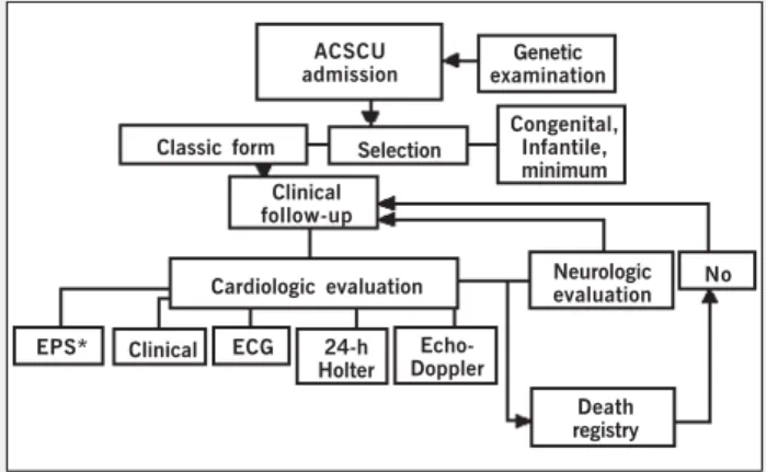
phy), or both, presence of myotonia, and measurement of the expansion of the CTG trinucleotide. The genetic diagnosis was obtained through analysis of the DNA extracted from peripheral blood leukocytes. On admission at the clinical unit of artificial cardiac stimulation, all patients with classic myotonic dystrophy underwent cardiologic evaluation. The sequence of evaluations performed during this study are shown in figure 1.
Annual neurologic evaluation determined the degree of peri-pheral neuromuscular dysfunction. A classification to correlate the neuromuscular alterations with cardiac impairment was crea-ted for this study, because the classic classifications by Walton and Gardner-Medwin 4 were considered to be mainly directed to
other dystrophies and to be difficult to manage. The classification criteria adopted are shown in table I.
The genetic examination comprised the following stages: DNA extraction; electrophoresis of the fragments; transference of the fragments (Southern method 23); and determination of the size of
the unknown fragment.
On clinical cardiologic evaluation, the following symptoms were investigated: palpitation, syncope, and precordial pain. The mani-festations of functional capacity were also considered for assessing the functional class (FC) of heart failure (HF) according to the New York Heart Association (NYHA) criteria 10.
During follow-up, recurrence of symptoms, evolutionary behavior of the arrhythmias and of conduction disturbances were assessed. Mortality rate, causes and types of death were also assessed ac-cording to the criteria of Hinkle 11, and sudden death was assessed
according to the criteria of Kuller 12.
Beginning of follow-up corresponded to the date of the first assessment, and end of follow-up was considered the last clinical observation or death.
Evolutionary data of at least one year of all patients beginning the study were obtained, and, therefore, no losses occurred.
Prevalence was studied through the registration of the absolute
values of the occurrences and their percentages. The evolutionary behavior was assessed by using the chi-square test (qualitative parameters) and analysis of variance of the repeated measurements complemented by the Bonferroni test (quantitative parameters). The analyses of correlation were performed by using the Pearson correlation test. The Kaplan-Meier methodology was used to cal-culate the survival curves, and the differences between them were evaluated by using the log-rank test. The search for predictive factors of worse prognosis occurred initially through univariate analysis (Fisher exact and chi-square tests for qualitative parame-ters, and the Student t test for quantitative parameters). The parameters with P < 0.1 on univariate analysis were assessed by using the Cox proportional hazards model (multivariate analysis) aiming at identifying independent prognostic factors. In all evalua-tions, the significance level of 5% was adopted.
Results
All patients underwent at least 1 year of clinical and electro-cardiographic follow-up, and, after 6 years, 23 patients had com-pleted the entire study protocol. The follow-up duration ranged from 12 to 124 months (mean=42.17±30.63). Because of these expressive findings of evolutionary analysis, the 6-year follow-up was established as the pattern for analyzing the variables studied. The electrophysiological study was performed in 59 patients: 20 due to palpitation, syncope, or pain, and 39 for risk assessment. The findings of prevalence and natural evolution at the beginning and end of follow-up are shown in table II.
In the first year, 3 patients became asymptomatic, 2 after reversion of sustained atrial tachyarrhythmia and one after pace-maker implantation.
At the beginning of follow-up, one patient had type I second-degree atrioventricular block (AVB), and 2 patients had advanced intermittent AVB. Of the 24 patients with AVB, one evolved to advanced AVB, and 9 (15.25%) patients who did not have AVB evolved to first-degree AVB. Of the 54 (65.06%) patients with no intraventricular conduction disturbance (IVCD), 8 (14.81%) had some type of intraventricular disturbance, and of the 29 patients with any type of AVB, 4 (13.79%) evolved with worsening of the intraventricular conduction disturbance.
Of the arrhythmias occurring at the beginning of follow-up, sustained atrial arrhythmia manifested in 4 (4.82%) patients as follows: 3 (3.61%) patients had atrial flutter, and one (1.20%) patient had atrial fibrillation. No case of sustained ventricular tachycardia was documented.
The number of cytosine-thymine-guanine trinucleotide (CTG) repeats referring to the entire case series ranged from 100 to 2,333 (mean, 791.15; standard deviation, 439.92).
The findings referring to the case series subgroup “relatives”, comprising 8 parents and 12 children, are shown in table III.
Table I - Criteria adopted for classifying the neuromuscular impairment in myotonic dystrophy
Activity degree Running Walking Stair climbing Standing up Eating Mild No No support required (abnormal posture) No support required No support required Alone Moderate No Support required Support required Support required Alone
Severe No No No No With help
ACSCU admission
Genetic examination
Classic form Selection
Congenital, Infantile, minimum Clinical
follow-up
Cardiologic evaluation Neurologicevaluation No
EPS* Clinical ECG 24-h Holter
Echo-Doppler
Fig. 1 - Study design - Sequence of evaluations performed during the study in a schematic form. Annual clinical cardiovascular and neurological assessment and ECG; Holter and echo every 2 years. * The electrophysiological study (EPS) was performed on the occasion of symptoms of low cerebral flow or on deciding about risk evaluation. ACSCU- Artificial Cardiac Stimulation Clinical Unit.
3
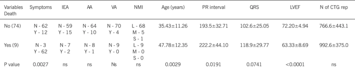
Nine (11%) patients died during follow-up. Sudden death ac-counted for 44% of the deaths (4 patients: 2 patients with first-degree AVB, LBBB, and definitive pacemaker; one patient with first-degree AVB; and one patient with left bundle-branch con-duction disturbance). Two patients died due to congestive heart failure (2 siblings with first-degree AVB, LBBB, definitive pace-maker, and myocardial dysfunction), and 3 others had noncardiac death (one due to melanoma, and 2 due to pneumonia). Table IV shows the univariate analysis of qualitative and quantitative varia-bles in regard to the outcome death.
In regard to multivariate analysis, the LVEF on Doppler echo-cardiography was the only independent predictive factor of evolution to death (P<0.0001).
For defining the rhythm of death and factors of influence, survival curves were elaborated (fig 2). Curve 1 shows the survival rate of the entire case series, and curve 2 shows the survival rate according to LVEF (≤ 60% and > 60%).
Fifty-nine patients underwent electrophysiological study, 14 (23.73%) of whom had H-V interval ≤ 55 ms; 25 patients (42.37%) had H-V between 55 and 70 ms; and 20 patients (33.90%) had H-V > 70 ms.
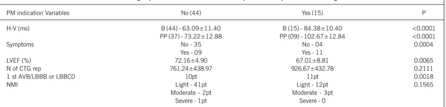
The H-V interval at baseline was measured in all patients who underwent electrophysiological study. However, under pharmaco-logical stress (postprocainamide), the H-V interval was measured in 46 patients, and the mean values of that interval were 74.37±13.16 (for patients with H-V < 100 ms) and 112±6.16 (for patients with H-V > 100 ms). Procainamide could not be used in 13 patients because of hypotension or marked bradycardia. Because of these findings associated with the clinical mani-festations and functional clinical behavior, pacemakers were im-planted in 15 patients. The characteristics of these 15 patients compared with those of the 44 patients who underwent electro-physiological study, and for whom no artificial stimulation was indicated, are shown in table V.
The electrophysiological study was also very important for as-sessing tachyarrhythmias and for establishing the clinical correla-tions. No relevant induced tachyarrhythmias occurred. Sustained ventricular tachycardia was induced in 5 patients, 3 of whom had rapid and poorly tolerated tachycardia, one had monomorphic ta-chycardia, and one had branch-to-branch tachyarrhythmia.
Concomitant analysis of the findings of the H-V interval and induction of tachyarrhythmias on the electrophysiological study
al-Table III - Distribution of the subgroups of 8 families according to the mean age of symptom onset and number of CTG repeat, neuromuscular impairment, mean age of appearance of the eletrocardiographic alterations, tachyarrhythmias, and conduction system impairment
Variables Pt Mean age Mean N of CTG repeat NMI Age of disease AA Or VA, Conduction disturbances subgroups (standard deviation) (standard deviation) onset or both Isolated AVB/IVCD Associated AVB/IVCD Pai 8 51.5 (6.1) 433.4 (326.5) 8L 41.75 ± 4.98 3 (37.5%) 3 (37.5%) 2 (25%) Filho 12 25.5 (7.7) 1041.6 (583.5) 11 L 1M 20.25 ± 3.93 4 (33.3%) 2 (16.67%) 6 (50%) N of CTG rep number of CTG repeats; NMI neuromuscular impairment; AA atrial arrhythmias; VA ventricular arrhythmias; AVB atrioventricular block; IVCD -intraventricular conduction disturbance; L - light; M - moderate.
Table II - Prevalence and natural evolution of the variables analyzed
Variables Start (83 pts) 6 years (23 pts) P value Symptoms n (% pt) 18 (21.68) 13 (56.52) < 0.0005 FC I of HF n (% pt) 82 (98.80) 22 (95.65) ns PR interval >200ms n (%) 24 (28.92) 10 (43.48) < 0.0001 Mean PR (ms) 196 ± 35 212 ± 31
QRS duration >100ms n (%) 29 (34.94) 11 (47.83)
Mean QRS (ms) 102 ± 26 111 ± 27 < 0.0001 Atrial arrhythmias n (% pt) 10 (12.05) - na Ventricular arrhythmias n (%pt) 14 (16.87) 1 (3.0) ns Inactive electric area n (% pt) 27 (32.52) 9 (39.13) ns LVEF (%) 71 ± 6.11 68 ± 6.29 ns Mild NMI n (% pt) 77 (92.77) 16 (69.57) ns FC - functional class; HF - heart failure; LVEF - left ventricular ejection fraction; NMI - neuromuscular impairment; ns - nonsignificant; na - not applicable.
Table IV - Univariate analysis of qualitative and quantitative variables for the groups of patients who survived or died
Variables Symptoms IEA AA VA NMI Age (years) PR interval QRS LVEF N of CTG rep Death
No (74) N - 62 N - 59 N - 64 N - 70 L - 68 35.43±11.26 193.5±32.71 102.6±25.05 72.20±4.94 766.6±443.1 Y - 12 Y - 15 Y - 10 Y - 4 M - 5
S - 1
Yes (9) N - 3 N - 7 N - 8 N - 9 L - 9 47.78±12.35 222.2±44.10 118.9±29.77 63.33±8.69 992.6±375.0 Y - 62 Y - 2 Y - 1 Y - 0 M - 0
S - 0
4
lowed the establishment of therapeutic management in 19 (32.20%) patients. Pacemaker implantation was indicated in 15 patients, and the implantable cardiodefibrillator (ICD) was indicated in 4.
The findings of the electrophysiological study in regard to the occurrence of symptoms had totally distinct implications in defining the therapy. Patients undergoing pacemaker implantation (bra-dyarrhythmias) more often had symptoms (P=0.0142) with a relative risk of 2.234 (0.986 – 5.061) and odds ratio = 5.833 (1.485 – 22.91). On the other hand, symptomatic patients un-dergoing ICD implantation (tachyarrhythmias) showed no difference as compared with the asymptomatic patients (P=0.3246).
No correlation (Pearson correlation analysis) was observed between the results of the genetic study (number of CTG repeats)
and the variables of the arrhythmic events and conduction distur-bances: PR interval (R=0.2406), QRS duration (R=0.2699), occur-rence of atrial (R=0.0003) and ventricular (R=0.0011) tachyar-rhythmias, and measurement of the H-V interval (R=0.2349).
Discussion
Of the presentations of myotonic dystrophy, the classic form is the most prevalent 3,24 and is associated with CTG expansion in
the chromosome region 19q13.3 5. It was the first single-locus
disease recognized with the anticipation phenomenon, the genetic phenomenon of increase in disease severity and earlier disease beginning in successive generations 24,25.
The findings in this study reliably confirm the concept proposed by Church 6 that, in myotonic dystrophy, the heart is sooner or
later impaired.
In this study, which assessed cardiac involvement through the behavior of arrhythmic events and conduction disturbances, the consecutive inclusion of 83 cases of the classic form of myotonic dystrophy essentially followed neurological and genetic criteria.
The clinical evaluation of our patients showed that, at the beginning of the study, only 22% of the patients had cardiovascular symptoms (10% palpitations, 6% presyncope or syncope, and 6% precordial pain). After a 6-year follow-up, 56% were symptomatic (17% palpitations, 13% presyncope or syncope, and 26% precordial pain), which is a significant evolutionary difference (P < 0.0005). This represents a contribution to the literature, which so far had reported no natural evolution of those manifestations.
At the beginning of the study, 99% of the patients were in FC I heart failure, and none of them had complaints or a physical examination characteristic of cardiac decompensation. With evo-lution, however, heart failure was responsible for 2 deaths.
Some studies have shown rates of cardiac impairment expressed as electrocardiographic abnormalities 17 ranging from 37 to 80%,
involving short case series 16,21,22. The overall prevalence of conduction
disturbances in our study was 50% in the initial assessment and the greatest prevalences were as follows: sinus bradycardia, 20% of the cases; first-degree AVB, 29%; isolated or associated ASRBBB, 23%; LBBB, 16%; left bundle-branch conduction disturbance (LBBCD), 9%, more prevalent than RBBB, 1%. The latter finding is not in accordance with the data in the literature that indicate a greater prevalence of RBBB. The discordances may result from differences in the profile of the population, the follow-up duration,
Table V - Profile of the groups with and without indication for pacemaker implantation according to the EPS
PM indication Variables No (44) Yes (15) P H-V (ms) B (44) - 63.09±11.40 B (15) - 84.38±10.40 <0.0001
PP (37) - 73.22±12.88 PP (09) - 102.67±12.84 <0.0001 Symptoms No - 35 No - 04 0.0004
Yes - 09 Yes - 11
LVEF (%) 72.16±4.90 67.01±8.81 0.0065 N of CTG rep 761.24±438.97 926.67±432.78 0.2111 1 st AVB/LBBB or LBBCD 10pt 11pt 0.0018 NMI Light - 41pt Light - 12pt 0.1565
Moderate - 2pt Moderate - 3pt Severe - 1pt Severe - 0
LVEF left ventricular ejection fraction; LBBCD left bundlebranch conduction disturbance; AVB atrioventricular block; LBBB left bundlebranch block; NMI -neuromuscular impairment; B - basal; PP - postprocainamide.
Fig. 2 - Curve 1 - Total survival in a 6-year follow-up; curve 2 - survival according to LVEF (≤ 60% and > 60%).
%
100
80
60
40
20
0 (83) 12
(53) 24
(44) 36
(33)
48 (29)
60 (23)
72 (13)
84 Curve 01 Follow-up (months)
%
100
80
60
40
20
0
0 12 24 36 48 60 72 84
LVEF > 60
LVEF ≤≤≤≤≤ 60
p<0.0001
5
and the well-known cardiac abnormalities studied, although not all studies specified such factors 13,14,16,17,18,22. In regard to LBBB, the
findings are surprising because that disturbance is usually associated with the systemic diseases that impair the cardiovascular system and with the presence of left ventricular dysfunction, which was not observed in our case series at the beginning of follow-up. However, of the patients who died during follow-up, 6 (66.7%) had LBBB or left bundle-branch conduction disturbances.
The evolutionary electrocardiographic findings showed a sig-nificant progression (P<0.0001) of the PR interval and QRS du-ration after 6 years of follow-up, corresponding to a mean increment of 0.1 ms and 0.03 ms, respectively, every 3 years. The PR interval behavior was not assessed on the electrophysiologic study for identifying the site of impairment of the conduction system; however, the electrocardiographic characteristics (IVCD or bundle-branch block with enlargement of the QRS in 24 of 35 patients with first-degree AVB and considerable temporal variations) sug-gested that a nodal atrioventricular impairment did not occur, revealing a worse prognosis.
Yet on the electrocardiogram, the presence of an inactive electric area (pathologic Q waves) was observed in 33% of the cases, which had inactive electric areas in some ventricular site with predominance of the septal and lateral regions. These alterations may be attributed to the disarray in myocardial fibers (alteration in the sequence of activation) and to impairment in the perivascular musculature. These electrical findings, in the absence of clinical evidence of coronary heart disease, suggest focal myocardial lesion 9,16,26-29. In addition, the evolutionary results
of some patients have shown the following: a reduction in pseu-donecrosis, probably due to worsening of the conduction distur-bances that may mask these aspects on the electrocardio-gram9,18; and an increase in the number of inactive areas that
suggest the existence of myocardial disease progression, although in a very insidious form as compared with the evolution of the conduction system impairment.
No episode of paroxysmal sustained ventricular tachycardia occurred or was identified on 24-hour Holter. However, ventricular arrhythmia (more than 200 ventricular extrasystoles per hour or NSVT) was present in 17% of our patients in the initial evaluation, and in only 3% after 6 years of follow-up. Atrial tachycardia was present in 12% of the patients (5% sustained, of which 75% were atrial flutter and 25% were atrial fibrillation). After reversion of arrhythmia, all our patients with sustained atrial tachycardia were maintained on antiarrhythmic medication and without anti-coagulation, because no patient remained in atrial fibrillation.
In addition to intra-atrial and intraventricular conduction dis-turbances, the modification in refractoriness and disarray of the muscle fibers may participate in the arrhythmogenic substrate of those arrhythmias 9,13,19,22,26,30,31. The abnormalities documented
on the electrocardiogram and on Holter, which were transient at the beginning but established throughout the follow-up, represented instability in stimulus conduction or generation, or both.
Colleran et al 32 reported that the prevalence of atrial
tachyar-rhythmias, mainly atrial fibrillation, is greater in patients with the severe form of the disease. Our results are in accordance with that statement when they show lower rates in a group of patients, of whom 93% had the mild form of the disease on the first assessment. They also agree when they demonstrate the induction
of atrial tachyarrhythmia in 20% (12/59) of the patients undergoing electrophysiologic study. Nine of those 12 patients were on a follow-up longer than 3 years, and, of them, 55% evolved to more severe forms of the neuromuscular disease.
Unlike that which happens with the skeletal muscle, myo-cardial impairment is rarely identified in myotonic dystrophy. Few cases of ventricular failure have been reported. However, mild abnormalities in systolic and diastolic function have been docu-mented on echocardiography 9,29,33. Similarly to those findings,
we observed that, in 95% of the cases, LVEF was > 60%, and, in only one, it was < 50% (LVEF=47%). In a 3-year follow-up, 4 of 6 patients who had a reduction in LVEF (< 60%) died (2 sudden deaths and 2 deaths due to heart failure).
In the period between the first and last assessment, 19% of the case series had an insidious worsening in their neuromuscular disease, 56% of whom also had worsening in their conduction disturbances or arrhythmic events, or both. The low prevalence of cardiovascular symptoms at the beginning of the disease is due to the rare myocardial impairment in this phase. However, with the progression and worsening in the neuromuscular disease, sub-clinical myocardial dysfunction may occur 9,29,33.
In our case series, the presence of the anticipation phenomenon in regard to the neuromuscular disease was once again demons-trated. In 8 parent-child pairs, the size of the genetic defect is at least 2.4 times greater in the children than that in parents. Our results showed that, for the first time, the anticipation phenomenon was also present in the conduction system impairment, because in 5 of the 8 parent-child pairs (62%) the alterations related to the conduction system were much more severe than those present in the parents. The age of appearance of the alterations related to cardiac conduction occurred at least 20 years before in 80% of the children when compared with the age of appearance in parents (P<0.0001).
We believe that in the classic form, the earlier (second decade of life) the manifestations of neuromuscular disease occur, even when only myotonia, the greater the degree of impairment of the His-Purkinje system in young adults, who usually have the greater numbers of repeats. On the other hand, when symptom onset of the neuromuscular disease occurs late (third and fourth decades), the manifestations of the cardiac conduction disturbances should correlate with the time of evolution and the degree of phenotypic variation of the disease.
6
1. Batten FE, Gibb HP. Myotonia atrophica. Brain 1909;32:187-205.
2. Steinert H. Myopathologische Beitäge: uber das klinische und anatomische bild des muskelschwunds der myotoniker. Deutsch Z Nervenheilk 1909;37:58-104. 3. Harper PS. Myotonic dystrophy: the clinical picture. In. HARPER, P.S. Myotonyc
Dystrophy, 2nd edn. W.B. Saunders Co., London, Philadelphia, Toronto, Sydney,
Tokyo; 1989:13-36.
4. Walton JN, Gardner-Medwin D. Progressive muscular dystrophy and myotonic di-sorders. In. WALTON, Disorders of voluntary muscle (Livingstone, London), 1981. 5. Brook JD, McCurrach ME, Harley HG et al. Molecular basis of myotonic dystro-phy: expansion of a trinucleotide (CTG) repeat at 3’ end of a transcript encoding a protein kinase family member. Cell 1992;68:799-808.
6. Church S. The heart in myotonia atrophica. Arch Intern Med. 1967; 119, 176-81. 7. Phillips MF, Harper PS. Cardiac disease in myotonic dystrophy. Cardiovasc Res
1997; 33:13-22.
8. Cohen MB, Snow J, Merkatz KA et al. Supression of ventricular tachycardia by sotalol in myotonic dystrophy. Am Heart J1996;132:446-9.
9. Hartwing GB, Rao KR, Radoff FM et al. Radionuclide angiocardiographic analysis of myocardial function in myotonic muscular dystrophy. Neurology 1983;33:657-60. 10. Critéria Committee, New York Heart Association. Inc. Diseases of the heart and
blood Vessels. Nomenclature and Critéria for diagnosis., 6th ed Boston, Little,
Brown and co. 1964; 114.
11. Hinkle LE Jr, Thaler HT. Clinical classification of cardiac deaths. Circulation 1982; 65: 457-64.
12. Kuller LH. Sudden Death: definition and epidemiologic considerations. Prog Car-diovasc Dis 1980. Supplement 1;23:1-12.
References
13. Hiromasa S, Ikeda T, Kubota K et al. Ventricular tachycardica and sudden death in myotonic dystrophy. Am Heart J 1988;115:914-15.
14. Komajda M, Frnak R, Vedel J et al. Intracardiac conduction defects in dystrophia myotonica. Br Heart J 1980;43:315-24.
15. Lazarus A, Varin J, Ounnoushene Z et al. Relationships among electrophysiologic findings and clinical status, heart function, and extent of DNA mutation in myo-tonic dystrophy. Circulation 1999;99:1041-46.
16. Melacini P, Villanova C, Menegazzo E et al. Correlation between cardiac involve-ment and CTG trinucleotide repeat length in myotonic dystrophy. J Am Coll Cardiol 1995; 25:239-45.
17. Motta J, Guilleminault C, Billingham M et al. Cardiac abnormalities in myotonic dys-trophy: electrophysiologic and histopathologic studies. Am J Med 1979;67: 467-73. 18. Nguyen HH, Wolfe JT, Holmer DR Jr et al. Pathology of the cardiac conduction system in myotonic dystrophy: a study of 12 cases. J Am Coll Cardiol 1988; 11:662-71.
19. Pencic-Popovic B, Nagulic S, Cebasek R et al. Arrhythmias conduction defects in myotonic dystrophy. (Ambulatory Electrocardiographic Monitoring Study). Acta Cardiomiol 1992;6:119-26.
20. Perloff JK, Stevenson WG, Roberts NK et al. Cardiac disease in myotonic dystro-phy (Steinert’s disease): a prospective study of 25 patients. Am J Cardiol 1984; 54: 1074-81.
21. Prystowsky EN, Pritchett ELC, Roses AD et al. The natural history of conduction system disease in myotonic muscular dystrophy as determined by serial electrophy-siologic study. Circulation 1979;60:1360-64.
neuromuscular disease showed a clinical spectrum ranging from normal cardiovascular assessment to high risk of sudden death, both for conduction disturbances (18%) and arrhythmic events (6%). Twenty-one (25.3%) patients with mild impairment of the neuromuscular disease had, on follow-up, an important conduction system impairment, and, in 56% of those who had progression of the neuromuscular disease, concomitant worsening of the cardiac impairment (conduction system or LVEF, or both) was observed.
Total mortality was 11%. Cardiac death occurred in 6%, 2 siblings with first-degree AVB associated with LBBB, left ventricular dysfunction, and definitive pacemaker. Their cause of death was heart failure. Four sudden deaths occurred as follows: one patient had antecedent anteroseptal fibrosis and probable arrhythmic death; 2 patients had LBBB associated with first-degree AVB, 1 with a baseline H-V interval of 100 ms, and another with a pacemaker; one asymptomatic patient had a normal electrocar-diogram. This association of sudden death and the presence of areas of fibrosis and conduction disturbances shows the importance of the known physiopathological mechanism of fatal arrhythmia probably implicated in these cases.
Univariate analysis of the factors related to prognosis revealed the following predictive factors of cardiac death: age (P=0.0029); presence of symptoms (P=0.0027); measurement of the PR inter-val (P=0.0191); duration of the QRS complex (P=0.0741); and left ventricular ejection fraction on echocardiography (P=0.0001). Multivariate analysis through the Cox proportional hazards model showed that left ventricular ejection fraction was the only independent predictive factor of evolution to death.
Some studies aimed at identifying predictive risk factors by using electrophysiological study and correlating the results of the measurements of the H-V interval with the electrocardiographic findings of patients with myotonic dystrophy 34-36.
Our patients underwent electrophysiological study for symptom clarification (34%) or for risk assessment (66%). The H-V interval was increased in more than 50% of the cases and was the finding
responsible for the therapeutic indication of the bradyarrhythmias (pacemaker implantation) in 15%. Of those, 73% had first-degree AVB associated with LBBB or LBBCD, and these findings were statistically different (P<0.0001) in regard to the group with no indication for pacemaker implantation. Therefore, a group at risk for third-degree AVB and sudden death was identified for the first time in the literature.
Induction of clinically important ventricular tachyarrhythmias on the electrophysiological study defined some cases whose pa-thophysiological mechanism was clarified and the therapy defined: implantable cardiodefibrillator (ICD), ablation of the arrhythmogenic focus, and use of antiarrhythmic agents. Of the symptomatic patients with induced SVT, one was of the fascicular type (ablation) and 2 were rapid and poorly tolerated SVT (ICD implantation).
In conclusion, in patients with the classic form of myotonic dystrophy, the prevalence of arrhythmic events or conduction dis-turbances was 50% and worsened after a 6-year follow-up (80%), and a significant occurrence of first-degree AVB and intraventricular block was observed. Atrial flutter was the most frequent sustained arrhythmia. Cardiac impairment increased with the severity of the neuromuscular impairment, but the cardiac disease worsened more rapidly than the neuromuscular disease. Total and cardiac mortality were low (11% and 7%, respectively), sudden death accounted for half of it. The predictive factors for cardiac mortality were clinical manifestations, age, PR interval, QRS duration, and LVEF, the latter being the only independent predictive factor of prognosis. The electrophysiological study defined the therapy for bradyarrhythmias and identified the group at risk for pacemaker implantation.
Acknowledgments
7
22. Fragola PV, Luzi M, Caló L et al. Cardiac involvement in myotonic dystrophy. Am J Cardiol 1994;74:1070-72.
23. Farah SB. DNA segredos e mistérios. São Paulo; Sarvier, 1997.
24. Howeler CJ, Busch HF, Geraedts JP et al. Anticipation in myotonic dystrophy: fact or fiction? Brain 1989;112:779-97.
25. Zatz M, Passos-Bueno MR, Cerqueira A et al. Analysis of CTG repeat in skeletal muscle of myotonic dystrophy young and adult patients: when does the expansion occur? Hum Molec Genet 1995;4:401-6.
26. Badano L, Autoro C, Fragola P et al. Left ventricular myocardial function in myo-tonic dystrophy. Am J Cardiol 1993;71:987-91.
27. Gottdiener JS, Hawley RJ, Gay JA et al. Left ventricular relaxation, mitral valve prolapse and intracardiac conduction in myotonia atrophica: assessment by digi-tized echocardiography and noninvasive His bundle recording. Am Heart J 1982;104:77-85.
28. Moffa PF, Sanches PCR. O eletrocardiograma normal. In. Moffa PF, Sanches PCR. Eletrocardiograma Normal e Patológico. Roca 2001; 99-126.
29. Venco A, Saviotti M, Besana D et al. Noninvasive assessment of left ventricular function in myotonic muscular dystrophy. Br Heart J 1978;40:1262-66.
30. Grigg LE, Chan W et al. Ventricular tachycardia and sudden death in myotonic dystrophy: clinical, electrophysiologic and pathologic features. J Am Coll Cardiol 1985; 6: 254-58.
31. Merino JL, Carmona JR, Fernandez-Lozano I et al. Mechanisms of sustained ven-tricular tachycardia in myotonic dystrophy: implications for catheter ablation. Circulation 1998; 98:541-6.
32. Colleran JA, Hawley RJ, Pinnow EE et al. Value of the electrocardiogram in deter-mining cardiac events and mortality in myotonic dystrophy. Am J Cardiol 1997; 80: 1494-7.
33. Moorman JR, Coleman RE, Packer DL et al. Cardiac involvement in myotonic mus-cular dystrophy. Medicine 1985;64:371-87.
34. Andrade JCS, Ávila Neto V, Braile DM et al. Diretrizes para o Implante de Marca-passo Cardíaco Permanente: Consenso DECA / SBCCV. Arq Bras Cardiol 1999; 74: 475-80.
35. Babuty D, Fauchier L, Tena-Carbi D et al. Is it possible to identify infrahissian cardiac conduction abnormalities in myotonic dystrophy by non-invasive methods. Heart 1999; 82:634-7.