Bárbara Albertini Roquim Alcantara(a)
Marina Lara de Carli(a) Luiz Alberto Beijo(b)
Alessandro Antônio Costa Pereira(c)
João Adolfo Costa Hanemann(a)
(a) Department of Clinics and Surgery, School of Dentistry, Universidade Federal de Alfenas - UNIFAL, Alfenas, MG, Brazil.
(b) Institute of Exact Sciences, Universidade Federal de Alfenas - UNIFAL, Alfenas, MG, Brazil.
(c) Institute of Biomedical Sciences,
Universidade Federal de Alfenas - UNIFAL, Alfenas, MG, Brazil.
Corresponding Author: Marina Lara de Carli
E-mail: [email protected]
Correlation between inflammatory
infiltrate and epithelial lining in 214
cases of periapical cysts
Abstract: The aim of this study was to evaluate the prevalence of peri-apical cysts, identify their clinical and microscopic features and corre-late their microscopic features with the inlammatory iniltrate present in the lesion site. A total of 214 cases were collected over a 10-year period. Clinical data, including gender, age, race, symptoms and location of the lesion, were recorded. Two independent examiners with no prior knowl-edge of the patients’ clinical data conducted the microscopic evaluations. Statistical analyses were performed using Fisher’s or chi-square tests at a 5% level of signiicance. The results showed that periapical cysts were more prevalent in white women, with a mean age of 35 years, and in the anterosuperior region. The majority of the lesions were lined by atrophic cystic epithelium, which was associated with moderate inlammatory in-iltrate in the cystic capsule (p < 0.01), with a diffuse localization pattern (p = 0.03) and absence of neutrophils (p = 0.01). Our indings suggest that periapical cysts lined by atrophic epithelium are related to the pres-ence of moderate mononuclear inlammatory iniltrate.
Descriptors: Inlammation; Radicular Cyst; Biopsy.
Introduction
Periapical cysts—also known as radicular, periradicular or apical periodontal cysts—are inlammatory jaw cysts on teeth with infected and necrotic pulp. The cysts are a direct sequela of apical granulomas, although not every apical granuloma will develop into a cyst.1
Radio-graphically, a periapical cyst has an imaging appearance similar to that of a periapical granuloma. Although cysts are often larger than granu-lomas, the size of the lesion cannot be used as a deinitive criterion for diagnosis. As the lesion becomes larger, it is more likely to be a cyst.2
Cystic transformation occurs owing to inlammatory stimulation of the epithelial rests of Malassez in the periodontal ligament, secondary to a root canal infection, in which the bacteria and their products, that were previously conined to the dental pulp, go beyond the root canal system and reach the periapical tissues.1,3
Periapical cyst formation seems to be induced by an acute inlamma-tory reaction,4 although mononuclear cells are commonly found in the
inlammatory iniltrate.5 T- and B-lymphocytes and macrophages
consti-tute most of the inlammatory cells involved.6 Macrophages are observed
primarily in active inlammatory sites, mainly in the subepithelial region
Declaration of Interests: The authors certify that they have no commercial or associative interest that represents a conflict of interest in connection with the manuscript.
Submitted: Feb 12, 2013
Accepted for publication: Jul 31, 2013 Last revision: Aug 13, 2013
and seldom in the surrounding area.7 Additionally,
the thin and thick cystic capsules present differenc-es with regard to the inlammatory cells involved. Thin capsules have a predominance of lymphocytes, whereas thick capsules are predominantly composed of plasma cells and macrophages.8 Inlammatory
cell count and degree of vascularization in periapi-cal cysts have shown a strong association with in-creased expression of tumor necrosis factor-alpha (TNF-α),8 indicating that inlammatory cells may
have an important role in the development of these lesions.
The pathogenesis and growth mechanisms of in-lammatory odontogenic cysts of the jaws are not fully understood.9 The inlammatory iniltrate in
the cystic lining appears to play an important role in the pathogenesis of periapical cysts.10 Therefore, the
purposes of the present study were to evaluate the prevalence of periapical cysts, identify their clini-cal and microscopic features and correlate their mi-croscopic features with the inlammatory iniltrate present in the lesion.
Methodology
Records of all the cases diagnosed as periapical cysts in the 2000 to 2010 period in the Anatomic Pathology Laboratory of our institution were re-trieved. Periapical cysts were deined as follows:
1. a lesion seated on the periapical region of a non-vital tooth, and
2. histologic evidence of stratiied non-keratinized squamous epithelium completely or partially lin-ing a cystic cavity or tissue fragments.
Clinical data, including gender, age, race, symp-toms, duration of the lesion up to the dental con-sultation and location were recorded. The study was carried out after the approval of the institutional Ethics Committee (protocol #140/2010) on research in human beings.
Qualitative and quantitative microscopic analyses
The material used in this study consisted of peri-cystic biopsy tissues from routine cyst surgeries sent to our institution’s Anatomic Pathology Laboratory
and immediately ixed in 10% neutral buffer forma-lin at room temperature. Five-µm parafin-embed-ded specimens were mounted on microscope slides and stained with hematoxylin and eosin (HE). Two independent examiners (J.A.C.H. and A.A.C.P.) with no prior knowledge of the patients’ clinical data conducted the microscopic evaluations using an optical binocular microscope (Axiostar Plus, Carl Zeiss, Jena, Germany) equipped with a 40×/0.65 objective (Achroplan, Carl Zeiss, Jena, Germany), thus achieving a 400× magniication. Whenever a disagreement arose, a consensus approach was ad-opted.
The following tissue structures were evaluated:
• cystic cavity;
• cystic epithelial lining and its thickness;
• characterization, distribution and intensity of inlammatory iniltrate;
• presence of congested and hyperemic blood vessels; and
• presence of cholesterol crystals.
The quantitative microscopic analysis was per-formed according to the method described by Jurisic
et al.8 Each specimen was graded under 400×
mag-niication as:
• slight, < 10 inlammatory cells per ield;
• moderate, 10–50 inlammatory cells per ield, and
• intense, more than 50 inlammatory cells per ield.
Grading of each specimen was based on the av-erage inlammatory cell number in three consecu-tive microscopic ields starting from the epithelial-connective tissue border and proceeding gradually deeper into the lamina propria. The thickness of the epithelial lining was evaluated in respect to the number of cell layers and described as:
• atrophic, < 6 epithelial cell layers;
• normal, 6–10 epithelial cell layers; and
• hyperplastic, more than 10 epithelial cell layers.
The lesions with a variable number of layers were classiied as follows:
phils associated with slight inlammatory iniltrate was observed in the cystic capsule (Table 2), and, when present, they were associated with congested and hyperemic blood vessels (p < 0.01). Further-more, cholesterol crystals were seen in 13.6% (29) of the lesions (Figure 2).
Discussion
The clinical diagnosis of oral lesions can be tricky owing to the similar appearance of many conditions presenting different etiology. Therefore, there is a risk of misdiagnosis and delayed treatment if the therapeutic choice is based solely on clinical aspects.11 A study in the New Zealand population
revealed that a correct clinical provisional diagno-sis of periapical cyst was given in only 36% of the
• atrophic and hyperplastic; or
• normal and hyperplastic.
Statistical analysis
Statistical analyses were performed using SPSS 17.0 (SPSS Inc., Chicago, USA) for Windows soft-ware (Microsoft, Mountain View, USA). Fisher’s or chi-square tests were used for the analyses, and p
values lower than 0.05 were considered signiicant.
Results
Periapical cysts corresponded to 6.21% (214 cases) of all the cases diagnosed over a 10-year pe-riod. Patient age ranged from 7 to 77 years old, with a mean age of 35 years. Regarding gender, 46.7%
(110) of the periapical cysts were diagnosed in men, and 53.3% (114) in women. Additionally, most of the patients (78.7%) were white. Concerning loca-tion, the lesions occurred mainly in the anterosupe-rior (41.7%) and posterosuperior (31%) regions. The majority of the lesions (56.8%) had a maximum di-ameter of 1 cm, and 71% of them were asymptom-atic. Recurrence was reported in 6.54% of all cases. Microscopic analysis revealed that 83.6% (179) of the lesions presented partial epithelial lining, and 66.4% (142) presented atrophic lining. Atrophic epi-thelial lining was associated with moderate inlam-matory iniltrate in the cystic capsule (Table 1), with diffuse location (p = 0.03) and with the absence of neutrophils (p = 0.01). The inlammatory iniltrate was found to be moderate in 54.7% (117) of the cas-es, it had a diffuse location in 65.1% (136) and was predominantly composed by lymphocytes, plasma cells and macrophages (Figure 1). Lack of
neutro-Inflammatory infiltrate
Thickness of epithelial lining
Normal Atrophic Hyperplastic Normal and atrophic
Normal and hyperplastic
Atrophic and hyperplastic
Absent 1 0 0 0 0 0
Slight 7 36 2 6 1 0
Moderate 7 80 4 20 4 2
Intense 1 26 5 5 1 6
p value* 0.001
* Chi-square test at 5% significance level.
Table 1 - Intensity of inflammatory infiltrate according to thickness of epithelial lining in periapical cysts.
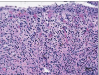
Figure 1 - Periapical cyst. Cystic wall covered by atrophic stratified squamous epithelial lining exhibiting moderate mononuclear inflammatory infiltrate (H&E; original magni-fication, 400×).
cases,12 which highlights the importance of knowing
the clinical, radiographic and microscopic aspects of the lesion. Diagnostic conirmation can only be as-certained after anatomic pathology examination.13
According to Becconsall-Ryan et al.,12
inlam-matory lesions corresponded to 72.8% of radiolu-cent lesions of the jaws. Among them, 29.2% were periapical cysts. Among Brazilians, periapical cysts were the most frequent odontogenic cysts in adults (66.5%).14 In the present study, periapical cysts were
more prevalent in white women, with a mean age of 35 years, and in the anterosuperior region, cor-roborating data found in the related literature.5,14
However, other authors have reported a similar prevalence in both genders12 and a mean age of 44.3
years.15 These discrepancies may be attributed to
differences in sample selection and size, as well as the diagnostic criteria adopted by the authors of dif-ferent studies.
There is continuing controversy regarding the kind of inlammatory iniltrate present in periapi-cal cysts. Marçal et al.5 found that mononuclear
in-iltrate was signiicantly more frequent than mixed iniltrate, and that the latter was present in lesions with istulae. Lin et al.,15 however, found a mixed
iniltrate in 52.1% of the lesions. Microscopic analy-sis of our samples revealed that the presence of con-gested and hyperemic blood vessels was associated with intense inlammatory iniltrate in the cystic capsule (p < 0.01) and with the presence of neutro-phils (p < 0.01), features which characterize lesions in the acute inlammatory phase. However, most of the lesions presented moderate inlammatory in-iltrate, which was associated with the presence of
plasma cells (p = 0.01) and macrophages (p < 0.01). These results are in agreement with those of Lin et al.,15 and may justify the fact that 71% of the lesions
in our study were asymptomatic, which is compat-ible with chronic inlammation (mononuclear inil-trate). The presence of plasma cells in cysts suggests a local humoral immune reaction, and indicates that the majority of the lesions were in a developing stage.6
Another histopathological feature evaluated was the presence of cholesterol crystals, which were ob-served in 13.6% of periapical cysts, a rate similar to that found by Santos et al.16 The major source of
cholesterol may be from locally dying inlammatory cells, and a result of the disintegrating membranes of these cells in long-standing lesions.17
Accumula-tion of cholesterol crystals can prevent healing in apical periodontitis lesions,17 but this accumulation
does not seem to be associated with the maintenance of periapical cysts inasmuch as the frequency of its occurrence is low in these lesions.
In the present study, almost all periapical cysts presented stratiied squamous epithelial lining, similarly to the indings of Lin et al.15 It is believed
that epithelial status may be related to the growth of periapical cysts.18 The majority of the lesions
evaluated in our study had atrophic epithelial lin-ing, which was associated with moderate inlamma-tory iniltrate in the cystic capsule (p < 0.01), with a
Table 2 - Intensity of inflammatory infiltrate according to presence of neutrophils in periapical cysts.
Inflammatory infiltrate Neutrophils
Absent Present
Slight 49 3
Moderate 70 47
Intense 15 28
p value* < 0.001
* Chi-square test at 5% significance level.
Figure 2 - Periapical cyst. Cholesterol crystals and foreign body-type reaction with giant cells present in the capsule (H&E; original magnification, 200×).
diffuse location pattern (p = 0.03), and absence of neutrophils (p = 0.01). Nevertheless, according to Moreira et al.,18 no difference can be seen in the
in-tensity of the iniltrate in lesions with atrophic or hy-perplastic epithelium. These authors found different expression patterns of CD57, in which lesions with atrophic epithelium presented a higher percentage of CD57-positive cells. As the expression of CD57 is indicative of immunosuppression, it may constitute a negative immunomodulator of cystic growth.
Among the periapical lesions, periapical cysts presented the worst prognosis, and the larger le-sions presented the worst evolution.19 It has been
suggested that the tissues of periapical cysts are self-sustaining because they do not depend on the pres-ence or abspres-ence of root canal infection. Therefore, because periapical cysts are less prone to heal after conventional endodontic therapy, surgical inter-vention is needed.13 For the treatment of periapical
cysts, enucleation is the most commonly used tech-nique,20 although Carrillo et al.19 performed cyst
enucleation in combination with apicoectomy and retrograde illing. To achieve satisfactory periapical healing, surgical removal of a periapical cyst must include elimination of root canal infection.13
Conclusion
Periapical cysts with atrophic epithelial lining were related to moderate mononuclear inlamma-tory iniltrate in the cyst capsule. In addition, our indings demonstrated that the majority of the peri-apical cysts were asymptomatic, chronic lesions. It is imperative that the endodontist be well acquainted with the clinical, radiographic and microscopic fea-tures of periapical cysts in order to perform early diagnosis and establish proper treatment, thus in-creasing the success rates of endodontic therapy.
Acknowledgments
The authors wish to thank FAPEMIG (Fundação de Amparo à Pesquisa do Estado de Minas Gerais) for supporting this study.
References
1. Nair PNR. New perspectives on radicular cysts: do they heal?. Int Endod J. 1998 May;31(3):155-60.
2. White SC, Sapp JP, Seto BG, Mankovich NJ. Absence of radio-metric differentiation between periapical cysts and granulo-mas. Oral Surg Oral Med Oral Pathol. 1994 Nov;78(5):650-4. 3. Meghji S, Qureshi W, Henderson B, Harris M. The role of
endotoxin and cytokines in the pathogenesis of odontogenic cysts. Arch Oral Biol. 1996 Jun;41(6):523-31.
4. Nair PNR, Sundqvist G, Sjögren U. Experimental evidence supports the abscess theory of development of radicular cysts. Oral Surg Oral Med Oral Pathol Oral Radiol Endod. 2008 Aug;106(2):294-303. DOI: 10.1016/j.tripleo.2008.04.009. Epub 2008 Jun 13.
5. Marçal JRB, Samuel RO, Fernandes D, Araujo MS, Napimoga MH, Pereira SAL, et al. helper cell type 17/regulatory T-cell immunoregulatory balance in human radicular cysts and periapical granulomas. J Endod. 2010 Jun;36(6):995-9. DOI: 10.1016/j.joen.2010.03.020.
6. Liapatas S, Nakou M, Rontogianni D. Inflammatory infiltrate of chronic periradicular lesions: an immunohistochemical study. Int Endod J. 2003 Jul;36(7):464-71.
7. Rodini CO, Lara VS. Study of the expression of CD68+ macro-phages and CD8+ T cells in human granulomas and periapical cysts. Oral Surg Oral Med Oral Pathol Oral Radiol Endod. 2001 Aug;92(2):221-7.
8. Jurisic V, Terzic T, Colic S, Jurisic M. The concentration of TNF-α correlate with number of inflammatory cells and de-gree of vascularization in radicular cysts. Oral Dis. 2008 Oct;14(7):600-5. DOI: 10.1111/j.1601-0825.2007.01426.x. Epub 2008 Jan 21.
9. Teronen O, Hietanen J, Lindqvist C, Salo T, Sorsa T, Eklund KK, et al. Mast cell-derived tryptase in odontogenic cysts. J Oral Pathol Med. 1996 Aug;25(7):376-81.
10. Cury VC, Sette PS, Silva JV, Araújo VC, Gomez RS. Immu-nohistochemical study of apical periodontal cysts. J Endod. 1998 Jan;24(1):36-7.
11. Kondori I, Mottin RW, Laskin DM. Accuracy of dentists in the clinical diagnosis of oral lesions. Quintessence Int. 2011 Jul-Aug;42(7):575-7.
12. Becconsall-Ryan K, Tong D, Love RM. Radiolucent inflam-matory jaw lesions: a twenty-year analysis. Int Endod J. 2010 Oct;43(10):859-65. DOI: 10.1111/j.1365-2591.2010.01751.x. Epub 2010 Aug 3.
13. Lin LM, Ricucci D, Lin J, Rosenberg PA. Nonsurgical root ca-nal therapy of large cyst-like inflammatory periapical lesions and inflammatory apical cysts. J Endod. 2009 May;35(5):607-15. DOI: 10.1016/j.joen.2009.02.012.
profile in a Brazilian population over a 38-year period. Med Oral Patol Oral Cir Bucal. 2010 Jul 1;15(4):e583-90. 15. Lin H-P, Chen H-M, Yu C-H, Kuo R-C, Kuo Y-S, Wang
Y-P. Clinicopathological study of 252 jaw bone periapi-cal lesions from a private pathology laboratory. J Formos Med Assoc. 2010 Nov;109(11):810-8. DOI: 10.1016/S0929-6646(10)60126-X.
16. Santos LCS, Vilas Bôas DS, Oliveira GQV, Ramos EAG, Gurgel CAS, Santos JN. Histopathological study of radicu-lar cysts diagnosed in a Brazilian population. Braz Dent J. 2011;22(6):449-54.
17. Nair PNR. On the causes of persistent apical periodontitis: a review. Int Endod J. 2006 Apr;39(4):249-81.
18. Moreira PR, Santos DFM, Martins RD, Gomez RS. CD57+ cells in radicular cyst. Int Endod J. 2000 Mar;33(2):99-102. 19. Carrillo C, Peñarrocha M, Bagán JV, Vera F. Relationship
Between histological diagnosis and evolution of 70 periapi-cal lesions at 12 months, treated by periapiperiapi-cal surgery. J Oral Maxillofac Surg. 2008 Aug;66(8):1606-9. DOI: 10.1016/j. joms.2007.12.014.