Asdrubal Falavigna
1, Jorge Luiz Kraemer
2ABSTRACT - We report a case of infrasellar craniopharyngioma in a 34 year-old woman who presented with progressive headache and diplopia. Computed tomographic and magnetic resonance images showed a heterogeneous tumor originating from the sphenoid bone with ethmoid sinus and sella turcica extension. A sublabial rhinoseptal transsphenoidal surgery was performed. Craniopharyngiomas with infrasellar development are very rare. Infrasellar craniopharyngioma is uncommon, thirty-five cases has been reported in literature. The embryology, clinical features and radiographic investigation of these tumors are discussed.
KEY WORDS: craniopharyngioma, sphenoid sinus, skull base tumor.
Craniofaringioma infra-selar: relato de caso
RESUMO – Relatamos um caso de craniofaringioma infra-selar em uma paciente de 34 anos com sintomas de cefaléia e diplopia. A investigação radiológica com tomografia computadorizada e ressonância magnética de encéfalo demonstrou um tumor heterogêneo localizado no osso esfenoidal e com extensão para o seio etmoidal e sela turcica. Realizada cirurgia pela via transesfenoidal. A ocorrência de craniofaringioma com topografia infra-selar é incomum, havendo relato na literatura de 35 casos. A literatura é revisada, sendo discutidas a embriologia, a apresentação clínica e características radiológicas do tumor.
PALAVRAS-CHAVE: craniofaringioma, seio esfenoidal, tumor de base de crânio.
1Professor Assistente da Disciplina de Neurologia da Faculdade de Medicina da Universidade de Caxias do Sul, Pós-Graduando em
Neurocirurgia da Universidade Federal de São Paulo - Escola Paulista de Medicina, São Paulo SP, Brasil (UNIFESP-EPM); 2Professor do
curso de Pós-Graduação em Cirurgia da Universidade Federal do Rio Grande do Sul (UFRGS) e da Fundação Faculdade de Ciências Médicas de Porto Alegre (FFCMPA-ISCMPA), Mestre pela UFRGS e Doutor em Neurocirurgia pela UNIFESP- EPM.
Received 14 November 2000, received in final form 18 January 2001. Accepted 29 January 2001.
Dr. Asdrubal Falavigna - Rua Dr. Moreira César 271/sala.1 - 95034-000 Caxias do Sul RS - Brasil. E-mail: [email protected] Craniopharyngioma is a rare tumor1. It arises
wi-thin the sella turcica and expands mainly into the suprasellar region2. Occasionally, the tumor can
oc-cur without sellar involvement. These tumors rarely extend below the sellar floor into the sphenoid si-nus or invade the pharynx and the nasal cavities3-34.
The infrasellar craniopharyngioma may then origi-nate anywhere along the tract of the obliterated craniopharyngeal duct, which would include the sphenoid bone, vomer, and nasopharynx35,36.
In this study we report a case of infrasellar cranio-pharyngioma with the epicenter situated in the sphe-noid sinus and review the 35 other reported cases in the literature of this unusual localization since 1924.
CASE
A 34 years-old woman presented with frontal head-ache with progression of 7 months and diplopia that ap-peared in the last month. Clinical evaluation revealed left abducens nerve palsy. No signs and symptoms of
pitu-itary dysfunction were evident. The laboratory studies, including hypophyseal function were normal.
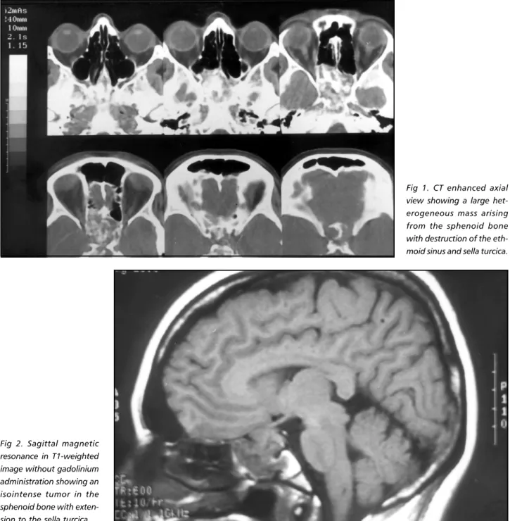
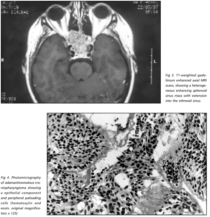
Coronal computed tomographic (CT) scans after intra-venous contrast showed a large sphenoid sinus heteroge-neous mass with destruction of the ethmoid sinus and sella turcica (Fig 1). T1-weighted (plain and gadolinium-enhanced 600/11/2 [repetion-time/echo time/excitations]) and T2-weighted (3000/108/1) magnetic resonance sho-wed isointense image (Fig 2). Post-contrast the brightly en-hancing, heterogeneous mass was clearly visible in the sphe-noid bone (Fig 3). There was extension of the tumor to ethmoid sinus and to the sella turcica. Carotid and verte-bral angiography revealed that the tumor was avascular.
Arq Neuropsiquiatr 2001;59(2-B) 425
Fig 1. CT enhanced axial view showing a large het-erogeneous mass arising from the sphenoid bone with destruction of the eth-moid sinus and sella turcica.
Fig 2. Sagittal magnetic resonance in T1-weighted image without gadolinium administration showing an isointense tumor in the sphenoid bone with exten-sion to the sella turcica.
and the nasal packing removed on the third postoperative day. The patient was discharged from the hospital on the eight postoperative day with recovery of the abducens ner-ve palsy and headache-free. Histologic examination re-vealed an adamantonomatous craniopharyngioma (Fig 4). The patient was referred for radiotherapy. Conforma-tional radiotherapy consisting of a total of 55Gy (30 frac-tions) was administered 1 month later. At clinical follow-up, 2 year after irradiation, her neurological examination and hormonal studies were normal.
DISCUSSION
Craniopharyngiomas are benign but aggressive epithelial neoplasms, which comprises approximately
3% of all intracranial tumors1. They are generally
found intracranially with a similar frequency in chil-dren and adults but with a slight preponderance in children between 5 and 15 years of age2. Equal sex
incidence has been noted in groups of children2.
Craniopharyngiomas are most commonly located ex-traaxially in the sellar or suprasellar area in 90% of cases2. They can extend to the anterior (2-5% of
cases), middle (2%), or posterior (1-4%) cranial fossa, and infrasellar extension is found in about 5% of cases34. Rarely, craniopharyngiomas arise primarily
fis-sure and cerebellopontine angle3-34,37-43. The growth
of a craniopharyngioma in the infrasellar region is extremely rare, only 36 cases were found in the lit-erature (Table 1).
The infrasellar craniopharyngioma reported in lit-erature has a similar sex distribution, being 3 times more frequent above 15 years of age. The micros-copic appearance of most craniopharyngiomas shows an external layer of high columnar epithe-lium, a variable amount of polygonal cell, and a cen-tral network of epithelial cells. Two clinicopathologic varieties have been delineated microscopically: the
papillary squamous type, which is seen almost ex-clusively in adults and carries a better prognosis; and the classical adamantinomatous variant, which de-velops mainly in children and has a worse overall outcome41.
Treatment of these tumors is mostly excisional, through a craniotomy or transsphenoid approach. Complete removal of the tumor is the preferred pro-cedure2,29. When vital structures are involved and
excision is compromised by significant risks of mor-bidity and mortality, subtotal removal of the tumor followed by supplemental radiotherapy is the pre-Fig 3. T1-weighted gado-linium enhanced axial MRI scans, showing a heteroge-neous enhancing sphenoid sinus mass with extension into the ethmoid sinus.
Arq Neuropsiquiatr 2001;59(2-B) 427
ferred treatment29. Radiotherapy, once considered
ineffective, is currently the most often used adju-vant treatment. Craniopharyngiomas have been found to be radiosensitive, and radiotherapy plays a major role in preventing recurrence and improving survival42.
Embryology - According to Warwick and Will-iams43, during the fourth week of gestation an
ectodermally lined diverticulum (Rathke’s pouch) develops in the roof of the stomodeum just anterior to the oropharyngeal membrane. This pouch ascends cranially traversing the mesenchyme to meet the neuroectoderm of the infundibulum (neurohypo-physis), which descends as a neural outgrowth from the floor of the third ventricle (diencephalon) of the embryonic brain. Rathke’s pouch differentiates into the anterior lobe of the pituitary, the adenohypo-physis. This course traversed by Rathke’s pouch forms a cord of cells joining the stomodeal ectoderm to the future adenohypophysis in embryo. Later in embryonic life this cord disintegrates, leaving an obliterated craniopharyngeal canal. It is a tract that runs from the anterior part of the hypophyseal fossa of the sphenoid bone to the junction of the poste-rior septum of the nose with the palate, which is the stomodeal end of the recess. The pharyngeal hypo-physis (functioning adenohypophyseal tissue), which remains in adults, is a caudal remnant of this cord.
Types of origin - There are several theories regard-ing the origin of infrasellar craniopharyngioma. Most of them are based on the embryologic development of the adenohypophysis. Mott and Barret35 in 1899
were the first to postulate that these tumors might arise from the remnants of the pharyngeal hypophy-sis. Erdheim36 proposed in 1904 that the
craniophar-yngioma originated from the remnants of the oblit-erated craniopharyngeal duct, suggesting that these tumors can arise anywhere along the tract of migra-tion of Rathke’s pouch from the vomer, the roof of the nasopharynx, through the midline sphenoid bone beneath the floor of the sella turcica.
Clinical presentation - Symptoms and clinical find-ings are related to the craniopharyngioma’s local-ization and mass effect with compression of the sur-rounding structures (Table 1). The symptomatology of suprasellar tumor is often characterized by de-fects in the visual fields (bitemporal or homonimus hemianopsia), varying signs of pituitary insufficiency (diabetes insipidus, amenorrhoea, diminished libido and cachexia), and the symptoms of increased
in-tracranial pressure (headache, vertigo and cranial nerve deficit) that occur relatively late in the course according to Carmel et al.2 and Sato quoted by
Fujitani et al.16. The signs of pituitary dysfunction
appear early in the cases in which the tumor expands within the sella. The tumor located at the sphenoid sinus usually presents with headache and cavernous sinus syndrome6-8,11,12,15,17,20,23,24,27. The
craniopharyn-giomas located in the nasopharyngeal region usu-ally present with frontal headache, nasal obstruction, epistaxis, nasopharyngeal and/or nasal fossa mas-ses3,5,9,10,13,15,18,19,22,25,26,29,32,34. Our case had symptoms
of headache, diplopia due to left abducens nerve palsy, without pituitary insufficiency, which suggests that the tumor had appeared in the sphenoid bone and, later, extended to the sella turcica region.
Localization - Craniopharyngiomas usually origi-nate intracranially, grow along the pituitary stalk, on the axis of the sella-infundibulum and located below the brain, above the pituitary, behind the optic chiasma, and within the circle of Willis. Occasionally there is an intrasellar one and, rarely, one within the body of the sphenoid or nasopharynx in the tract of the former craniopharyngeal canal proper. Since 1924, 35 cases of infrasellar involvement by cranio-pharyngioma have been described in the medical lit-erature. In these cases the tumors are situated at the nasopharyngeal region, the sphenoid sinus, the maxillary sinus and usually involved the sella turcica and the supra sellar region. The most common loca-tion of infrasellar craniopharyngioma has been the sphenoid sinus either alone, 4 cases, or combined with other sites, 28 cases (Table 1). The tumor loca-tion in the case presented could have been exten-sions from the sella turcica or derivatives of pharyn-geal canal remnants. In the former situation, the tumor could be a typical craniopharyngioma that arose in the sella turcica and then went downwards into the sphenoid sinus. In the latter, the tumor origi-nated in the infrasellar region, sphenoid sinus, and then grew extradurally and superiorly. We suspect that this tumor was a craniopharyngioma that origi-nated in the infrasellar region because it is situated mainly in the sphenoid sinus, the pituitary hormones were always normal and we could not find the dura mater of the sellar floor during the transsphenoidal approach.
A
rq
N
e
u
ro
p
siq
u
ia
tr
2
0
0
1
;5
9
(2
-B
)
to left side
Drummond (5) 1938 14/F NPX, SS, ST Nasal obstruction, headache, loss or decreased Skull x-ray vision, NPX and NF mass
Sato acc. Futitani 1944 56/F NPX, SS, ST, S Visual loss Skull x-ray et al. (16) 6)
Northfield (6) 1957 54/F SS Headache, vomiting, homonymous, hemianopia (L) Skull x-ray Hamberger et al. (7) 1960 25/M SS Headaches, face hypoesthesia in territory Skull x-ray
of infraorbital nerve (R)
Johnson (8) 1962 39/M SS, ST, S Headache, diplopia, vision loss (R) Skull x-ray, tomogram Podoshin et al. (9) 1970 15/F NPX, SS, ST Nasal obstruction, NPX and NF mass Skull x-ray
Isayama (11) 1970 62/F SS, ST, S Facial pain, diplopia Skull x-ray Trible (13) 1970 71/M NPX, SS, ES, ST Headaches, diplopia, nasal obstruction, epistaxis Skull x-ray Cooper & Ransohoff (14) 1972 16/M NPX, SS, S (middle fossa) Headache, epistaxis, CSF rhinorrhea, diplopia, lack of Skull x-ray
hearing (L), palpable mass in the zygomatic area (L)
Prasad & Kwi (10) 1975 55/F NPX, SS Nasal obstruction, NPX and NF mass Skull x-ray, tomogram, angiogram 48/M NPX, SS, ST Nasal obstruction, headache, diplopia with Skull x-ray, tomogram
lateral rectus weakness (L), facial hypoesthesia in territory of infraorbital nerve (L), NPX and NF mass
Ishiyama (12) 1977 41/F SS, ST, S Facial pain Skull x-ray, tomogram 25/M SS Headache, diplopia Skull x-ray, tomogram Illum et al. (15) 1977 14/F NPX, SS, ST Headache, diplopia with abducens nerve palsy (L), Tomogram, angiogram,
temporal hemianopsia (L) pneumo-encephalography Majlessi et al/ (18) 1978 17/M NPX, SS, ST, S Headache, bitemporal hemianopsia, Skull x-ray
A
rq
N
e
u
ro
p
siq
u
ia
tr
2
0
0
1
;5
9
(2
-B
)
4
2
9
Fujitani et al . (16) 1979 18/F NPX, SS, ES, MS, orbit (R) Exophthalmos, visual disturbance Skull x-ray, angiogram, scintigraphy, cystography CT Pheline et al. (20) 1981 12/M SS Headache, diplopia with abducent nerve palsy Skull x-ray, angiogram pneumogram Mukada et al. (21) 1984 13/M NPX, SS, ST, S Visual disturbances, panhypopituitarism Skull x-ray, tomograms, CT,
angiogram, pneumogram
Lewin et al (20) 1984 27/F NPX Epistaxis, NPX mass Skull x-ray, tomogram, angiogram, CT Maier (23) 1985 77/M NPX, SS, ST Headache, CSF rhinorrhea Skull x-ray, tomogram
Maiuri et al. (22) 1987 25/F NPX, SS, ST Headache, nasal obstruction Skull x-ray, tomograms, CT Hillman et al. (24) 1988 64/M SS, ST, S Visual disturbances, abducent nerve palsy (L) CT, MRI
Benitez et al. (26) 1998 29/M NPX, SS, ES Nasal obstruction, pressure in the nose Skull x-ray, CT, MRI, angiograms and behind the eyes
Ortiz et al. (25) 1998 20/M NPX Epistaxis CT Pharaboz et al. (26) 1989 27/F NPX, SS Nasal obstruction CT, MRI
Akimura et al. (28) 1989 12/F NPX, SS, ES, ST S, MS Visual disturbance CT, MRI, angiogram Byrne & Sessions (29) 1990 29/M NPX, SS, ES, MS Nasal obstruction, NPX na NF mass Skull x-ray, CT, MRI Gili & Garcia (17) 1991 9/M NPX, SS Nasal obstruction, diplopia with Skull x-ray, CT, MRI
abducent nerve palsy (R)
Bret & Beziat (30) 1993 16/F NPX, SS Nasal obstruction CT, MRI Sener ((34) 1994 8/M NPX, SS, ES, S Headache, decreased vision, nasal obstruction CT, MRI
Kanungo et. al (32) 1995 40/F NPX, SS, S Headache, nasal obstruction Skull x-ray, CT, MRI Cheddadi et al. (31) 1996 Premature/F NPX Respiratory insufficiency Nasal endoscopy, CT
Jiang et al. (33) 1998 7/M ES Epistaxis CT
Chakrabarty et al. (40) 1998 46/M NPX, SS, ST Nasal obstruction, double vision CT, MRI
NPX, nasopharynx; SS, sphenoid sinus; ST, sella turcica; S, suprasellar; ES, ethmoid sinuses; MS, maxillary sinus; NF, nasal fossa; CSF, cerebrospinal fluid; MRI, magnetic resonance imaging; CT, computed tomography; [L],
and cystic components, calcification, multicysts, lyctic lesions, irregular enhancement. Multiple calcifica-tions are observed within the tumor, especially in younger patients. In adults, craniopharyngiomas are often not calcified17,19,21-23,26,27,29,30,32,35,40. Magnetic
resonance image clearly showed the tumor exten-sion, cystic portions, mixed intensity signal, inhomo-geneous or heteroinhomo-geneous enhancement 17,23,25-30,32,34,40.
CONCLUSION
Craniopharyngiomas with infrasellar develop-ment are very rare. Up to now, thirty-five cases has been reported in literature since 1924. The most common location of infrasellar craniopharyngioma has been the sphenoid sinus either alone or com-bined with other sites. The infrasellar craniopharyn-gioma may then originate anywhere along the tract of the obliterated craniopharyngeal duct, which would include the sphenoid bone, vomer, and na-sopharynx.
REFERENCES
1. Kernohan JW. Tumors of congenital origin. In Minckler J. Pathology of the nervous system. New York:Mc-Graw-Hill, 1971:1927-1937. 2. Carmel PW, Antunes JL, Chang CH. Craniopharyngiomas in children.
Neurosurgery 1982;11:382-389.
3. Bock E. Beitrag zur pathologie der hypophyse. Virchow Arch Pathol Anat 1924;252:98-112.
4. Zeitlin H. Adamantinomas of the hypophyseal stalk and sphenoid bone. Am J Cancer 1935;23:729-740.
5. Drummond WAD. Infrasellar adamantinoma. Proc R Soc Med 1938;32:200-207.
6. Northfield DWC. Rathke-pouch tumours. Brain 1957;80:293-312. 7. Hamberger CA, Hammer G, Norlén , Sjögren B. Surgical treatment of
craniopharyngioma: radical removal by the transantrosphenoidal ap-proach. Acta Oto-Laryngol 1960;52:285-292.
8. Johnson NE. Craniopharyngioma: review with a discussion of transpalatal approach. Laryngoscope 1962;72:1731-1749.
9. Podoshin L, Rolan L, Altman MM, Peyser E. Pharyngeal craniopha-ryngioma. J Laryngol Otol 1970;84:93-99.
10. Prasad U, Kwi NK. Nasopharyngeal craniopharyngioma. J Laryngol Otol 1975;89:445-452.
11. Isayama Y. Changes in visual field associated with changes of tension in large cyst, which is craniopharyngioma, extending to the middle cranial fossa. Acta Soc Ophthalmol Jpn 1970;74:596-604.
12. Ishiyama R, Yasue S, Sanada S, Yuki K, Nakamura N. Rare cases of craniopharyngioma extending into the nasopharynx: two cases report. Neurol Med Chir 1977;17.
13. Trible WM. Destructive lesions of the sphenoid. South Med J 1970;63:849-852.
14. Cooper PR, Ransohoff J. Craniopharyngioma originating in the sphe-noid bone. J Neurosurg 1972;36:102-106.
case report. J Neurosurg 1978;49:119-120.
19. Lewin F, Ruffolo E, Saraceno C. Craniopharyngioma arising in the pharyngeal hypophysis. South Med J 1984;77:1519-1523.
20. Pheline C, Jamois Y, Engel PH, Leguyarder B, Penot JC. Craniopha-ryngiome ectopique basi-sphénoidal: abord par voie basse, naso-septale, puis antro-ethmoïdale. Neurochirurgie 1981;27:221-224.
21. Mukada K, Mori S, Matsumura S, Uozomi T, Goishi J. Infrasellar cran-iopharyngioma. Surg Neurol 1984;21:565-571.
22. Maiuri F, Corriero G, Elefante R, Cirillo S, Giamundo A. Craniophar-yngioma of the cranial base and nasopharynx. Surg Neurol 1987;27:191-194.
23. Maier HC. Craniopharyngioma with erosion and drainage into the nasopharynx. An autobioghaphical case report. J Neurosurg 1985;62:132-134.
24. Hillman TD, Peyster RG, Hoover ED, Nair S, Finkelstein SD. Infrasellar craniopharyngioma:CT and MR studies. J Comput Assist Tomogr 1988;12:702-704.
25. Ortiz RM, Sola JJM, Martinez FA, Garcia JA, Prado JH. Craneofaringio-ma de cavum: aportatión de un caso. Acta Otorrinolaring Esp 1988; 39:119-121.
26. Pharaboz C, Merran S, Cordoliani Y, Rizzoli P. Craniopharyngiome “rhinopharyngé”. J Radiol 1989;70:573-575.
27. Benitez WI, Sartor KJ, Angtuaco EJC. Craniopharyngioma presenting as a nasopharyngeal mass: CT and MR findings. J Comput Assist Tomogr 1988;12:1068-1072.
28. Akimura T, Kameda H, Abiko S, Aoki H, Kido T. Infrasellar craniophar-yngioma. Neuroradiology 1989;31:180-183.
29. Byrne MN, Sessions DG. Nasopharyngeal craniopharyngioma: Case report and literature review. Ann Otol Rhinol Laryngol 1990;99:633-639.
30. Bret PH, Beziat JL. Crâniopharyngiome sphénoïdo-naso-pharyngé: un cas avec exérèse radicale par maxillotomie de le fort I. Neurochirurgie 1993;39:235-240.
31. Cheddadi D, Triki S, Gallet S, et al.. Obstruction rhinopharyngée néonatale par craniopharyngiome. Arch Pediatr 1996;3:348-351. 32. Kanungo N, Just N, Black M, Mohr G, Glikstein R, Rochon L.
Nasopha-ryngeal craniopharyngioma in an usual location. AM J Neuroradiol 1995;16:1372-1374.
33. Jiang RS, Wu CY, Jan YJ, Hsu CY. Primary ethmoid sinus craniopharyn-gioma:a case report. J Laryngol Otol 1998;112:403-405.
34. Sener RN. Giant craniopharyngioma extending to the anterior cranial fossa and nasopharynx. AM J Roentgenol 1994;162:441-442. 35. Mott FW, Barret JOW. Three cases of tumor of the third ventricle. Arch
Neurol 1899;1:417-440.
36. Erdheim J. Ueber hypophysengangsgeschwulste und hirncholeste-ratome. Akad Wiss Wien 1904;113:537-726.
37. Altinörs N, Senveli E, Erdogan A, Arda N, Pak I. Craniopharyngioma of the cerebellopontine angle, case report. J Neurosurg 1984;60:842-4. 38. Ragoowansi AT, Piepgras DG. Postoperative ectopic
craniopharyngio-ma: case report. J Neurosurg 1991;74:653-655.
39. Long DM, Chou SN. Transcallosal removal of craniopharyngioma within the third ventricle. J Neurosurg 1973;39:563-567.
40. Chakrabarty A, Mitchell P, Bridges L. Craniopharingioma invading the nasal and paranasal spaces, and presenting as nasal obstruction. Br J Neurosurg 1998;12:361-363.
41. Adamson TE, Wiesler OD, Kleihaus P, Yasergil MG. Correlation of clini-cal and pathologiclini-cal features in surgiclini-cally treated craniopharyngiomas. J Neurosurg 1990;73:12-17.
42. Wen DY, Seltjeskog EL, Haines SJ. Microsurgical management of cran-iopharyngioma. Br J Neurosurg 1992;6:467-474.