1. Perfusionist. São José do Rio Preto Pediatric Cardiovascular Surgery Service – Hospital de Base – São José do Rio Preto Medical School, São José do Rio Preto, SP, Brazil.
2. Professor Livre Docente; São José do Rio Preto Medical School and Unicamp. Editor of Brazilian Journal of Cardiovascular Surgery. 3. Pediatric Cardiovascular Surgeon. São José do Rio Preto Pediatric Cardiovascular Surgery Service – Hospital de Base – São José do Rio Preto Medical School, São José do Rio Preto, SP, Brazil. 4. Resident physician. São José do Rio Preto Pediatric Cardiovascular
Surgery Service – Hospital de Base – São José do Rio Preto Medical School, São José do Rio Preto, SP, Brazil.
5. Professor Livre Docente. Deputy Director of the São José do Rio Preto Medical School, São José do Rio Preto, SP, Brazil. 6. Cardiovascular Surgeon. Cardiovascular Surgery Service; Instituto
Domingo Braile, São José do Rio Preto, SP, Brazil.
7. Perfusionist. São José do Rio Preto Pediatric Cardiovascular Surgery Service – Hospital de Base – São José do Rio Preto Medical School, São José do Rio Preto, SP, Brazil.
Renata Geron FINOTI1, Domingo Marcolino BRAILE2, Ulisses Alexandre CROTI3, Marcos Aurélio Barboza de OLIVEIRA4, Moacir Fernandes de GODOY5, João Carlos Ferreira LEAL6, Sebastião Rodrigues POLICARPO7, Marden Leonardi LOPES8
RBCCV 44205-1000
Avaliação de oxigenador de membrana infantil em ovinos
Evaluation of infant membrane oxygenator in
sheep
Abstract
Objective: To analyze the security and efficacy of a new
membrane oxygenator, the so-called OXM – 1500.
Methods: From May 2005 to September 2006, six sheep
of Santa Inês breed (5 male and 1 female, respectively) were studied. The average body weight was 14.1 (±5) kg, body surface 0.6 (±0.2) m2 and a mean age 3.8 (±1.5) months. All
of them were submitted to extracorporeal circulation (CEC) with evaluation at 10, 30, 60, 120, 180 and 240 minutes. The following values were obtained: values of oxygen transference (TTO2) and carbon dioxide transference (TTCO2), haemoglobin (HBS) and free haemoglobin (HBL), the score of platelets and of leucocytes, and heat transference rate.
Results: TTO2 and TTCO2 were adequate. Lesion of the majority formed blood elements was insignificant; there no modifications in HBS, HBL levels; platelets and leucocytes decreased over time. Heat exchange was effective (p < 0.05).
Conclusions: The membrane OXM – 1500 infant
oxygenator, tested in sheep, showed adequate oxygenation capacity, CO2 removal capacity, and small alteration of haemoglobin and platelets without significant decrease of
leucocytes, as expected. Heat exchanger connected to the oxygenator was efficient in temperature changes.
Descriptors: Extracorporeal membrane oxygenation.
Extracorporeal circulation. Sheep.
Resumo
Objetivo: Analisar a segurança e a eficácia de um novo
oxigenador de membrana denominado OXM -1500.
Métodos: No período de maio de 2005 a setembro de 2006,
foram estudados seis ovinos da raça Santa Inês, sendo cinco machos e uma fêmea, com peso corpóreo médio de 14,1 (±5) kg, superfície corpórea de 0,6 (±0,2) m2 e idade média de 3,8
(±1,5) meses. Todos foram submetidos a circulação extracorpórea (CEC) com avaliação nos tempos 10, 30, 60, 120, 180 e 240 minutos, obtendo-se os valores de taxa de transferência de oxigênio (TTO2) e de taxa de transferência de gás carbônico (TTCO2), hemoglobina sérica (HBS) e livre (HBL), plaquetometria, leucometria e taxa de transferência de calor.
Resultados: Houve adequadas TTO2 e TTCO2. A lesão da maioria dos elementos figurados do sangue foi
8. Engineer; Empresa Braile Biomédica S/A®, São José do Rio Preto, SP, Brazil.
CONFLICT OF INTEREST STATEMENT: The author declares that she has competing interests. This study was funding by Braile Biomédica S/A®, from which the author is an employee.
This study was carried out at the São José do Rio Preto Medical School Biotherium, São José do Rio Preto, SP, Brazil.
Correspondence adress: Renata Geron Finoti
Avenida São Judas Tadeu, 790 - apto 34, bloco C - São José do Rio Preto – SP – Brasil. CEP: 15075-290
E-mail: [email protected]
INTRODUCTION
The membrane oxygenator was used for the first time, in 1958, in an experiment using dogs undergoing cardiac surgical procedures performed by Clowes and Neville [1]. In 1970s, the membrane oxygenator becomes discarded and miniaturized to be used in children [2].
In Brazil, a new generation of membrane oxygenators manufactured especially for pediatric perfusion, allowed a dramatic perfusate volume reduction and, consequently, better surgical outcomes [3-5].
In a constant search for improvements in pediatric cardiovascular surgeries, a new oxygenator, the OXM-1500, was developed by Braile Biomédica S/A®, being evaluated in sheep for safety and efficacy.
METHODS
From May 2005 to September 2006, six membrane oxygenators OXM-1500 were tested in 26 sheep of the Santa Inês sheep breed. Five animals were male, age of 3.8 (±1.5) months, mean body weight of 14.1 (±5) kg, and body surface of 0.6 (±0.2) m2. All animals received care according to the guidelines of the USA Committee of the Institute of Laboratory Animal Resources (ILAR) do National Research Council [6].
Tests were performed at the São José do Rio Preto Medical School (FAMERP) Biotherium. The project was approved by the FAMERP Animal Experimentation Ethics Committee, number 001 00 5067/2006.
The oxygenation system OXM-1500 consists of a membrane oxygenator, a venous reservoir, and a heat exchanger coupled to the oxygenator.
The oxygenator circuit was filled with a 65 mL of non-blood prime solution, and consisted of a hollow-fiber microporous polypropylene oxygenation chamber. The fibers were woven in a fashion to keep the spacing among them and the coiled tissue steady under constant tension. A computer microprocessor was used to control the circuit in order to keep gas exchange in a predictable fashion.
The venous reservoir stored a maximum volume of 2000 mL and consisted of polycarbonate.
The heat exchanger manufactured with microtubes made of polyester, allowing an excellent heat transfer performance and low-volume fulfillment.
Initially, the animal was weighted and blind-folded with a crepe bandage in order to avoid stress. The external jugular vein was punctured with a 14-gauge peripheral intravascular catheter (Jelco Medex® - São Paulo - SP).
Anesthesia was performed using Ketamine chlorhydrate, 7 mg/kg, and atropine sulfate, 0.02 mg/kg.
Next, the animal was intubated using an endotracheal cannula (Portex® - USA), which caliber was according to the trachea diameter; mechanical ventilation was established using a device Samurai III modelo 674 (Takaoka® - São Paulo - SP). A 22Fr nasogastric feeding tube (Sondoplast® - São Paulo - SP) was also used.
Anesthetic induction was continued with propafenone, 0.02 mg/kg; thiopental sodium, 0.4 mg/kg; midazolam maleate, 0.15 mg/kg; and atracurium, 0.04 mg/kg. The maintenance was performed using sevorane inhalation associated with propafenone and atracurium endovenously according to the needs and the anesthetic plan desired. In addition, as an antibiotic prophylaxis, amicacine sulphate, 7 mg/kg, and cephalothin sodium, 25 mg/kg were administered endovenously.
For monitoring purposes, the femoral artery was dissected in order to control the mean arterial pressure and connected to the pressure monitor model BPM 2000 (Braile Biomédica S/A® - São José do Rio Preto - SP). Once electrocardiogram was performed, peripheral saturation was measured through a Dixtal monitor model DX-2010. Using an esophageal sensor, animal temperature was monitorized through Braile Biomédica Measurement Unit, model 62009 – TAG-UM-002L.
After placing the animal in a right lateral recumbent position, chest trichotomy and antisepsis using polyvinylpyrolidone iodine (PVPI) were performed aiming at to prepare the animal to be incised. Left thoracotomy was started and heparin sodium, 4 mg/kg, was administered aiming at preventing coagulation.
At that time, the first laboratory exams, such as gasometry, hematometry, leukometry, platelet count, and activated clotting time (ACT) were performed. Animal temperature was also checked.
insignificante, sem alterações dos níveis de HBS, HBL, plaquetas e o número de leucócitos diminuíram com o tempo. A troca de calor foi efetiva (p < 0,05).
Conclusão: O oxigenador de membrana infantil
OXM-1500, testado em ovinos, mostrou-se com capacidade adequada de oxigenação, remoção de gás carbônico e pequena alteração
da hemoglobina e plaquetas, com diminuição do número de leucócitos de forma esperada. O trocador de calor acoplado ao oxigenador foi eficaz nas variações de temperatura.
Descritores: Circulação extracorpórea com oxigenador de
Then, an arterial cannula was introduced into the aorta e venous cannulae in both superior and inferior vena cava (Braile Biomédica® - São José do Rio Preto - SP). The diameter was chosen according to the animal weight.
Cardiopulmonary bypass (CPB) circuit with a new oxygenator on trial (OXM-1500) was filled with sodium Ringer Lactate intravenous solution (Baxter® São Paulo -SP), in a dose of 340 to 800 mL according to the animal weight; 5 ml/kg of 20% intravenous mannitol, and sodium heparin, 0.6 mL.
Aiming at measuring the pressures in the oxygenator, 3/16-inch Luer connectors were placed in the oxygenator entrance and exit. Immediately after CPB was started and venous drainage stabilization achieved, blood flow was standardized in 2.6 L/min/m2.
With the assistance of a chronometer from the Braile Biomédica Measurement Unit, model 62009 TAG UM -0001L, CPB times and animal temperatures were recorded. Blood harvesting was performed at 10, 30, 60, 120, 180, and 240 minutes of CPB.
At the beginning of CPB, gas mixture (FiO2) was fixed in 80% and O2 flow in 1 liter to each liter of blood flow, being posteriorly altered according to temperature variation and the needs of the living organism, which were based on the venous and arterial gasometry results.
The gas analyzer Roche® model OMNIC TAG G5 -0011L, calibrated to be used in sheep was employed throughout the procedure to measure gasometric data (pH, PCO2, PO2, BE) of venous and arterial blood.
Arterial blood hematocrit, hemoglobin, leukometry and platelet count were measured in the ordinary way, analyzing serum hemoglobin and free hemoglobin.
The expired fraction carbon dioxide (ETCO2) was measured using a Dixtal® Capnograph model DX-1265.
Medicinal compressed air/oxygen concentration used in the OXM-1500 was accomplished using a calibrated gas mixer (Blender - Braile Biomédica® - São José do Rio Preto - SP).
After 120 minutes of CPB, the animals were submitted to hypothermia at 25ºC and kept in this temperature for up to 10 minutes; posteriorly, they were warmed in order to test the efficacy of the heat exchanger coupled to the oxygenator OXM-1500.
Perfusion interruption was performed at 240 minutes of CPB; arterial and venous cannulae were removed and heparin sodium neutralized with protamine sulphate in a ratio of 1:1.
The animal’s thorax was drained with a ¼-inch diameter tube and sutured in planes.
Muscle relaxant was reverted with neostigmine methylsulfate in doses of 0.01 to 0.02 mg/kg. The animal was placed in a supine position until being completely awakened, when it was extubated. After thorax drain was
removed, the animal was sent back to its bail, remaining there for a 24-hour observation period.
In data analysis, the oxygen transfer rate (OTR) and the carbon dioxide transfer rate (CDTR) was calculated using the following formulas: OTR = Qs*(PO2-A/760)-(PO2-V/ 760))*aO2)+((SaO2-SvO2)/100)*(1,34*12/100)), where “Qs” = the flow rate (mL/min), “A” = arterial, “V” = venous, “Sa” = arterial oxygen saturation, and “a” = plasma oxygen solubility (fixed value of 0.0031); CDTR = 10*BF(L/ mim)*ETCO2(%)*(R G/B), where “BF” = blood flow, “ET” = expired carbon dioxide fraction, “RG/B” = the ratio between gas and blood.
Statistical data considered a p value ≤ 0.05 as significant. The Kruskal-Wallis test was used to analyze nonparametric data. Analysis was performed with statistical software StatDirect 1.6.0 for Windows®.
RESULTS
After cardiopulmonary bypass all the animals remained alive.
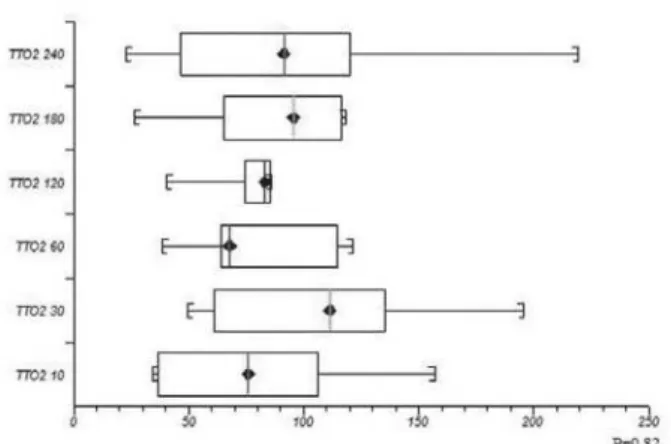
OTR was not shown to be statistically altered (p=0.82) in different times, presenting the following measurements: 75.8 mL/min; 111.3 mL/min; 67.7 mL/min; 82.9 mL/min; 95.6 mL/min; and 91.3 mL/min at the respective times of 10, 30, 60, 120, 180, and 240 minutes, according to Figure 1.
CDTR was not shown to be statistically altered (p=0.50) in different times, presenting the following measurements: 36.5 mL/min; 24.5 mL/min; 33.5 mL/min; 25.5 mL/min; 32.5 mL/min; and 35.0 mL/min at the respective times of 10, 30, 60, 120, 180, and 240 minutes, according to Figure 2.
There was no statistically significant alteration between serum and free hemoglobin levels (p=0.94 and p=0.85, respectively). Serum hemoglobin level prior to CPB was 7.1 mg/dL, with the following values: 6.5 mg/dL; 6.5 mg/dL; 6.5 mg/dL; 6.6 mg/dL; 6.4 mg/dL; and 6.0 mg/dL. Free hemoglobin level prior to9 CPB was 19.6 mg/dL; after CPB was 17.2 mg/dL; 18.3 mg/dL; 16.6 mg/Dl; 19.1 mg/dL; 19.1 mg/dL; and 20.0 mg/dL at the respective times of 10, 30, 60, 120, 180, and 240 minutes, according to Figures 3 and 4.
Platelet count prior to CPB was 42800 and it did not change with a longer CPB time (p=0.07), presenting the following measurements: 33200; 27400; 25300; 22800; 17350; and 21200 at the respective times of 10, 30, 60, 120, 180, and 240 minutes, according to Figure 5.
Leukocyte dosage prior to CPB was 5745; however, after CPB it was found to be significantly altered (p=0.02), presenting the following values: 3185; 3200; 5605; 4860; 4855; 3840 at the respective times of 10, 30, 60, 120, 180, and 240 minutes, according to Figure 6.
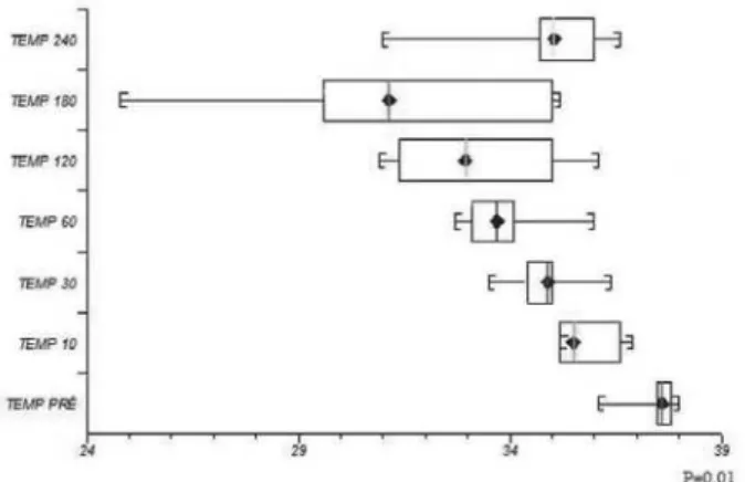
Heat exchange was effective when observing that the median temperature prior to CPB was 37.6ºC, presenting a significant change during CPB (p=0.01) with the following Fig. 2 – Carbon dioxide transfer rate in relation to cardiopulmonary
bypass times
Fig. 3 – Earlier doses of serum hemoglobin and at different cardiopulmonary bypass times
Fig. 4 – Earlier doses of free hemoglobin and at different cardiopulmonary bypass times
Fig. 5 – Earlier dosages of platelets and at different cardiopulmonary bypass times
measurements: 35.5ºC; 34.9ºC; 33.7ºC; 33.0ºC; 31.2ºC; and 35.1ºC at the respective times of 10, 30, 60, 120, 180, and 240 minutes, according to Figure 7.
Fig. 7 – Earlier temperature and during cardiopulmonary bypass
DISCUSSION
Membrane oxygenators, since their development, have shown to be superior to bubble oxygenators. The evolution of medicine associated to that of Engineering has allowed great improvement, reducing the size of the products and significantly improving their performance, which allowed using smaller and smaller children.
Volume of blood (volemia) and performance capacity were always difficulties to be overcome, thus there is a large range of people and companies in a constant search to develop and improve the oxygenators to be used in pediatric cardiovascular surgery.
Considering the facility to the extent which the organic systems can be affected during pediatric infusion, the OXM-1500 was tested in sheep, because these animals mimic similar conditions to those ones found in humans during CPB. There were no accidents or technique complications during our procedures with the sheep.
During the “open heart” surgeries, the use of oxygenators is of critical importance in blood oxygenation and carbon dioxide removal. In attempt to promote an oxygenation as close as possible to the physiological oxygenation, the membrane oxygenators were idealized and built [7,8].
These types of oxygenators allow an independent control of the oxygen transfer to the blood and the removal of the carbon dioxide.
The oxygen transfer occurs by diffusion and depends on both membrane permeability and gas pressure difference between both sides of the membrane. Preferably, capillary membrane oxygenators are currently used in which the blood flows externally to the bundle of fibers, aiming at to reduce the resistance of gas transfer to the blood [9].
In order the blood oxygenation to occur, the oxygen should pass over the oxygenator membrane, mix to the plasma, diffuse into the red blood cells, and thus bind to the hemoglobin. Regarding the gasometric exams, we can observe that the OXM-1500 showed throughout the procedure high oxygenation capacity with satisfactory OTR during CPB time.
The removal of CO2 from the tissues to the oxygenator occurs in a simpler manner due to its solubility being 20 times over that of the oxygenator. The ideal oxygenator should avoid CO2 retention, which triggers the process of respiratory acidosis or its excessive elimination, which leads the organism to a process of respiratory acidosis. It should also keep partial blood pressure compatible with temperature and level of existing metabolism, what can be observed in the OXM-1500 when it showed an excellent level of CDTR, demonstrating to be simple and safe.
Because the organism understands CPB as a great aggressor agent, many reactions of hemodynamic, physical, and chemistry character occur during the perfusion process. Blood cells are subjected to several and different actions from the normal circulation. Red blood cells, leukocytes, and platelets are profoundly affected by physical trauma and by blood contact with the circuit surface [10,11]. For the purpose of measuring these lesions, cellular behavior was analyzed, as shown in Figures 3 to 6. Closely related to the red blood cell, the hemoglobin carries oxygen from the lungs to the tissues, and carries carbon dioxide from the tissues to the lungs by means of a number of chemistry reactions. It consists of four heme radicals bound to the protein globulin, which plays an important role in gas changes performed by the oxygenator during CPB.
When a red blood cell is injured, serum hemoglobin is released into the plasma, then being called free hemoglobin. The amount of free hemoglobin in the normal human plasma is of 8 mg% and during perfusion reaches up to 50 mg%, with a certain frequency. The value achieved depend on the quality of the equipment, technique used, and duration of perfusion. Reaching values close to 100-150 mg%, free hemoglobin starts being eliminated by urine, and the production of hemoglobinuria suggests intense cellular trauma [12,13]. The results we obtained were within the normal patterns expected for an adequate performance of a pediatric oxygenator.
during the perfusion due to CPB circuit, and even by the planned anticoagulation performed prior to CPB. Just in the first two minutes of CPB, there is a platelet decrease by 20% of its baseline value [14,15]. This platelet variation is well represented in Figure 5, which shows a marked platelet deletion when the number of platelets before CPB is compared to that found after 180 minutes of perfusion.
Moreover, the platelet lesion releases inflammatory mediators, such as the thromboxane A2 and the interleukins, which promote tissue vasoconstriction and hypoxemia, leading to tissue acidosis. The membrane oxygenators present lower platelet lesion when compared to the bubble oxygenators due to the interface between blood and gas, suggesting a better behavior in relation to platelet sequestration and inactivation [9].
Important organ protective agent, the leukocytes are units capable of migrating to sites where their action is needed. The activation of the complementary system leads to leukocyte activation, causing, a number of times, the interruption of the normal endothelial barrier, tissue edema, and organ failure [16,17]. Of the blood elements, the one which is most affected by the destructive influence of CPB circuit is the leukocyte due to the direct trauma, followed by cellular destruction or functional deficit. During CPB, the leukocytes migrate towards the lungs, releasing toxic substances that cause increased vascular patency and interstitial edema. CPB is often associated to early neutropenia, followed by neutrophilia in the immediate postoperative period [16,17].
Generally, the results reported for the leukocytes did not present statistical significance, but when compared alone, it was observed leukocyte activation when compared to the number of leukocytes prior to CPB and after 180 minutes of perfusion.
The only variable presenting statistical significance was the temperature, which was already expected, because since the beginning of CPB, the sheep looses heat to environment radiation through the tubes (isothermia) and short before 180 minutes, the animal cooling was performed in order to trigger hypothermia, reaching a temperature of 24.8ºC, with the purpose of testing the heat exchanger coupled to the oxygenator, then the animal was warmed up to 240 minutes. These data are confirmed and easily visualized in Figure 7. Heat exchangers are devices coupled to the oxygenators allowing the performance of thermal exchanges between the blood and the circulating environment (water) by convection. Moreover, the oxygenators are built using good heat conduction materials and with a large contact surface with the purpose of favoring thermal exchanges [18]. In the described oxygenator, the exchanger was built of polyester hollow-fiber, as well as the existing oxygenators, which presents an excellent heat transfer performance with a low filling volume.
REFERENCES
1. Clowes GHA, Neville WE. Membrane oxygenator. In: Alle JG, ed. Extracorporeal circulation. Springfield:CC Thomas;1958.
2. Peirce EC 2nd, Thebaut AL, Kent BB, Kirkland JS, Goetter WE, Wright BG. Techniques of extended perfusion using a membrane lung. Ann Thorac Surg. 1971;12(5):451-70.
3. Novello WP. Dispositivo para oxigenação e remoção de dióxido de carbono do sangue em circuitos de circulação extracorpórea [Tese de doutorado]. Campinas:Universidade Estadual de Campinas;1996. 102p.
4. Moscardini AC, Godoy MF, Braile DM, Godoy JMP, Soares MJ, Brandi AC, et al. Oxigenação extracorpórea por membrana e alterações hematológicas em estudo experimental. Rev Bras Hematol Hemoter. 2002;24(2):97-104.
5. Gandolfi JF, Braile DM. Perspective of clinical application of pumpless extracorporeal lung assist (ECMO) in newborn. Rev Bras Cir Cardiovasc. 2003;18(4):359-63.
6. Committee on Care and Use of Laboratory Animals - Institute of Animal Resources - Commission on Life Sciences - National Research Council. Guide for the Care and Use of Laboratory Animals;1996. p.125.
7. Björk VO, Sternlieb JJ, Davenport C. From the spinning disc to the membrane oxygenator for open-heart surgery. Scand J Thorac Cardiovasc Surg. 1985;19(3):207-16.
8. Gaylor JD. Membrane oxygenators: current developments in design and application. J Biomed Eng. 1988;10(6):541-7.
9. Souza MHL, Elias DO. Fundamentos da circulação extracorpórea. 2ª ed. Rio de Janeiro:Centro editorial Alfa Rio;2006.
CONCLUSION
10. Siderys H, Herod GT, Halbrook H, Pittman JN, Rubush JL, Kasebaker V, et al. A comparison of membrane and bubble oxygenation as used in cardiopulmonary bypass in patients. The importance of pericardial blood as a source of hemolysis. J Thorac Cardiovasc Surg. 1975;69(5):708-12.
11. Ward BD, Beny GL. Comparative platelet function during prolonged extracorporeal bubble and membrane oxygenation. Amsect Proc. 1975;7(1):1-14.
12. Fleisch H. Quantitative determination of methemoglobin and of methemalbumin in the blood. Helv Physiol Pharmacol Acta. 1959;17:318-28.
13. Heide K, Haupt H, Stoeriko K, Schultze HE. On the heme-binding capacity of hemopexin. Clin Chim Acta. 1964;10:460-9.
14. Addonizio VP Jr, Macarack EJ, Niewiarowski S, Colman RW, Edmunds LH Jr. Preservation of human platelets with
prostaglandin E1 during in vitro simulation of cardiopulmonary bypass. Circ Res. 1979;44(3):350-7.
15. Addonizio VP Jr, Strauss JF 3rd, Colman RW, Edmunds LH Jr. Effects of prostaglandin E1 on platelet loss during in vivo and in vitro extracorporeal circulation with a bubble oxygenator. J Thorac Cardiovasc Surg. 1979;77(1):119-26.
16. Butler J, Rocker GM, Westaby S. Inflammatory response to cardiopulmonary bypass. Ann Thorac Surg. 1993;55(2):552-9.
17. Asimakopoulos G. Mechanisms of the systemic inflammatory response. Perfusion. 1999;14(4):269-77.