Heart Institute, Hospital das Clínicas, Medical School, São Paulo University (InCor HC-FMUSP.).
Correspondence address: Dr. Luís Alberto Dallan. Av. Dr Enéas Carvalho de Aguiar, 44, 2º andar, Bloco II sala 11 – Jd. América. São Paulo – SP – CEP: 05403-000. Phone: (11) 3069-5234 - Fax: (11) 3069-5014. E-mail: dcidallan@incor.usp.br
Luís Alberto O. DALLAN, Ayumi A. MIYAKAWA, Luiz Augusto LISBOA, Carlos Alberto ABREU FILHO, Luciene CAMPOS, Thaiz BORIN, José Eduardo KRIEGER, Sérgio Almeida de OLIVEIRA
Article received in April, 2004 Article accepted in July, 2004
RBCCV 44205-680
Abstract
Objective: The saphenous vein (SV) used in coronary artery bypass grafting is submitted to elevated and continuous shear stress. Occlusion of the grafts can occur in response to the new hemodynamic conditions. The aim of this study is to compare the precocious structural and molecular (cDNA) changes in saphenous veins grafts submitted to low pressure hemodynamic conditions versus systemic hemodynamic conditions.
Method: Forty sections of SV were cultivated “ex-vivo” under venous hemodynamic conditions (VHC) (without pressure, flow: 5 mL/min) and under arterial hemodynamic conditions (AHC) (pressure: 80 mmHg, flow: 50 mL/min). The following variables were analyzed: cellular viability (MTT assay) cellular density (Hoechst 33258 staining) and apoptosis (TUNEL assay), before the procedure and one, two and four days after the procedure. “cDNA microarray” analysis of the SV sections was used to determine the precociously changed molecular targets in the veins cultivated under arterial conditions. The identification of these targets was achieved using a RNA homogenized pool of these vein sections, interacting on slides with 16,000 pre-determined human genes (Agilent Technologies slide). The genes with changed expressions were verified by real time PCR in the veins of 16 patients.
Results: There was a gradual reduction in the cellular
density and in the tissue viability in the saphenous veins cultivated under AHC, whereas no alterations were observed in the saphenous veins cultivated under VHC of up to four days. In the AHC group there were signs of a cellular apoptotic process (positive – TUNEL) from the first day after cultivation. In the VHC group these alterations were not observed. Although the cellular density was the same in the veins submitted to arterial conditions, after 24 hours of cultivation, many cells already showed signs of the apoptotic process. The Oncogene 3 and the Interleukin 1ß were the most common sites with alterations identified in this research. The Oncogene 3 expression was elevated in 11 (68.7%) of the veins cultivated under AHC, and the Interleukin 1ß expression was elevated in 9 (56.2%) of these vein sections (p<0.05).
Conclusion: The “ex vivo” study model was able to mimic events that occur “in vivo” by SVs utilized in the coronary artery bypass grafting. In the AHC group precocious loss of cellular viability (apoptosis) and significant elevation in the Oncogene 3 and Interleukin 1ß gene expressions were observed. The long-term follow up of these patients is important to determine the real effect of these immediate changes in the patency of the vein grafts.
Descriptors:Myocardial revascularization. Saphenous Vein. Gene Expression.
Alterações estruturais e moleculares (cDNA) precoces em veias safenas humanas cultivadas sob regime
pressórico arterial
Precocious structural and molecular (cDNA)
INTRODUCTION
Coronary artery bypass grafting is one of the most common surgical procedures owing to its efficiency in the treatment of coronary insufficiency, improving the symptoms of angina and increasing the life expectancy of the patients.
Autogenic segments of saphenous veins are the most frequent grafts utilized in coronary artery bypass grafting with the aim of re-establishing the coronary blood flow. Despite of the great effectiveness of the procedure, these vessels can, with the time, suffer from a degenerative process, especially of an atherosclerotic origin, compromising the surgical results. During the first year after coronary artery bypass grafting, as many as 15% of the saphenous vein grafts can become occluded. In the subsequent five years, this rate drops to 1% to 2% per year; at the end of 10 years, only 60% of the grafts remain patent, with only 50% of these free of atherosclerotic injury [1].
The high rates of evolution to atherosclerotic disease of the saphenous vein grafts and the native coronary arteries cause significant clinical repercussions. As many
as 20% of the patients can present with anginal events during the first year after the operation and 4% of these present with recurrence of the symptom during the subsequent five years [2]. As a consequence, approximately 31% of the patients need to be submitted to new CABG procedures either using percutaneous or surgical procedures within 12 postoperative years of the first operation.
The principal factors related to the failure of the SV grafts are: surgical trauma during the dissection and preparation of the graft, an adaptation response of the graft to the arterial blood flow and the activation of inflammatory and coagulation responses [1].
The venous graft is submitted to high and continuous shear stress due to the effect of the systemic arterial blood pressure, leading to a greater radial deformation and a greater shear stress force. This hemodynamic variation acts on the endothelium function, influencing the vascular tonus, hormonal secretion and the gene regulation of several proteins [3].
In this work, a system that enables the SV to be submitted to different pressure regimes was developed that imitates situations of internal venous and arterial
Resumo
Objetivo: A veia safena (VS) empregada na revascularização do miocárdio (RM) fica submetida a estresse tênsil elevado e contínuo. Sua resposta adaptativa à nova condição hemodinâmica pode predispor à oclusão do enxerto. Este trabalho visou verificar as alterações estruturais precoces e moleculares (cDNA) entre VS humanas submetidas a baixo regime pressórico versus condições hemodinâmicas sistêmicas.
Método: Quarenta segmentos de VS foram cultivados “ ex-vivo” sob condição hemodinâmica venosa (CHV) (sem pressão, fluxo:5 ml/min) e sob condição hemodinâmica arterial (CHA) (pressão: 80mmHg, fluxo:50mL/min). Foram analisadas: viabilidade celular (coloração MTT), densidade celular (coloração hoechst 33258) e apoptose (ensaio TUNEL), antes e um, dois e quatro dias após o procedimento. Determinamos alvos moleculares alterados precocemente nas veias cultivadas sob condição arterial, através da análise “cDNA microarray” de segmentos das VS. A busca desses alvos foi realizada através de pool homogeneizado do RNA desses segmentos venosos, interagindo por homologia em lâmina contendo 16000 genes humanos pré-determinados (Agilent Technologies slide). Os genes com expressão alterada foram certificados por PCR em tempo real, em veias de 16 diferentes indivíduos.
Resultados: Houve diminuição gradual da densidade celular e da viabilidade tecidual nas VS cultivadas mediante
CHA, enquanto nenhuma alteração ocorreu quando a veia foi cultivada até quatro dias na CHV. No grupo sob CHA houve sinais de processo apoptótico celular (TUNEL-positivo) já a partir do 1º dia de cultivo, o que não ocorreu no outro grupo. A densidade celular das veias sob regime arterial, decorridas 24h de cultivo, era similar à das amostras frescas das mesmas, mas inúmeras células já apresentavam indícios de processo apoptótico. Os alvos moleculares mais alterados(de acordo com o PCR em tempo real) e selecionados para pesquisa foram o Oncogene 3 e a Interleucina 1ß. A expressão do Oncogene 3 estava elevada em 11 (68,7%) das veias cultivadas sob regime arterial, enquanto observou-se aumento da expressão da Interleucina 1ß em nove (56,2%) desses segmentos venosos (p<0,05).
Conclusão: O modelo de estudo “ex vivo” permitiu mimetizar os eventos iniciais sofridos “in vivo” pela VS utilizada na RM. No grupo CHA houve perda de viabilidade precoce das células (apoptose) e elevação significativa nas expressões gênicas do Oncogene 3 e da Interleucina 1ß. O seguimento em longo prazo desses pacientes poderá esclarecer o real papel dessas alterações precoces na perviabilidade desses enxertos venosos.
pressures. The objective of this study was to investigate the occurrence of cellular and gene (cDNA) alterations owing to the different hemodynamic regimes in force.
METHOD
Saphenous vein
Forty segments of SV were obtained from patients who were electively submitted to CABG. Histological sections were taken and extraction of the RNA and proteins was achieved from these samples. This research was approved by the Ethics Committee of the Hospital das Clinicas, FMUSP, Brazil.
Culture of the saphenous vein
The method of culture of the SV was adapted from the artificial capillary system (Cellmax – Spectrum Laboratories), substituting the cartridge containing the capillaries for a cube containing the SV sample (Figure 1). This was attached to a pressure regulator that allowed independent control of the pressure and of the flow (Figure 2).
The 40 SV segments were cultivated ex vivo under venous hemodynamic conditions (without pressure, flow = 5 mL/min) and under arterial hemodynamic conditions (pressure = 80 mmHg, flow = 50 mL/min).
The cellular viability (MTT stain), cellular density (Hoechst 33258 stain) and apoptosis (TUNEL test) were analyzed before initiation of the trial and one, two and four days after the procedure. After this period, venous segments were taken for the histological analysis. The cDNA microarray was obtained utilizing RNA of the tissues cultivated for one day.
Histological analysis
The SV segments were prepared in 4% formaldehyde buffer for a period of 24 to 48 hours. After this period, the samples were embedded in paraffin and cut at 5 µm using a microtomy. The sections were then stained using hematoxylin-eosin, enabling evaluation of the tissue integrity with blue stain in the nucleus and pink in the cytoplasm and with Verhoeff-van Gienson (VVG) to see the elastic laminas.
The cellular nuclei were also studied using Hoechst stain to assess the cellular density and TUNEL test to verify apoptotic processes.
Nuclear staining with Hoechst
The cellular nuclei were stained with Hoechst 33258 (Sigma Chemical CO, St. Louis, MO). The sections of tissue were deparaffinized and treated with 0.5% triton X-100 for 15 minutes at 20 ºC. The staining reaction was achieved with 20 µg/mL of Hoechst 33258 diluted in a reaction buffer (mmol/L): NaCl 137, KCl 5, Na2HPO4 0.4, NaHCO3 4, glucose 5.5, MgCl2 2, EGTA 2 PIPES, pH 6.1. Visualization of the sections was performed utilizing a fluorescence microscope.
TUNEL test
The fragmentation of the DNA was observed using In
Situ Cell Death Detection kit, AP (Roche Molecular
Biochemicals, Mannheim, Germany) according to the instructions of the manufacturer. The sections were deparaffinized and permeablized with 0.5% triton X-100 in 0.1% sodium citrate. The reaction with deoxynucleotide terminal transferase and the conversion of alkaline phosphatase were performed and the sections were studied under a light microscope.
Staining with MTT
To verify their viability, the SV sections were incubated for 1 hour at 37 ºC with 0.5 mg/mL of methyltiazol tetrazolio (MTT). MTT has the property of staining the active mitochondria of live cells dark blue.
Fig. 1 - Ex vivo culture system of the saphenous vein
cDNA microarray
The whole RNA was extracted from the samples using the method that utilizes the reagent Trizol (Gibco BRL). The sample was treated with DNase aiming at eliminating possible contamination of genomic DNA. From the RNA samples cDNA was produced, marked (Cy3 and Cy5) and hybridized on slides with a panel of human cDNAs. The fluorescent signals were read and the differences in intensities were calculated for the two populations of transcripts.
The 3DNATM SubmicroTM EX Array Detection Kit
(Genishere Inc.) was utilized to obtain the marked cDNAs according to the instructions of the manufacturer. First, reverse transcription of 4 ìg of whole DNA was made utilizing dT oligo containing a specific capture sequence for each fluorescent 3DNA molecule. The resulting cDNAs were hybridized on a slide with two arrays with 16000 human cDNAs (Agilent Technologies) at 55 ºC for 15 hours. Subsequently, hybridization of the fluorescent 3DNA was performed for the formation of the cDNA/3DNA complex.
Measurement of the gene expression utilizing RT-PCR RT-PCR is a combination of PCR with fluorescent techniques. Its real time determination was achieved utilizing the SYBR® Green PCR Master Mix kit (Applied Biosysystems). Thus, the quantity of the PCR product in the reaction is directly proportional to the generation of a fluorescent signal. The intensity of the fluorescence is monitored after each PCR cycle, which enables the analysis during the exponential phase of the reaction. To obtain accurate reproducible results, the efficiency of the reaction should be close to 100%, that is, the quantity of DNA duplicates during each cycle of the exponential phase. The standard curve was constructed and the slope of the straight line enabled the calculation of the efficiency of the reaction. Additionally, a dissociation curve was drawn to verify the purity of the product formed, that is, if there was more than one type of amplification formed within the reaction. The dissociation curve evaluated the dissociation of the double ribbon of DNA with the increase of the temperature and the product present in the reaction is considered pure when only one temperature of dissociation is evidenced.
Aiming at identifying molecular targets altered at an early stage in the cultivated veins under arterial conditions, a microarray analysis of the cDNA of the SV segments cultivated for one day was performed. Identification of these targets was achieved using a homogenized pool of RNA of these venous segments, interacting by homology on a slide containing 16000 predetermined human genes (Agilent Technologies slide). The most altered expressions of the cDNA of the veins of each group were then identified by pre-established staining and researched in the veins of 16 different individuals.
RESULTS
Characterization of the ex vivo system of the human saphenous vein culture
Staining by hematoxylin-eosin showed that the number of stained nuclei gradually diminished in the SV cultivated for one to four days in the arterial conditions. On the other hand, no alteration was seen in the segments cultivated in the venous conditions (Figure 3). This same result was verified by staining of the nuclei with Hoechst 33258. The cellular density gradually reduced in the segments cultivated for one to four days in the arterial conditions, whilst the cellular density remained unaltered in those cultivated in the venous conditions (Figure 4).
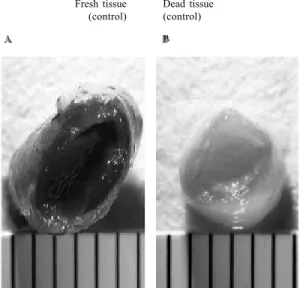
Aiming at evaluating the tissue viability, staining with MTT was performed as a control in fresh tissue segments (taken before the start of the culture process) and in dead segments (exposed to low temperatures). The fresh segments were stained completely and the dead fragments did not present any staining by MTT whatsoever (Figure 5).
Based on this property of MTT staining, our SV samples were stained to test the tissue viability. The SV segments cultivated for one, two and four days in venous
Fig. 3 - Sections of saphenous vein cultivated for 1 (A,B) and 4 (C,D) days in venous (A,C) and arterial (B,D) hemodynamic conditions (magnification 100x). The staining using hematoxylin-eosin demonstrates a gradual reduction in the number of stained nuclei in the group submitted to the arterial conditions which did not occur in the venous conditions.
Venous
1 day
Fig. 5 - Control staining of the saphenous vein with MTT to establish the tissue viability: A – fresh saphenous vein that acquires homogenous staining. B – Dead tissue of the saphenous vein (after refrigeration) without staining by MTT
hemodynamic conditions presented viable cells over the entire vascular wall. The segments cultivated for four days in arterial conditions, however, presented with an absence of staining by MTT in some portions of the vascular wall, suggesting cellular death in these regions (Figure 6).
Finally, the TUNEL test was performed to verify if the
reduction in cellular density and cellular viability observed in the SV samples cultivated for four days in arterial conditions occurred due to the apoptotic process. Significant fragmentation of the DNA of the SV cultivated in venous conditions was not confirmed. The veins cultivated in arterial conditions, however, presented with a large quantity of apoptotic cells when cultivated for one or two days (Figure 7).
Study of the molecular alterations of the saphenous vein cultivated in arterial hemodynamic conditions – expression of interleukin-1 beta (IL-1 â) and of the GRO3
Fig. 4 - Staining of the saphenous vein with Hoechst 33258 for 1 (A,B) and 4 (C,D) days in venous (A,C) and arterial (B,D) hemodynamic conditions (magnification 100x). Note the maintenance of the cellular density in the saphenous veins of the group submitted to venous conditions (A,C), while there is a reduction of this density in the group submitted to arterial conditions (B,D)
Fig. 6 - Study of the cellular viability by MTT staining of saphenous vein segments cultivated for 1 (A,B) and 4 (C,D) days. Note the presence of viable cells in all the vascular wall when cultivated in venous conditions (A,C). The veins of the group cultivated in arterial conditions (B,D) present with portions without stain suggesting cellular death in these regions
Fig. 7 - TUNEL test for the study of cellular apoptosis in saphenous veins cultivated for 1 (A,B) and 4 (C,D) days in venous (A,C) and arterial (B,D) hemodynamic conditions (magnification 100x). Only the veins cultivated in arterial conditions presented with a great amount of cellular apoptosis
Venous
1 day
4 days
Fresh tissue (control)
Dead tissue (control)
Venous
1 day
4 days 1 day
4 days
1 day
4 days Venous
Conditions
oncogene
The molecular alterations of the SV cultivated for one day under arterial hemodynamic conditions observed using cDNA microarray are presented in Table 1.
Among the genes the present with a differentiated expression in the SV cultivated for one day in arterial conditions, basically the cytokines are the most evidenced,
oncogene expression in 11 (68.7%) of these vein segments (p< 0.05).
Fig. 8 - Analysis of the gene expression by RT-PCR. The graph represents values of the expression of the saphenous vein cultivated in arterial hemodynamic conditions compared to saphenous vein cultivated in venous hemodynamic conditions (dashed line). Note the increased expression of interleukin 1ß and of oncogene 3 in the veins cultivated in arterial conditions.
G e n e
colony stimulating factor 3 (CSF3) Interleukin 1, beta (IL 1ß) Interleukin 8 (IL 8) Oncogene GRO3 Oncogene GRO2 Oncogene GRO3 Interleukin 8 (IL 8) tumoral necrosis factor, alpha-induced protein 3 Oncogene GRO1
Interleukin 6 – interferon, beta 2 Interleukin 1, beta (IL 1ß)
serine (or cysteine) proteinase inhibitor Interleukin 1, beta (IL 1ß)
tumoral necrosis factor, alpha-induced protein 3 tumorogenicity suppressor 16 tumorogenicity suppressor 16
“Array” A (A/V)
1.9 2.2 2.3 2.1 2.1 2.0 2.7
1.6 2.4 2.2 2.4 1.7 2.1
1.9 3.9 1.9
“Array” B (A/V)
2.1 2.5 1.8 2.5 2.6 2.4 2.2
1.7 2.6 1.99
2.5 2.2 2.0
1.7 2.5 3.1 Table 1. Genes selected in the cDNA microarray experiment
performed on saphenous veins cultivated in venous (flow = 5 mL/min) and arterial (flow = 50 mL/min; pressure = 80 mmHg) conditions for 1 day. The slide had two arrays (A and B) containing 16,000 human cDNAs.
(A/V) = Fluorescence of the vein cultivated under arterial conditions/ Fluorescence of the vein cultivated under venous conditions
COMMENTS
Failure of the SV grafts utilized in CABG involves many factors. Early occlusion, within the first 30 days after the operation is normally the result of thrombosis, which could be caused by factors such as: reduced diameter or unfavorable distal bed of the revascularized coronary artery. This generates a reduction in the blood flow and an increase in the risk of thrombosis of the graft [4].
The development of diffused and concentric hyperplasia of the intimal layer is observed in SV grafts of patients who evolve with relapse of the anginal symptoms after CABG and require reoperations. RATLIFF & MYLES [5] detected the presence of hyperplasia in 99% of SV grafts analyzed in a series of 100 patients submitted to reoperation of CABG.
The surgical manipulation during the dissection and preparation of the venous graft may cause endothelial injury and alterations of the thromboregulation. The loss of endothelial integrity may cause aggregation of the sanguineous cells and platelets and alter the homeostasis of the vascular wall of the graft. The injury of the endothelial cells leads to a reduction in the local liberation of nitrous oxide and prostacyclins, increasing the adhesion and the platelet aggregation. The platelet activation leads to the all of which are involved in tumoral processes.
Among the most changed molecular targets, interleukin-1ßand the GRO3 oncogene were chosen for investigation (Figure 8). The expression of interleukin-1ß was increased by nine fold (56.2%) in the veins cultivated under arterial conditions, while there was a three-fold increase of the GRO3
G
row
th of ge
ne
e
xpre
ss
liberation of pro-coagulant factors and vasoconstrictors, which stimulate the proliferation of the medial vascular smooth muscle cells (VSMC) and the cellular migration to the intimal layer of the graft [6].
The elevated hydrostatic pressure experienced during the preparation of the graft may cause necrosis of the VSMC of the medial layer. The periods of ischemia/reperfusion during the implantation of the graft promote infiltration of inflammatory cells in the wall of the graft. These inflammatory cells secrete cytokines and growth factors, stimulating the proliferation and migration of the VSMC to the medial layer of the graft. The loss of endothelial integrity is the central factor of the degeneration of the venous graft. Even after the reconstitution of the endothelial integrity, the process of vascular remodeling is maintained at a lesser degree [7]. On the other hand, the success of the graft also depends on the capacity of adaptation of the vessel that passes from low pressure venous conditions to arterial conditions with a higher pressure and pulsating flow. This new pressure condition brings to the venous tissue hemodynamic forces that start to have a direct influence on the vascular tone and the gene regulation of diverse proteins. The vascular behavior is very dependent on the endothelial function and among the hemodynamic factors shear stress is the main force acting on the endothelium [3]. The mechanism by which the endothelium detects shear stress is not completely understood, but there is evidence demonstrating that the shear stress actively participates in vascular homeostasis through the regulation and liberation of endothelial proteins. One example of this is that the atherosclerotic lesions frequently appear in regions of vascular bifurcation, where the flow pattern is altered by turbulence and with a reduction of the shear stress [8]. Reduced shear stress seems to favor the appearance of atherosclerosis through the regulation of the expression of genes that participate in the mechanism of structural and vascular function control. It is speculated that low shear stress enables the adhesion of leukocytes through the increased expression of adhesion molecules such as VCAM-1 (vascular cellular adhesion molecule), as well as the transmigration through the expression of chemokines proteins such as MCP-1 (monocyte chemoattractant protein) [9].
Reduced shear stress also increases the production of mitogenic factors such as endothelin-1. Additionally, reduced shear stress diminishes, in these regions, the secretion of the vascular proliferation inhibitors such as nitrous oxide (NO) and TGF-ß (transforming-ß growth factor) [10] and provides favorable conditions for the proliferation of muscle cells.
REIDER et al. [11] demonstrated that shear stress diminished the activity and expression of ECA and that this response is mediated by two regulatory sequences,
Barbie-box and GAGA-Barbie-box, located in the interval between -288 and -232 pb of the promoter rat ECA [12]. This supports the idea that in vascular bifurcation regions, where the shear stress is reduced and an increase of the production of ECA occurs, increasing the local availability of Ang II. Ang II may trigger or increase vascular injury through the mitogenic effect or by the increase of oxidization of LDL and the expression of its receptor LOX-1 in endothelial cells. Thus, the ECA can be stipulated as a gene with susceptibility in the development of atherosclerosis and, corroborating with this idea several studies have shown its presence in atherosclerotic lesions, mainly in macrophages and in endothelial cells of micro-vessels found in the lesions, very efficient to reduce the development of atherosclerosis [13]. In spite of not understanding the mechanism of its action, it has been postulated that the action of these inhibitors of ECA may occur through the antiproliferative effects in the vascular muscle layer [14], inhibition of platelet aggregation [15] and the attenuation of the oxidization of the LDL [16].
In the same manner that the appearance of the atherosclerosis seems to be related to the local flow pattern, occlusion of saphenous grafts may be related to the sudden hemodynamic alteration imposed on the vein. There are ex vivo studies demonstrating that the arterial flow in the saphenous vein reduces the concentration and the functional action of thrombomodulin [17], a protein which confers an antithrombogenic characteristic to the endothelium, alters the expression of eNOS (endothelial NO synthase) and of the adhesion molecules ICAM-1 and VCAM-1 [18], influencing the adhesiveness of the endothelium to platelets and monocytes.
An interesting fact that occurs in the venous graft interposed in an artery is the induction of the apoptotic process. The decrease of blood flow in the venous graft may be caused by an accentuated reduction in the caliber of the coronary artery in relation to the diameter of the venous graft and by the unfavorable distal bed of the revascularized coronary artery, not allowing adequate out flow of blood [4].
flow and higher pressure cause a stretching of the wall of the vein, whose structure is not prepared to receive such a tension. This can be confirmed when a polytetrafluoroethylene membrane is positioned surrounding a venous graft, reducing the tension and the stretching on the wall of the vessel, with a significant reduction of the rate of apoptosis in the graft [20].
In the ex vivo system presented in this work, the cellular density and the tissue viability gradually diminished when the saphenous vein is cultivated in the arterial hemodynamic conditions, whilst no morphological or viability changes were observed in the vein cultivated under venous conditions for up to 4 days. It is interesting to note that the saphenous vein cultivated in arterial conditions for 1 day presented with cellular density similar to the fresh fragments, although several of these cells presented with fragmentation of the DNA, indicating the presence of the apoptotic process (Figures 3 and 5). This event may explain the cellular loss observed in the fragments of saphenous vein cultivated in the arterial conditions.
Vascular remodeling results from the balance between cellular death and proliferation. In the venous graft in an arterial bed, apoptosis occurs in response to the increase in mechanical stress and is counterbalanced by a subsequent process of cellular proliferation. In our ex vivo system, the absence of cellular proliferation in the saphenous vein in arterial conditions may be explained by the necessity components originating in the blood circulation, that are not present in the culture medium, or maybe, by the necessity of a greater time than four days of culture. Even though the proliferation event was not observed, we verified that the phenomena that occur in vivo were being reproduced in our
ex vivo culture system, demonstrating that this system is a good model to study the changes that occur in the saphenous graft.
It is evident that the stretching of the wall of the venous graft in the arterial bed provokes a series of structural and molecular alterations that contribute to hyperplasia of the intima, which frequently evolves to an atherosclerotic process. This process may be related to the regulation of growth factors such as PDGF and TGF-ß components of the MAP kineses pathway such as p38, transcription factors such as E2F and proteins that compose and break down the extracellular matrix such as tenascin-C and MMPs (matrix metalloproteinases) respectively [21]. The occlusion process that occurs in the saphenous grafts involves the participation of several proteins and the study of how these proteins jointly participate is an important approach to understanding the vascular physiology and physiopathology.
Currently, we arranged the strategies for the global analysis of thousands of genes simultaneously. Among these ‘differential display’, random sequencing of the cDNA
libraries, SAGE (serial analysis of gene expression) and cDNA microarray stand out. The greater knowledge in respect to the genes involved in the atherosclerotic process of grafts has improved the possibility of gene therapy development. In this work we developed an ex vivo system for the culture of saphenous veins that enables the analysis of vascular behavior in controlled hemodynamic conditions for periods of up to four days. The study was performed cultivating the saphenous vein in arterial hemodynamic conditions to simulate the condition that the vein is submitted after the process of coronary artery bypass grafting. This system was adequate as a model for the study of the changes that occur in the saphenous graft, as seen by the fact that the phenomena that occur in vivo were being reproduced. Additionally, using the technology of the cDNA microarray we identified two genes, IL-1ß (interleukin-1ß) and GRO3 (oncogene 3), that may be participating in the transformation process of venous grafts to arteries. A future action on the genes could contribute to the greater patency of the graft.
Molecular alterations of the saphenous veins cultivated for one day under arterial hemodynamic conditions, observed using the cDNA microarray are presented in Table 1. Among the genes that present with a differentiated expression in the saphenous vein cultivated for one day under arterial hemodynamic conditions are the cytokines, all of which are involved in the tumoral process. In this context, the response to cellular proliferation on the vessel wall imitates common aspects to those observed in the development of neoplasias with activation of the growth factors due to the increased stress. Unhappily, this is a process which is little understood and that evolves to the development of atherosclerosis and occlusion of the graft in a significant number of patients.
Elucidation of the physiopathologic basis of the atherosclerotic process of saphenous grafts is of fundamental importance. Studies in respect to the histologic and molecular changes involved may provide the development of alternative therapies efficacious at reducing the number of reoperations after coronary artery bypass grafting.
The success of gene therapy depends on the following factors: the correct choice of the therapy to act on the vascular wall, the availability of a safe and efficient vector for the transference of the gene and the availability of an adequate mechanism for the gene transference.
There are studies under way in respect to gene therapy for saphenous vein grafts. MANNION et al. [23] obtained an 82% reduction in the proliferation of cells of the medial layer of porcine saphenous veins submitted to gene therapy with antisense c-myc oligonucleotides.
GEORGE et al. [24] performed a study with the culture of saphenous veins in humans, utilizing tissue inhibitors of metalloproteinase (TIMP-1). The authors reported a 54% reduction in neointima and a 78% reduction in the migration of smooth muscle cells after 14 days of therapy.
MANN et al. [25] utilized CDC2 and PCNA antisense oligonucleotides in rabbit jugular veins interposed in carotid arteries. The vector used was the herpes virus. The authors demonstrated a reduction in the hyperplasia of the neointima and an increase in the resistance of the venous grafts against atherosclerosis induced by diet.
In our study, confirmation of the gene expression was achieved using a measurement method known as RT-PCR. Until now, an increase in the expression of the IL-1ß and GRO3 genes has been confirmed. The role in the atherosclerotic process of venous grafts and the therapeutic potential of these two genes need to be evaluated. For this, future models of venous grafts in the arterial bed of experimental animals and interference methods such as fusion protein and interference by RNA will be used.
CONCLUSIONS
The ex vivo study model associated with analysis using the cDNA microarray system of saphenous graft segments utilized in coronary artery bypass grafting allowed the identification of the apoptotic process and a greater gene expression of interleukin 1ß and oncogene 3 in veins cultivated under arterial hemodynamic conditions.
Elucidation of these gene alterations in saphenous vein grafts utilized in coronary artery bypass grafting is of fundamental importance for the development of new alternative therapies such as gene therapy for the prevention of the atherosclerotic process in venous grafts.
BIBLIOGRAPHIC REFERENCES
1 Akowuah EF, Sheridan PJ, Cooper GJ, Newman C. Preventing saphenous vein graft failure: does gene therapy have a role? Ann Thorac Surg 2003;76:959-66.
2 Cameron A, Davis K, Rogers W. Recurrence of angina after coronary artery bypass surgery: predictors and prognosis (CASS Registry). Coronary Artery Surgery Study. J Am Coll Cardiol 1995;26:895-9.
3 Davies PF. How do vascular endothelial cells respond to flow? News Physiol Sci 1989;4:22-5.
4 Paz MA, Lupon J, Bosch X, Pomar JL, Sanz G. Predictors of early saphenous vein aortocoronary bypass graft occlusion. The GESIC Study Group. Ann Thorac Surg 1993;56:1101-6.
5 Ratliff NB, Myles JL. Rapidly progressive atherosclerosis in aortocoronay saphenous vein grafts: possible immune-mediated disease. Arch Pathol Lab Med 1989;113:772-6.
6 Groves HM, Kinlough-Rathbone RL, Mustard JF. Development of non-thrombogenicity of injured rabbit aortas despite inhibition of platelet adherence. Arteriosclerosis 1986;6:189-95.
7 Angelini GD, Passani SL, Breckenridge IM, Newby AC. Nature and pressure dependence of damage induced by distension of human saphenous vein coronary artery bypass grafts. Cardiovasc Res 1987;21:902-7.
8 Glagov S, Zarins C, Giddens DP, Ku DN. Hemodynamics and atherosclerosis: insights and perspectives gained from studies of human arteries. Arch Pathol Lab Med 1988;112:1018-31.
9 Shyy YJ, Hsieh HJ, Usami S, Chien S. Fluid shear stress induces a biphasic response of human monocyte chemotactic protein 1 gene expression in vascular endothelium. Proc Natl Acad Sci USA 1994;91:4678-82.
10 Ohno M., Cooke JP, Dzau VJ, Gibbons GH. Fluid shear stress induces endothelial transforming growth factor beta-1 transcription and production: modulation by potassium channel blockade. J Clin Invest 1995;95:1363-9.
11 Rieder MJ, Carmona R, Krieger JE, Pritchard Jr. KA, Greene AS. Suppression of angiotensin-converting enzyme expression and activity by shear stress. Circ Res 1997;80:312-9.
12 Miyakawa AA, Junqueira ML, Krieger JE. Identification of two novel shear stress responsive elements in rat angiotensin-I converting enzyme promoter. Physiol Genomics. 2004. In press.
14 Daemen MJ, Lombardi DM, Bosman FT, Schwartz SM. Angiotensin II induces smooth muscle cell proliferation in the normal and injured rat arterial wall. Circ Res 1991;68:450-6.
15 Keidar S, Oiknine J, Leiba A, Shapira C, Leiba M, Aviram M. Fosinopril reduces ADP-induced platelet aggregation in hypertensive patients. J Cardiovasc Pharmacol 1996;27:183-6.
16 Hayek T, Attias J, Smith J, Breslow JL, Keidar S. Antiatherosclerotic and antioxidative effects of captopril in apoliprotein E-deficient mice. J Cardiovasc Pharmacol 1998;31:540-4.
17 Gosling M, Golledge J, Turner RJ, Powell JT. Arterial flow conditions downregulate thrombomodulin on saphenous vein endothelium. Circulation 1999;99:1047-53.
18 Golledge J, Turner RJ, Harley SL, Springall DR, Powell .JT. Circumferential deformation and shear stress induce differential responses in saphenous vein endothelium exposed to arterial flow. J Clin Invest 1997;99:2719-26.
19 Yamamura S, Okadome K, Onohara T, Komori K, Sugimachi K. Blood flow and kinetics of smooth muscle cell proliferation in canine autogenous vein grafts: in vivo BrdU incorporation. J Surg Res 1994;56:155-61.
20 Moore MM, Goldman J, Patel AR, Chien S, Liu SQ. Role of tensile stress and Strain in the induction of cell death in experimental vein grafts. J Biomech 2001;34:289-97.
21 Leville CD, Dassow MS, Seabrook GR, Jean-Claude JM, Towne JB, Cambria RA. All-trans-retinoic acid decreases vein graft intimal hyperplasia and matrix metalloproteinase activity in vivo. J Surg Res 2000;90:183-90.
22 Nabel EG, Plautz G, Boyce FM, Stanley JC, Nabel GJ. Recombinant gene expression in vivo within endothelial cells of arterial wall. Science 1989;244:1342-4.
23 Mannion JD, Ormont ML, Shi Y, O'Brien JE Jr, Chung W, Roque F et al.. Saphenous vein graft protection: effects of c-myc antisense. J Thorac Cardiovasc Surg 1998;115:152-61.
24 George SJ, Lloyd CT, Angelini GD, Newby AC, Baker AH. Inhibition of late vein graft neointima formation in human and porcine models by adenovirus-mediated overexpression of tissue inhibitor of metalloproteinase-3. Circulation 2000;101:296-304.