Major Article
Journal of the Brazilian Society of Tropical Medicine Vol.:52:e20180254: 2019
doi: 10.1590/0037-8682-0254-2018
Corresponding Author: Dr.Sri Winarsih.
e-mail: [email protected]
Orcid: 0000-0003-0860-0716
Received 11 August 2018
Accepted 20 December 2018
INTRODUCTION
Typhoid fever, which is caused by Salmonella Typhi, is still considered a global health burden. Approximately 21 million cases occur worldwide each year, causing 200 thousand deaths;1 almost 80% of these cases are from Asia.2 Antimicrobial
medication for typhoid fever kills S. Typhi even if the bacteria survive inside phagocytes;3 however, S. Typhi has developed
resistance to antimicrobials.4 Because of this phenomenon,
many countries like Indonesia and China have faced Multidrug-Resistant Typhoid Fever (MDRTF) for the last two decades.2
β-Glucan of
Candida albicans
Cell Wall Extract Inhibits
Salmonella
Typhimurium Colonization by Potentiating
Cellular Immunity (CD8
+and CD4
+T Cells)
Sri Winarsih
[1], Tomson Kosasih
[1], Marvin Anthony Putera
[2], Nayla Rahmadhiani
[2],
Erlien Lindawati Poernomo
[1], Kresna Septiandy Runtuk
[2], Melissa Valensia Oswari
[2][1]. Pharmacy Study Program, Faculty of Medicine, Universitas Brawijaya, Indonesia. [2]. Medicine Study Program, Faculty of Medicine, Universitas Brawijaya, Indonesia.
Abstract
Introduction: Antimicrobial resistance has been reported in the drugs used for the treatment of typhoid fever. The
immunomodulatory substance β-glucan can be used as an alternative therapy as it potentiates host immunity. The aims of this
study are to observe the effect of Candida albicans cell wall (CCW) extract towards host immunity (TCD8+ and TCD4+ cells
in spleen, intestinal sIgA) and its capacity to kill Salmonella in the intestine and liver of typhoid fever mice models. Methods:
Typhoid fever mice models were created by infecting mice with S. Typhimurium orally. Mice were divided into four groups: the Non-Infected, Infected, CCW (infected mice treated with 300 µg CCW extract/mouse once a day), and Ciprofloxacin groups
(infected mice treated with 15 mg/kg BW ciprofloxacin twice a day). Results: Secretory IgA (sIgA) concentrations of mice
in the CCW group remained unchanged. However, their TCD4+ and TCD8+ cells increased substantially compared to those in
the Infected group. In the Ciprofloxacin group, sIgA concentrations increased markedly compared to those in the Non-Infected and CCW groups; TCD4+ and TCD8+ cells also increased significantly compared to those in the Non-Infected Group,
but not significant compared to those in the CCW group. Colonization of S. Typhimurium in the intestine and liver decreased
significantly in the CCW and Ciprofloxacin groups compared to that in the Infected group, with the lowest reduction being found
in the Ciprofloxacin group. Conclusions:The inhibition of S. Typhimurium colonization by CCW is associated with the increase
in TCD4+ and TCD8+ cells.
Keywords: β-glucan. Candida albicans cell wall extract. CD4+. CD8+. Salmonella. sIgA.
On the other hand, drug development takes 12 to 15 years to complete and costs around 802 million USD.5 This situation
has encouraged scientists to discover new therapies other than conventional antimicrobials to circumvent the development of resistance by these bacteria.
There are alternative approaches to overcome antimicrobial resistance and one of them is immunostimulation, which can indirectly kill pathogens by enhancing the host immune system.
β-glucan is an example of a potent immunostimulant.6 It is
contained in the cell wall of yeasts such as Candida albicans, along with other components.7 C. albicans cell wall (CCW) can
be easily collected in a microbiology laboratory as residue of laboratory specimens.8
S. Typhimurium may survive within both macrophages and non-macrophage cells and disseminates to various organs, including the liver and spleen. S. Typhimurium is classified as an intracellular pathogen that is eliminated mainly by cellular immunity composed of macrophages, CD4+ T cells, and CD8+ T
cells.10,11,12 Secretory IgA (sIgA) composes 80% of total mucosal
antibodies and is located at the intestinal mucosa to encounter pathogens entering through the oral route.13
This study used S. Typhimurium, which has been broadly
utilized in research models of typhoid fever in mice due to
the similarity in its pathogenesis with that of human typhoid fever.14,15 The aim of this research is to examine the effect
of CCW extract in modulating the host’s immune response (intestinal sIgA, CD4+ T cell, and CD8+ T cell) against
Salmonella infection in relation to its colonization pattern in the intestine and liver.
METHODS
Animal
Twenty four male BALB/c mice aged 6–8 weeks were divided randomly into four groups: Non-Infected group (non-infected mice that were only given standard animal nursing care), Infected group (untreated infected mice), CCW group (infected mice treated with 300 µg C. albicans cell wall extract/mouse
once a day), and Ciprofloxacin group (infected mice treated with 15 mg/kg BW ciprofloxacin twice a day). Ciprofloxacin
therapeutic regimen in mice was based on standard therapy of laboratory animal. Mice were orally infected with 300 µL of S. Typhimurium at a concentration of 108 cells/mL orally on
days 1 and 3. Mice in the Ciprofloxacin and CCW groups were treated from days 5 to 9. Mice were sacrificed at day 10. The
experiment was conducted according to the regulation of animal laboratory care of Universitas Brawijaya Indonesia as stated in the Ethical Clearance of this study (No. 427/EC/KEPK/08/2015 03 Aug 2015).
C. albicans Culture and Identification
C. albicans used in this study was isolated from a clinical vaginal specimen. It was cultured in Sabouraud Dextrose Agar
(SDA) and incubated at 37°C for 18–24 h. Gram staining and observation under a microscope at 1000× magnification, and
germ tube test were performed to identify C. albicans. The C. albicans culture obtained was soft, beige, and had the typical odor of yeast. To ensure that C. albicans was correctly cultured, microscopic analysis was performed by observing
under a microscope at 1000× magnification; the presence of budding cells were noted. Gram staining and germ tube test
of the C. albicans culture were also performed and observed
under 1000× magnification. It was found to be gram positive
and pseudohyphae were also observed.
C. albicans Cell Wall Extraction
C. albicans culture was harvested and collected by centrifugation at 3000×g with phosphate-buffered saline (PBS), pH 7.4. The cell-pellet was collected and washed with lysis buffer (10 mM Tris-HCl combined with 1 mM phenylmethylsulfonyl
fluoride [PMSF]) five times, and then centrifuged at 3000×g for
10 min. The sediment was again collected and suspended with lysis buffer at 4°C, and then mechanically lysed with 1-mm
glass beads using an Omni Mixer Homogenizer at 6000×g
for 10 min. The lysed cells were separated subsequently by centrifugation at 3000×g for 10 min. The precipitate obtained was the cell wall fraction.
β-glucan Isolation
The cell wall fraction of C. albicans was washed once with distilled water at 4°C, 5 times with 1 mM PMSF containing 5% NaCL, 2% NaCl, and 1% NaCl, respectively, then centrifuged at 4°C at 6000×g for 10 min.8 Then, 1 M NaOH (1:1) was added
to the sediment, and the suspension was incubated at 90°C for 2 h and centrifuged thereafter. H3PO4 was added to the sediment, mixed using a vortex mixer and incubated at room temperature for 2 h before being centrifuged and washed with distilled water 3 times. Afterwards, 96% ethanol was added to the sediment
and it was homogenized using a vortex mixer. The mixture was strained with paper strainer, and β-glucan was obtained as a solid matter on the filter.16
β-glucan Identification
Fourier Transformed Infrared Spectroscopy (FTIR;
Shimadzu FTIR-8400) was used to analyze the β-glucan
extracted from the C. albicans cell wall. KBr was mixed with
the extract and then it was ground until it appeared fine and
homogenous. This mixture was then pressed using a hand press until it was thin enough to be measured. The same procedure was
also applied to the β-glucan standard. Baseline was determined
by measuring KBr only. The measurement was done by inserting the mixture into the sample holder of the FTIR to obtain the spectra.17 The spectra of β-glucan in CCW and the β-glucan
standard were compared.
S. Typhimurium Identification
This study used an S. Typhimurium reference strain obtained from the National Collection of Type Cultures with the catalogue number NTC12023. S. Typhimurium was cultured in Bismuth
Sulfite Agar (BSA) medium and incubated at 37°C overnight.
Identification of S. Typhimurium was performed using Gram
staining and Microbact System.
Ciprofloxacin Sensitivity Test in Vitro
Ciprofloxacin was used as a control to compare the effect of β-glucan on typhoid fever. The Kirby-Bauer disk diffusion
method was used to test the sensitivity of S. Typhimurium
toward ciprofloxacin. Based on the inhibition zone obtained
(32 mm), the Salmonella strain used in this study was sensitive.
Bacteria Culture from Intestine and Liver
three dilution groups (10-1, 10-2, and 10-3). Bacterial culture was
performed by the pour plate method using BSA medium. One mL of each specimen in every dilution concentration was mixed with 9 mL of liquid BSA. Bacterial cultures was incubated at 37°C for 48 h. Colonies were counted with a colony counter.
sIgA Measurement
Intestinal mucus sample in PBS was centrifuged at 1000×g for 5 min. The supernatant was collected and centrifuged at 7000×g for 5 min. Supernatant was collected and its sIgA was measured by indirect ELISA (Biolegend, USA). Whole cells of S. Typhimurium to be used as antigens were obtained by
sonication (SONICA ETH series) at 20 kHz, 10 times, 3 min
each. This was used to coat the wells overnight before ELISA was performed.
CD4+ T Cell and CD8+ T Cell Measurement
CD4+ and CD8+ T lymphocytes were measured using flow
cytometry.18 Spleen was ground and diluted in 2 mL PBS.
The homogenate was transferred to a 1.5-mL microtube and then centrifuged at 1500×g at 4°C for 5 min. The sediment was washed twice with PBS (pH 7.2). Then, 50 µL of staining solution (FITC anti-mouse CD4 and PE anti-mouse CD8; 1:100 dilution) and 350 µL of buffer were added and mixed into 50 µL of suspension. The mixture was incubated in a dark room
at room temperature for 30 min and analyzed using the flow
cytometer (BD FACS Calibur).
Data Analysis
Statistical analysis was performed using IBM SPSS
Statistics, version 21.0 with 95% confidence interval (α = 0.05).
Homogeneity of variances was tested using the Levene statistic, and normality was tested using the Shapiro-Wilk normality test. In the case of homogenous equal variance data, one-way ANOVA was performed. In the case of unequal variances or non-homogenous data, the Kruskal-Wallis test was performed.
RESULTS
β-glucan Identification
Two similar spectra were obtained for both substances. The
specific characteristics of β-glucan include peak transmittance
patterns at 1160, 1078, 1044, and 890 cm-1 wavelength bands.19 CCW
extraction was able to produce a β-glucan yield of around 90%.
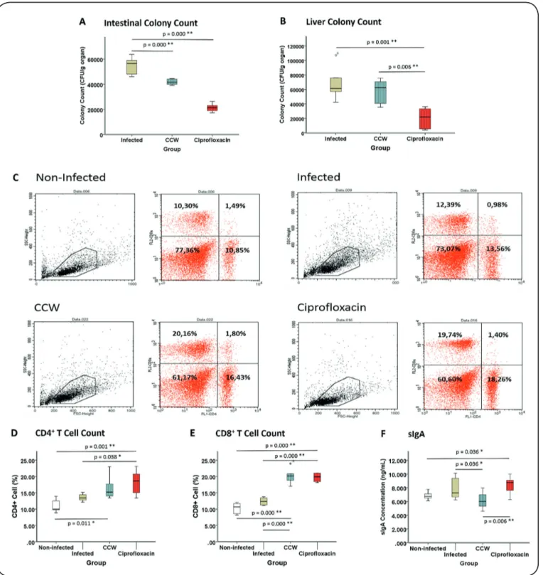
S. Typhimurium Colonization in Intestine and Liver
Intestinal specimen was obtained from the mucus scraped from the gut. Hence, only bacteria that survived the sIgA response was cultured. There were significant differences between the intestinal S. Typhimurium colony numbers among groups (p = 0.000). The mean intestinal colony number of S. Typhimurium in the CCW group was substantially lower than that in the Infected group (p = 0.000), but it was higher than
that in the Ciprofloxacin group (p = 0.000) (see Figure 1A).
In the liver, the colony count of the CCW group was lower than that of the Infected group, but it was not statistically
significant (p = 0.631); however, it was substantially higher
(p = 0.006) than that of the Ciprofloxacin group, which had the
lowest colony count (p = 0.001 compared to Infected group) as shown in Figure 1B.
Splenic CD4+ T Cell and CD8+ T Cell Count Figures 1C–1E depict CD4+ T cell and CD8+ T cell counts
obtained by flow cytometry. There was a substantial difference
in the number of CD4+ T cells among groups (p = 0.001).
Administration of ciprofloxacin resulted in a higher number
of CD4+ T cells than those in the Non-infected (p = 0.001) and
Infected groups (p = 0.038). The CCW group also had a higher number of CD4+ T cells than the Non-infected group (p = 0.011).
The CD8+ T cell counts of the Ciprofloxacin and CCW
groups were higher than those of the Non-infected (p = 0.000) and Infected groups (p = 0.000). There was no significant difference between the CD8+ T cell counts of the Non-infected
and Infected groups (p = 0.170). There was also no significant difference between the CD8+ T cell counts of the CCW and
Ciprofloxacin groups (p = 0.971).
sIgA Concentration
There was substantial difference in the sIgA concentrations among groups (p = 0.017). The sIgA concentration of the Ciprofloxacin group was higher than those of the Non-infected (p = 0.036) and CCW groups (p = 0.006), but the sIgA concentration of the CCW group was lower than that of the Infected group (p = 0,036) (see Figure 1F).
DISCUSSION
Typhoid fever antimicrobial resistance has encouraged scientists to develop alternative therapies to conventional
antimicrobials. β-glucan extracted from the cell wall of C.
albicans was studied in this research as a potential alternative.
According to FTIR analysis, β-glucan composed about 90% of CCW. β-glucan is a substance recognized by dectin-1 receptor,
complement 3 receptor, and Toll-like receptor (TLR), which are present on immune cells including monocytes, macrophages, dendritic cells, neutrophils, eosinophils, and natural killer cells; it is capable of inducing both innate and adaptive immunity.20,21
Our study found that oral administration of CCW reduced Salmonella colonization substantially in the intestine and slightly
in the liver. Although the reduction in colonization was not as good as that by ciprofloxacin, the inhibition of colonization
in intestine by CCW denoted its potency to interfere with the growth of Salmonella.22 The potency of β-glucan to inhibit the
colonization of intracellular pathogens (Mycobacterium bovis)
has been previously demonstrated as it stimulates respiratory burst and cytokine production in macrophages, as well as the secretion of antimicrobial substances that inhibit the growth of M. bovis, but not M. tuberculosis.23
Significant increments in the splenic and CD4+ T cell counts
of the CCW and Ciprofloxacin groups were followed by the raise
in CD8+ T cell counts of both groups. Intracellular pathogens
are eliminated mainly through CD8+ T cell-mediated immune
FIGURE 1: CCW Administration Enhances Cellular Immunity in Treating Salmonella. (A) Colony count of Salmonella in the intestine of Infected, CCW,
and Ciprofloxacin groups. (B) Salmonella colony count in the liver of Infected, CCW, and Ciprofloxacin groups. (C) Flow cytometry profile of spleen cells.
Gating was performed in lymphocyte area. Upper left quadrant: CD4- CD8+; upper right quadrant: CD4+ CD8+; lower left quadrant: CD4- CD8-; lower right
quadrant: CD4+ CD8-. (D) CD4+ splenic T cells of Non-Infected, Infected, CCW, and Ciprofloxacin groups based on CD4+ CD8- percentage. (E) CD8+ splenic
T cell count from CD4- CD8+ percentage. (F) sIgA concentration from mice gut mucus. *p < 0.05; **p < 0.005.
reported that β-glucan was able to enhance CD8+ and CD4+ T
cells by promoting dendritic cell maturation in Hepatitis B DNA vaccination via Th1.25 Interestingly, sIgA, the main antibody
of mucosal organs, declined with CCW treatment, indicating that the Th1 response was potentiated in this study. Th1 and
Th2 antagonize each other, while sIgA antibody synthesis is
mediated mainly by Th2 cytokines.26 This study observed that
an increase in CD8+ T cell was accompanied by a decline in sIgA
concentration, indicating that the cellular immunity pathway was potentiated via Th1.
by ciprofloxacin. To this end, several studies have demonstrated the immunomodulatory activity of fluoroquinolones, including ciprofloxacin, even though the underlying mechanism remains
unclear and needs to be elucidated further. A study by Bode et al.
(2014) using moxifloxacin showed a decrease in inflammatory cytokines (IL-1β, IL-6) modulated by TLR signaling in
immune cells.27 Another study also found that moxifloxacin and
ciprofloxacin were able to reduce interferon-gamma and IL-4
in helper T cells with an unchanged Th1/Th2 ratio.28 Another
study reported a contrasting result wherein ciprofloxacin in
human peripheral blood lymphocytes caused an increase in T cell function and macrophage-T cell interactions.29
In summary, our study indicates that CCW treatment reduced Salmonella colonization mainly in the intestine, likely through the potentiation of cellular immunity by CD8+ T cells
via Th1 pathway, as the decline in sIgA was also observed. The
colonization inhibition ability of ciprofloxacin was superior to
that of CCW, and this may be due to the synergistic dual role
of ciprofloxacin (anti-microbial and immunomodulatory) that was observed in this study. These findings indicated that the
immunomodulatory activity of CCW may contribute towards eliminating Salmonella during infection.
Acknowledgement: We wish to thank the laboratory assistants who were involved in this study, our Flow Cytometry assistant, Wahyudha Ngatiril Lady, animal care assistant Heni Endrawati, ELISA assistant Satuman, and bacterial culture assistant Slamet Riyanto.
Conflict of Interest: The authors state that there was no conflict of interest
to declare.
Financial Support: This research was funded by DPP/UPP Universitas Brawijaya.
REFERENCES
1. Centers for Disease Control and Prevention. Typhoid Fever [Internet]. Atlanta: National Center for Emerging and Zoonotic Infectious Diseases; c2014 [updated 2016 Dec 5; cited 2017 Dec 9]. Available from http://www.cdc.gov/typhoid-fever/.
2. Zaki SA, Karande S. Multidrug-resistant typhoid fever: a review. J Infect Dev Countries. 2011;5(5):324-37.
3. Steele D, editor. Background document: The diagnosis, treatment
and prevention of typhoid fever [Internet]. Geneva (CH): World Health Organization; 2003 [cited 2017 Dec 12]. Available from: http://www.who.int/rpc/TFGuideWHO.pdf.
4. Blair JMA, Webber MA, Baylay AJ, Ogbolu DO, Piddock LJV. Molecular mechanisms of antibiotic resistance. Nat Rev Microbiol. 2015;13(1):42-51.
5. Gomes CM, Abrunhosa AJ, Ramos P, Pauwels EK. Molecular imaging with SPECT as a tool for drug development. Adv Drug Delivery Rev. 2011;63(7):547-54.
6. Vetvicka V. Glucan-immunostimulant, adjuvant, potential drug. World J Clin Oncol. 2011;2(2):115-9.
7. Chan GC, Chan WK, Sze DM. The effects of beta-glucan on human immune and cancer cells. J Hematol Oncol. 2009;2:25.
8. Pitarch A, Sánchez M, Nombela C, Gil C. Sequential fractionation and two-dimensional gel analysis unravels the complexity of the dimorphic fungus Candida albicans cell wall proteome. Mol Cell Proteomics. 2002;1(12):967-82.
9. Jones BD, Ghori N, Falkow S. Salmonella Typhimurium Initiates
Murine Infection by Penetrating and Destroying the Specialized
Epithelial M Cells of the Peyer’s Patches. J Exp Med. 1994;180(1): 15-23.
10. Chiu HC, Kulp SK, Soni S, Wang D, Gunn JS, Schlesinger LS, et al. Eradication of Intracellular Salmonella enterica Serovar Typhimurium with a Small-molecule, Host Cell-Directed Agent. Antimicrob Agents Chemother.2009;53(12):5236-44.
11. Abbas AK, Lichtman AH, Pillai S. Immunity to Microbes. In: Abbas AK, editor. Cellular and Molecular Immunology. 8th ed. Philadelphia: Elsevier Saunders; 2014. p. 339-57.
12. McSorley SJ. Immunity to intestinal pathogens: lessons learned from Salmonella. Immunol Rev. 2014;260(1):168-82.
13. McGhee JR, Mestecky J, Dertzbaugh MT, Eldridge JH, Hirasawa M, Kiyono H. The mucosal immune system: from fundamental concepts to vaccine development. Vaccines. 1992;10(2):75-88. 14. Eisenstein TK. Mucosal immune defense: the Salmonella
Typhimurium model. In: Paterson Y, editor. Intracellular Bacterial Vaccine Vectors. New York: Wiley-Liss; 1999. p. 51-109.
15. Brumell JH, Finlay BB. Bacterial Adherence, Colonization, and Invasion of Mucosal Surfaces. In: Brogden KA, editor. Virulence Mechanisms of Bacterial Pathogens. 3rd ed. Washington: ASM Science; 2000. p. 3-13.
16. Zechner-Krpan V, Petravić-Tominac V, Gospodarić I, Sajli L,
Ðaković S, Filipović-Grčić J. Characterization of β-Glucans
Isolated from Brewer’s Yeast and Dried by Different Methods. Food Technol Biotechnol. 2010;48(2):189-97.
17. Song X, Hubbe MA. Cationization of oat β-D glucan as a dry-strength additive for paper. J Eng Fiber Fabr. 2014;13(7):57-64. 18. Sckisel GD, Mirsoian A, Minnar CM, Crittenden M. Curti B, Chen
JQ, et al. Differential phenotypes of memory CD4 and CD8 T cells in the spleen and peripheral tissues following immunostimulatory therapy. J Immunother Cancer. 2017;5:33.
19. Synytsya A, Novak M. Structural Analysis of Fungal Glucans. In:
Vetvicka V editor. Biology and Chemistry of Beta Glucan - Volume
2: Beta-Glucan, Structure, Chemistry and Specific Application.
Dubai: Bentham Science Publishers; 2013. p. 14.
20. Stier H, Ebbeskotte V, Gruenwald J. Immune-modulatory effects of dietary Yeast Beta-1,3/1,6-D–glucan. Nutr J. 2014;13(38):1-9. 21. Li X, Wang J, Wang W, Lie C, Gu J, Wang X, et al. Immunomodulatory
activity of a novel synthetic beta glucan (β-glu6) in murine
macrophages and human peripheral blood mononuclear cells. PLoS One. 2013;8(11):e80399.
22. Pucciarelli MG, García-Del Portillo F. Salmonella Intracellular Lifestyles and Their Impact on Host-to-Host Transmission. Microbiol Spectr. 2017;5(4).
23. Betz BE, Azad AK, Morris JD, Rajaram MVS, Schlesinger LS.
β-glucans inhibit intracellular growth of Mycobacterium bovis
BCG but not virulent Mycobacterium tuberculosis in human macrophages. Microb Pathog. 2011; 51(4):233-242.
24. Fresnay S, McArthur MA, Magder L, Darton TC, Jones C, Waddington CS, et al. Salmonella Typhi-specific multifunctional
CD8+ T cells play a dominant role in protection from typhoid fever in humans. J Transl Med. 2016;14:62.
by a DNA Vaccine Encoding Hepatitis B Virus Core Antigen. J Biomed Biotechnol. 2010:1-10.
26. Ren W, Wang K, Yin J, Chen S, Liu G, Tan B, et al.
Glutamine-Induced Secretion of Intestinal Secretory Immunoglobulin A: A Mechanistic Perspective. Front Immunol. 2016;7(503).
27. Bode C, Diedrich B, Muenster S, Hentschel V, Weisheit C, Rommelsheim K, et al. Antibiotics regulate the immune response in both presence and absence of lipopolysaccharide through
modulation of Toll-like receptors, cytokine production and phagocytosis in vitro. Int Immunopharmacol. 2014;18(1):27-34. 28. Williams AC, Galley HF, Watt AM, Webster NR. Differential
effects of three antibiotics on T helper cell cytokine expression. J Antimicrob Chemother. 2005;56(3):502-6.