Abstract
Submitted: May 9, 2018 Modification: August 8, 2018 Accepted: August 20, 2018
Effects of dodecacalcium
hepta-aluminate content on the setting time,
compressive strength, alkalinity, and
cytocompatibility of tricalcium silicate
cement
Objective: This study aimed to investigate the effects of dodecacalcium hepta-aluminate (C12A7) content on some physicochemical properties and cytocompatibility of tricalcium silicate (C3S) cement using human dental pulp cells (hDPCs). Material and Methods: High purity C3S cement was manufactured by a solid phase method. C12A7 was mixed with the cement in proportions of 0, 5, 8, and 10 wt% (C12A7-0, -5, -8, and -10, respectively). Physicochemical properties including initial setting time, compressive strength, and alkalinity were evaluated. Cytocompatibility was assessed with cell viability tests and cell number counts. Statistical analysis was performed by using one-way analysis of variance (ANOVA) and Tukey’s test (p<0.05). Results: The initial setting time of C3S-based cement was shorter in the presence of C12A7 (p<0.05). After 1 day, C12A7-5 showed significantly higher compressive strength than the other groups (p<0.05). After 7 days, the compressive strength of C12A7-5 was similar to that of C12A7-0, whereas other groups showed strength lower than C12A7-0. The pH values of all tested groups showed no significant differences after 1 day (p>0.05). The C12A7-5 group showed similar cell viability to the C12A7-0 group (p>0.05), while the other experimental groups showed lower values compared to C12A7-0 group (p<0.05). The number of cells grown on the C12A7-5 specimen was higher than that on C12A7-8 and -10 (p<0.05).
Conclusions: The addition of C12A7 to C3S cement at a proportion of 5% resulted in rapid initial setting time and higher compressive strength with no adverse effects on cytocompatibility.
Keywords: Calcium silicate. Dental pulp. Physicochemical analysis. Material testing.
Yoorina CHOI1,2 Jong-Lye BAE2 Hee-Jin KIM3 Mi-Kyung YU2,4,5 Kwang-Won LEE2,4,5 Kyung-San MIN2,4,5
1Wonkwang University Dental Hospital, Department of Conservative Dentistry, Iksan, Korea. 2Chonbuk National University, School of Dentistry and Institute of Oral Bioscience, Department of
Conservative Dentistry, Jeonju, Korea.
3Kosin University, College of Medicine, Department of Dentistry, Busan, Korea. 4Chonbuk National University, Research Institute of Clinical Medicine, Jeonju, Korea. 5Chonbuk National University Hospital, Biomedical Research Institute, Jeonju, Korea.
Introduction
Since its introduction in the 1990s, mineral
trioxide aggregate (MTA) has been widely used in the
endodontic field for purposes including retrograde filling, perforation repair, apexification, and vital pulp
therapy.1 Considerable evidence has demonstrated the
excellent biocompatibility and sealing ability of MTA
and promising outcomes for endodontic procedures.2,3
Nevertheless, because of drawbacks including long
setting time and initial wash-out possibility, poor
handling characteristics, discoloration of teeth, and
heavy metal content, the development of novel
MTA-like calcium silicate (CS) cement to overcome these
drawbacks and improve biocompatibility and clinical
convenience of MTA is an important goal.4
The composition of MTA is similar to that of Portland
cement except for the presence of
radiopacifier-like bismuth oxide, which is generally composed
of tricalcium silicate (C3S), dicalcium silicate
(C2S), tricalcium aluminates (C3A), tetracalcium
aluminoferrite, and other ingredients.5 Among these,
C3S is one of the major constituents of MTA.6 C3S
forms into calcium silicate hydrates (C-S-H) through
hydration reactions with water, which contribute to
the spontaneous development of strength.7 Despite
its excellent in vitro bioactivity and biocompatibility,8
C3S alone has limitations in clinical contexts due to its
long setting time and low mechanical strength during
the early stages of hydration.9
Tricalcium aluminate (C3A) is the most reactive
part of Portland cement.10 When C3A is mixed with
calcium silicate, it contributes to the initial hydration
process of the cement.10 In studies, C3A mixed with
calcium silicate cement showed a faster hydration rate
and higher initial mechanical strength than non-mixed
cement.7,9,10 Dodecacalcium hepta-aluminate (C12A7),
one of the stable phases of calcium aluminates (CA),
is also expected to react rapidly with water and may
provide beneficial properties during the early hydration
stages of calcium silicate. However, there have been
no attempts to mix C12A7 in C3S cement and evaluate
the effects of C12A7 on cement in dental contexts. This
study aimed to evaluate the effects of C12A7 on C3S
cement regarding some physicochemical properties
and cytocompatibility with hDPCs. We manufactured
high-purity cement with uniformly fine particles
to determine the optimal component ratio of C3S/
C12A7 for use as an endodontic biomaterial. The null
hypothesis was that cements with different C12A7
contents would not significantly differ in practical
properties.
Material and methods
Material preparation
For the manufacture of C3S cement powder,
calcium carbonate (Sigma-Aldrich, St. Louis, MO, USA)
and silicon dioxide (Junsei Chemical, Kyoto, Japan)
were uniformly mixed in a 3:1 molar ratio and stirred
by ball milling for 1 h in ethanol. The powder (10 g)
was put into a mold 15-mm in diameter and pressed
uniaxially at pressures of 2 to 3 t. After the
cylinder-type pelletized samples were desiccated at 40°C for
12 h, the sample was heated at a rate of 10°C/min
to the sintering temperature of 1400-1500°C, held
for 1 to 20 h, and then cooled. Sintering process was
repeated four times to achieve higher proportions of
C3S in the cement. The sintered material was ground
to powders less than 8 μm in diameter by using air
jet mill (CGS16, NETZSCH GmbH, Selb, Germany)
with 6000 rpm. The characteristics of prepared C3S
cement powder were evaluated with X-ray diffraction
(XRD) analysis (D/MAX 2500V/PC, Rigaku, Tokyo,
Japan), a laser diffraction particle size analyzer (LS
13 320, Beckman Coulter Inc., Miami, FL, USA),
and a scanning electron microscope (SEM; S-4300,
Hitachi, Tokyo, Japan). C12A7 was manufactured as
follows; calcium oxide (Sigma-Aldrich) and aluminum
hydroxide (Duksan pure chemical, Ansan, Korea) were
uniformly mixed at a 12:7 molar ratio. The mixed
compounds were heated at 1100°C and quenched
to fabricate a hydratable mixture of CA. Then
CaO-Al2O3-H2O was obtained by adding water to the
calcium aluminates, and the hydrates were heated at
1100°C and quenched, resulting in CaO-Al2O3 clinker,
which was ground to powder. To prepare the C3S/
C12A7 mixture, the resultant C3S powder was mixed
with C12A7 powder at proportions of 0, 5, 8, and 10
wt% of C12A7 (noted as C12A7-0, -5, -8, and -10,
respectively).
Initial setting time
We measured the initial setting time according to
the recommendations specified in ISO 6876:2012.
Each of the four powder groups was mixed with
mL/g and then inserted into Teflon molds of 1-mm
thickness and 10-mm diameter. The initial setting time
of the specimen (n=3) was measured using a Gilmore
apparatus at 30-s intervals. Then, a 1/4-pound
stainless steel indenter was applied vertically on to the
horizontal surface of the specimen for 5 s. The initial
setting time was determined as the time at which the
indenter ceased to leave a distinct indentation on the
surface of the specimen.
Compressive strength test
Compressive strength was determined according
to ISO 9917-1:2003. After the powder was mixed
with DW, the resulting cement was inserted into
Teflon molds of 6-mm thickness and 4-mm diameter
and allowed to set at 37°C (n=10). After 1 or 7
days, compressive strength was measured using a
universal testing machine (Z020, Zwick GmbH, Ulm,
Germany) with a 0.5-N load cell at a crosshead speed
of 1.0 mm/min. We calculated compressive strength
with following formula: C=4P/D2, where C is the
compressive strength (MPa), P is the maximum load
force before fracture (N), and D is the diameter (mm)
of the specimen.
Alkalinity
Alkalinity was evaluated by measuring pH according
to a previously published study.11 In brief, we prepared
specimen (1-mm thickness and 5-mm diameter) and
allowed to set completely. After setting, we inserted
one tablet into 10 mL of deionized water. Then, the pH
value was measured using a pH meter (Orion 3 Star;
Thermo Fisher Scientific, Singapore).
Primary culture of human dental pulp cells
(hDPCs)
The experimental procedures with hDPCs of this
study were approved by the institutional review
board (IRB No: 2018-02-013). From the sectioned
teeth, dental pulp tissue was obtained aseptically and
rinsed with phosphate buffered saline (PBS; HyClone
Laboratories Inc., Logan, UT, USA). The tissue was
minced into small fragments in a 60-mm dish (Nunc,
Roskilde, Denmark) and cultured in Minimum Essential
Medium-α (MEM-α; HyClone Laboratories Inc.)
containing 10% fetal bovine serum (FBS; Invitrogen,
Carlsbad, CA, USA), 100 U/mL penicillin and 100 U/
mL streptomycin (Invitrogen) at 37°C in 5% CO2. The
cells between the third and fifth passages were used
in this study.
Cell viability test
After the powder was mixed with DW, the cement
was allowed to set in disc shaped-paraffin wax molds
(2 mm × 10 mm) for 24 h at 37°C. The cement was
sterilized under ultraviolet light for 1 h. Then, the
specimen was immersed in MEM-α containing 10%
FBS at a ratio of 0.5 cm2/mL for 3 days to prepare
the material extracts. The cells (2 × 10⁴/well) were
inoculated in 24-well culture plates and incubated in
culture medium. After 24 h, the cells were treated
with 1 mL of the prepared extracts (n=4). The sample
which contained no material extract was used as
negative control, and C12A7-0 was considered as
positive control. After 24 or 48 h, cell viability was
measured by using the
3-(4,5-dimethylthiazol-2-yl)-2,5-diphenyltetrazolium bromide (MTT) assay.
Briefly, MTT solution (0.05%, 0.2 mL) was added
to each well, and the cells were incubated for 2 h.
Then, 0.2 mL of dimethyl sulfoxide (Amresco, Solon,
OH, USA) was inserted to each well. After the plates
were shaken for about 5 min, the optical density was
measured spectrophotometrically at 590 nm by using
a microplate reader (SPECTROstarNano, BMG Labtech,
Ortenberg, Germany).
Measurements of the numbers of hDPCs
Each powder sample for each of four groups was
mixed with DW at a liquid-to-powder ratio of 0.4 mL/g
and allowed to set in a disc shaped-paraffin wax mold
(1 mm × 5 mm) for 24 h at 37°C. Cells (1 × 10⁵) were
inoculated on each specimen in the 24-well plates and
cultured for 48 h in culture media (n=3). Then, the
cells were stained with 4’,6-diamidino-2-phenylindole
(DAPI) (Invitrogen) and scanned by a confocal laser
scanning microscope (LSM 510 META, Carl Zeiss, Jena,
Germany). Cell numbers were analyzed quantitatively
from acquired images with an image analysis program
(Image J ver. 1.37; National Institutes of Health,
Bethesda, MD, USA).
Statistical analysis
The results were statistically analyzed using
one-way analysis of variance (ANOVA) and Tukey’s tests
to detect any significance (p<0.05). Prior to the
analysis, the Kolmogorov-Smirnov test was used for
determining normal distribution. The analyses were
performed using SPSS software (SPSS ver. 12.0; SPSS
Results
Characterization of C3S and C12A7
The characteristics of prepared C3S and C12A7
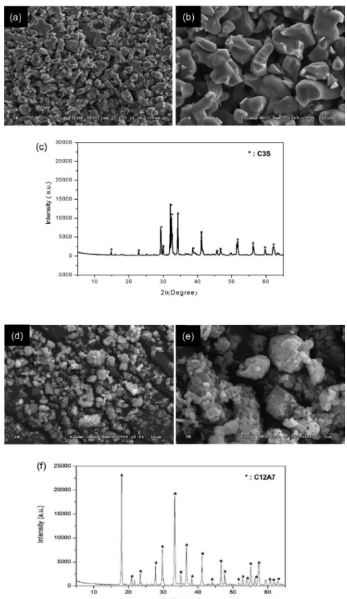
were identified by SEM and XRD (Figure 1). The
SEM images of fabricated C3S powders showed
homogeneous composition of particles (Figure 1A and
1B). Phase analysis results by XRD indicated that the
prepared powders were C3S and C12A7, respectively
(Figure 1C and 1F). Quantitatively, the proportion of
C3S was 100%, and particle size was less than 8 μm
(Table 1).
Figure 1- Characterization of prepared C3S and C12A7 powder; (A-B) Scanning electron microscope (SEM) images (x1000 and x3000,
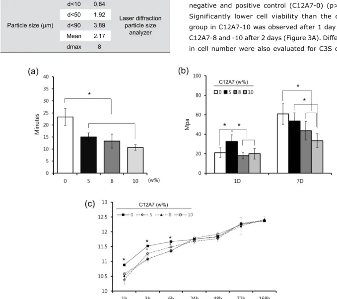
Measurement of initial setting time, compressive
strength, and alkalinity
Initial setting time and compressive strength
were measured to determine the physicochemical
properties of cement with different ratios of C12A7.
Regarding the initial setting time, there were
significant differences between control (C12A7-0)
and other samples (C12A7-5, -8, and -10) (p<0.05)
(Figure 2A). As shown in Figure 2B, the compressive
strength of C12A7-5 was significantly higher than
that of other groups 1 day after the setting (p<0.05).
However, there was no statistical difference between
the other groups (p>0.05). After 7 days, the strength
of C12A7-5 was similar to C12A7-0, whereas other
groups showed lower strength compared to C12A7-0
(Figure 2B) (p>0.05). Regarding alkalinity, samples of
all groups showed high pH values around 10 - 12 with
an increasing pattern for 7 days. Although pH values
of C12A7-0 were higher than other groups within 6 h
(p<0.05), there were no significant differences after
1 day (Figure 2C). Furthermore, the C12A7-5 group
showed higher pH value than C12A7-8 and -10 within
6 h (p<0.05).
Measurement of viability and the number of
hDPCs
Viability and number of hDPCs were measured
to investigate the cytocompatibiltiy of the cement.
Although the cell viability of C12A7-containing cement
decreased as the proportion of C12A7 increased, the
cell viability of C12A7-5 was similar to that of both
negative and positive control (C12A7-0) (p>0.05).
Significantly lower cell viability than the control
group in C12A7-10 was observed after 1 day and in
C12A7-8 and -10 after 2 days (Figure 3A). Differences
in cell number were also evaluated for C3S cement
Contents of evaluation
Results Methods
Proportion of C3S
(%) 100 refinement methodXRD-rietveld
Particle size (μm)
d<10 0.84
Laser diffraction particle size
analyzer
d<50 1.92
d<90 3.89
Mean 2.17
dmax 8
Table 1- Characteristics of prepared cement powder. "d<n" is
defined as the diameter at which n% of the sample's cumulative mass is comprised of particles with a diameter less than this value in the particle size distribution, and "dmax" is defined as the maximum diameter of particle size
Figure 2- The physicochemical properties of C3S cement containing the various proportion of C12A7; (A) Initial setting time, (B)
samples with various proportions of C12A7. While the
cell numbers of C12A7-containing groups showed no
statistical differences compared to the C12A7-0 group,
the cell numbers of C12A7-5 were higher than those
of C12A7-8 and -10 (p<0.05) (Figure 3B-F).
Discussion
Portland cement acquired from natural materials
can vary in composition and include impurities such
as heavy metals of leachable lead and arsenic.1
According to several studies, commercially available
MTAs may also contain heavy metals, although the
amount released by MTAs is less than that of Portland
cement.12 This contamination is of major concern in
the development of cements that have high-purity CS,
which leads to compositional stability and reliability,
and the exclusion of heavy metal elements.13 Most new
formulations of CS-based cements are currently based
on C3S.14 C3S-based cement can be manufactured
using pure raw materials, unlike Portland cement.
C3S-based cement exhibits adequate physical properties
and biologic performances, showing microstructure
and hydration characteristics similar to those of
Portland cement.15 In this study, we fabricated pure
C3S cement with fine particles in the laboratory to
exclude impurities and produce a stable composition
of the base material (Figure 1A-C and Table 1). The
processes of sintering, cooling and grinding were
repeated to enhance the reaction rate of CaO-SiO2 by
solid-state reaction, bringing about high proportions
of C3S. The highly purified C3S cement we produced
is expected to provide non-biased and stable results
when used as a base material for evaluation of other
additives in terms of effects on the base material.
Although C3S is largely responsible for initial setting
and early strength of concrete used in the building
industry, cement must have even more rapid setting
ability for clinical applications. Several authors have
Figure 3- The cytocompatibility of C3S cement with the various proportions of C12A7; (A) The effects of C3S cement containing the
shown that the addition of CA induces faster hydration
reactions and improvements of the early mechanical
strength of CS-based cement.7,10 C3A has been used
as a representative additive in CS-based cement.
However, the synthesis of non-blended C3A clinker
is an energy-consuming process, because sintering
must be repeated several times to avoid eutectic and
peritectic reactions on the CaO-Al2O3 phase diagram. In
contrast, C12A7 is more efficient to fabricate than C3A,
due to its favorable sinterability. In addition, C12A7,
known as mayenite, is one of the intermediary phases
of the CaO-Al2O3 binary system and is also known to
contribute to the first stage of strength development
in aluminous cements like C3A.16 Therefore, C12A7
was selected for use in this study and was fabricated
by sintering processes (Figure 1D-F).
Despite characteristics related to initial rapid
hydration, high concentrations of CA can result in
low final mechanical strength and negative effects
on biocompatibility.7 Hence, we prepared various
proportions of C12A7/C3S cement by mixing C12A7
with C3S cement to investigate the best proportions
of C12A7 in C3S cement. In clinical contexts, MTA is
surrounded by vital tissues like pulp and periodontal
ligaments, as well as tissue fluids and blood. Faster
initial setting and greater early strength can provide
better initial support in bioactive circumstances.
According to the previous report, the initial setting
time of Portland cement was around 25 min,17 which
is similar to our results of pure C3S (C12A7-0) around
23 min. On the other hand, commercially available
products showed shortened initial setting time
around 10-15 min.17-19 In C12A7-containing groups
(C12A7-5, 8 and 10) of this study, the setting time
was significantly shortened compared to pure the C3S
group (C12A7-0), resulting in a value less than 15 min
similar to that of commercial products (Figure 2A).17-19
In this respect, the addition of C12A7 in C3S cement
showed significant benefit regarding the reduction
of initial setting time. Furthermore, according to the
results of the compressive strength test after 1 day,
C12A7-5 showed higher early strength compared to
C12A7-0. The compressive strength values obtained
in this study were higher than those of MTA in another
previous study.20 The results indicated that the addition
of C12A7 might be beneficial for the early strength
of the cement. Although the values of C12A7-8 and
-10 decreased compared to control after 7 days,
C12A7-5 showed no significant difference compared
to the control group (Figure 2B). We also evaluated
the pH of hydrated samples, since alkalinity is one
of the most important characteristics of CS-based
cement, as it is related to antimicrobial effect and
dentin bridge formation near the pulp cells.1,20,21 The
pH values increased during the period until 7 days,
retaining high alkalinity at around 10 - 12, showing
similar pattern and values with other previous studies
using white MTA.18,21 Although the alkalinity of C12A7-0
was significantly higher in the early period within 6 h,
there were no differences among all groups after 24 h
(Figure 2C). Based on these findings from the initial
setting time, compressive strength, and alkalinity
test, we suggest that the addition of 5% C12A7 to
C3S cement provide suitable benefit in terms of the
clinical perspectives.
To e va l u a t e t h e e f f e c t o f C 1 2 A 7 o n t h e
cytocompatibility of C3S-based cement in our study, we
performed cell viability tests and measurements of cell
number with hDPCs. Previously, several cell lines,3,8,10
were used for cell viability tests of CS-based cement.
However, susceptibility to toxicity may vary between
human- and animal-derived cells. Moreover, in clinics,
as CS-based cement usually contacts exposed dental
pulp when used as direct pulp capping materials, using
the hDPCs can provide more exact informations.22
In our study, although cell viability decreased with
increasing C12A7 concentration, the cell viability of
C12A7-5 was similar to that of the control group after
1 and 2 days (p>0.05) (Figure 3A). Furthermore, the
C12A7-5 group showed higher cell count than other
groups (Figure 3B). In general, CS-based cement
has proven to be biocompatible and to stimulate cell
proliferation.23 It has been also reported that the
dissolution of calcium and silicate ions from CS-based
cements stimulates cell proliferation.8,24 However, the
addition of CA can also exhibit negative effects on
cytocompatibility in measures such as cell viability,
attachment, and growth.7 It was also reported that,
within the range of 10% of C3A in C3S, there was
non-cytotoxic in the L929 cell.10 According to our results,
we argue that adding 5% C12A7 to C3S cement does
not negatively affect the cytocompatibility of hDPCs.
Conclusions
In our study, we fabricated purified C3S cement
C3S cement, in order to identify a suitable proportion
of this combination used in clinical contexts.
Within the limitations of this study, we found that
C3S-based cement with 5% C12A7 exhibited
optimal characteristics, including faster initial setting
time, improved compressive strength, and optimal
alkalinity without adverse effects on cytocompatibility.
Therefore, C3S-based cement containing 5% C12A7
could be used as a base material for the further study
and development of new biomaterials for endodontic
procedures.
Acknowledgements
This research was financially supported by the
Ministry of Trade, Industry & Energy, Korea Institute
for Advancement of Technology, and Gangwon
Institute for Regional Program Evaluation through the
Leading Industry Development for Economic Region.
The study was supported by the Basic Science
Research Program through the National Research
Foundation of Korea, funded by the Ministry of Education,
Science, and Technology (2016R1D1A3B03933274).
This paper was supported by the Biomedical
Research Institute of the Chonbuk National University
Hospital in 2018.
References
1- Parirokh M, Torabinejad M. Mineral trioxide aggregate: a
comprehensive literature review - Part I: chemical, physical, and
antibacterial properties. J Endod. 2010;36(1):16-27.
2- Camilleri J. Mineral trioxide aggregate in dentistry: from preparation
to application. Wuerzburg: Springer-Verlag; 2014.
3- Mitchell PJ, Pitt Ford TR, Torabinejad M, McDonald F. Osteoblast
biocompatibility of mineral trioxide aggregate. Biomaterials.
1999;20(2):167-73.
4- Parirokh M, Torabinejad M. Mineral trioxide aggregate: a
comprehensive literature review - Part III: Clinical applications,
drawbacks, and mechanism of action. J Endod. 2010;36(3):400-13.
5- Camilleri J, Montesin FE, Brady K, Sweeney R, Curtis RV, Ford
TR. The constitution of mineral trioxide aggregate. Dent Mater.
2005;21(4):297-303.
6- Roberts HW, Toth JM, Berzins DW, Charlton DG. Mineral trioxide
aggregate material use in endodontic treatment: a review of the
literature. Dent Mater. 2008;24(2):149-64.
7- Liu W, Peng W, Zhu Y, Chang J. Physicochemical properties and in vitro biocompatibility of a hydraulic calcium silicate/tricalcium aluminate cement for endodontic use. J Biomed Mater Res B Appl Biomater.
2012;100(5):1257-63.
8- Zhao W, Wang J, Zhai W, Wang Z, Chang J. The self-setting
properties and in vitro bioactivity of tricalcium silicate. Biomaterials.
2005;26(31):6113-21.
9- Chang KC, Chang CC, Huang YC, Chen MH, Lin FH, Lin CP. Effect
of tricalcium aluminate on the physicochemical properties, bioactivity,
and biocompatibility of partially stabilized cements. PLoS One.
2014;9(9):e106754.
10- Liu WN, Chang J, Zhu YQ, Zhang M. Effect of tricalcium aluminate
on the properties of tricalcium silicate-tricalcium aluminate mixtures:
setting time, mechanical strength and biocompatibility. Int Endod J.
2011;44(1):41-50.
11- Lee JB, Park SJ, Kim HH, Kwon YS, Lee KW, Min KS. Physical
properties and biological/odontogenic effects of an experimentally
developed fast-setting alpha-tricalcium phosphate-based pulp capping
material. BMC Oral Health. 2014;14:87.
12- Chang SW, Shon WJ, Lee W, Kum KY, Baek SH, Bae KS. Analysis
of heavy metal contents in gray and white MTA and 2 kinds of Portland
cement: a preliminary study. Oral Surg Oral Med Oral Pathol Oral Radiol
Endod. 2010;109(4):642-6.
13- Yamamoto S, Han L, Noiri Y, Okiji T. Evaluation of the Ca ion
release, pH and surface apatite formation of a prototype tricalcium
silicate cement. Int Endod J. 2017;50 Suppl 2:e73-e82.
14- Camilleri J, Kralj P, Veber M, Sinagra E. Characterization and
analyses of acid-extractable and leached trace elements in dental
cements. Int Endod J. 2012;45(8):737-43.
15- Camilleri J. Characterization and hydration kinetics of tricalcium
silicate cement for use as a dental biomaterial. Dent Mater.
2011;27(8):836-44.
16- Matović B, Prekajski M, Pantić J, Bräuniger T, Rosić M, Zagorac D, et al. Synthesis and densification of single-phase mayenite (C12A7).
J Eur Ceram Soc. 2016;36(16):4237-41.
17- Massi S, Tanomaru-Filho M, Silva GF, Duarte MA, Grizzo LT,
Buzalaf MA, et al. pH, calcium ion release, and setting time of an
experimental mineral trioxide aggregate-based root canal sealer. J
Endod. 2011;37(6):844-6.
18- Natu VP, Dubey N, Loke GC, Tan TS, Ng WH, Yong CW, et al.
Bioactivity, physical and chemical properties of MTA mixed with
propylene glycol. J Appl Oral Sci. 2015;23(4):405-11.
19- Vivan RR, Zapata RO, Zeferino MA, Bramante CM, Bernardineli N,
Garcia RB, et al. Evaluation of the physical and chemical properties
of two commercial and three experimental root-end filling materials.
Oral Surg Oral Med Oral Pathol Oral Radiol Endod. 2010;110(2):250-6.
20- Lee BN, Chun SJ, Chang HS, Hwang YC, Hwang IN, Oh WM.
Physical properties and biological effects of mineral trioxide aggregate
mixed with methylcellulose and calcium chloride. J Appl Oral Sci.
2017;25(6):680-8.
21- Dubey N, Rajan SS, Bello YD, Min KS, Rosa V. Graphene nanosheets
to improve physico-mechanical properties of bioactive calcium silicate
cements. Materials (Basel). 2017;10(6). pii: E606.
22- Min KS, Kim HI, Park HJ, Pi SH, Hong CU, Kim EC. Human pulp
cells response to Portland cement in vitro. J Endod. 2007;33(2):163-6.
23- Laurent P, Camps J, De Méo M, Déjou J, About I. Induction of specific
cell responses to a Ca3SiO5-based posterior restorative material. Dent
Mater. 2008;24(11):1486-94.
24- Chou MY, Kao CT, Hung CJ, Huang TH, Huang SC, Shie MY, et al.
Role of the P38 pathway in calcium silicate cement-induced cell viability