Anatomo-pathological and epidemiological analysis of urinary tract lesions in dogs
Análise anatomopatológica e epidemiologica de lesões do trato urinário de cães
Carolina da Fonseca SapinI Luisa Cerqueira Silva-MarianoI
Jordana Nunes BassiI Fabiane Borelli GreccoI*
ISSN 1678-4596
ABSTRACT
In dogs, diseases of the urinary tract are common and can be caused by disorders of varied etiology. The objective of this study was to classify qualitatively and quantitatively urinary tract lesions of 363 dogs, which were classified according to its anatomical distribution and etiology. The data was obtained from the revision of 36 years of protocols from the Regional Laboratory of Diagnosis (LRD/UFPel) and it represents 4.0% of diagnoses from a total of 8980 for that period and species. Renal injury accounted for 93.1% of cases, with 309 being primary kidney lesions; from which the main lesions were the tubulointerstitial nephritis (142 cases) often associated with Leptospirosis (47). Injuries of lower urinary tract accounted for 6.9% of the cases where acute cystitis stands out (19). In this study, renal failure, acute or chronic, represented an important cause of death in dogs.
Key words: dogs, urinary tract, kidney, nefritis.
RESUMO
Em cães, as doenças do trato urinário são frequentes e podem ser causadas por desordens de etiologia variada. O objetivo deste trabalho foi classificar qualitativa e quantitativamente lesões do trato urinário de 363 cães, as quais foram classificadas de acordo com a distribuição anatômica e etiologia. Os dados foram obtidos em uma revisão de protocolos de 36 anos do LRD/UFPel e corresponderam a 4,0% do total de 8980 diagnósticos realizados no período para a espécie. As lesões renais representaram 93,1%, sendo 309 primárias do rim; dentre as principais lesões, está a nefrite tubulo-intersticial (142 casos), geralmente associada à Leptospirose (47). O trato urinário inferior representou 6,9% dos casos, destacando-se cistite aguda (19). Neste estudo, a insuficiência renal, aguda ou crônica, representou importante
causa mortis em cães.
Palavras-chave: cães, trato urinário, rim, nefrite.
INTRODUCTION
In dogs, diseases of the urinary tract are common (MAXIE & NEWMAN, 2007) and may be caused by disorders of varied etiology that induce structural and functional changes of the organs and which are diagnosed through its clinical aspects and histopathology (INKELMANN et al., 2012a). However, pathological studies of the urinary tract of small animals are rare and are mostly related to conditions of familial
kidney disease or specific diseases (RHA et al., 2000).
In the veterinary literature, urinary tract lesions have
mainly been classified according to their distribution and
etiopathogenesis (NEWMAN et al., 2013), associating them to clinical and epidemiological factors and to the resulting impact of these lesions occurrence (LULICH et al., 2008). Many injuries can be considered incidental
necropsy findings (MAXIE & NEWMAN, 2007) but
can be the cause of death or reason for euthanasia in this animal species, especially when associated with acute or chronic renal failure (KHAN et al., 2015).
The objective of this study was to classify qualitatively and quantitatively the urinary tract lesions of 363 dogs between 1978-2014 at the Regional Laboratory of Diagnosis (LRD) of the College of Veterinary in the Universidade Federal de Pelotas (UFPel).
MATERIALS AND METHODS
Protocols of biopsies and necropsies between 1978 and 2014 were analyzed and those protocols where
ISetor de Patologia Animal, Laboratório Regional de Diagnóstico, Faculdade de Veterinária, Universidade Federal de Pelotas (UFPel),
96010-140, Pelotas, RS, Brasil. E-mail: [email protected]. *Corresponding author.
and/or etiology. The lesions were separated and
quantified according to its description in the protocols, and were after classified in single and multiple, so the same animal, could have multiple lesions classified. It
was evaluated the clinical history, epidemiological data (breed, sex and age) and type of urinary tract injury.
Concerning the breed, the dogs were classified as pure
breed (PB) or mixed breed (MB). Regarding age, the method used was described by FIGHERA et al. (2008) which ranks as puppies (under one year old animals), adults (one to nine years) and elderly (over 9 years). Those lesions were grouped regarding its distribution in the urinary tract as kidney lesions and/or lower urinary tract lesions (LUT) (MAXIE & NEWMAN, 2007; NEWMAN et al., 2013).
Kidney lesions were classified according to the
distribution and nature in tubule-interstitial, glomerular, renal pelvis injuries, circulatory disorders, neoplasms,
birth defects or kidney fibroplasia. Tubulointerstitial lesions were classified as tubulointerstitial nephritis,
granulomatous nephritis, acute tubular necrosis (nephrosis), tubular dilation and presence of bile pigment in the tubular epithelium. Glomerular lesions were divided into: membranoproliferative glomerulonephritis, membranous glomerulonephritis, proliferative glomerulonephritis and glomerulosclerosis. Pelvis injuries were clustered into: pyelonephritis, hydronephrosis and kidney injuries caused by parasites. Circulatory disorders included: infarct, congestion/ hyperemia, hemorrhage and edema. Neoplasms were
classified as primary or metastatic. In kidney fibroplasia cases of renal fibrosis were allocated. In other lesions
were included kidney stones and renal amyloidosis.
In the LUT, lesions were classified as inflammatory, obstructive, acquired anatomical
variations, circulatory disorders, neoplasms and
congenital diseases. Inflammatory lesions included
acute cystitis, chronic cystitis and acute urethritis. Obstructive lesions were divided into urolithiasis and hydroureter. The rupture of the bladder, urethral
rupture and urethral stricture were classified as
acquired anatomical variations. In the group of circulatory disorders were considered cases of
bleeding and congestion. Neoplasms were defined as
primary or metastatic and congenital diseases were
classified as dysplasia, renal cysts and agenesis.
RESULTS
A total of 8980 samples were obtained from dogs, including necropsies, biopsies, and cultures, at
tract, representing 4.04% of the total diagnoses over this time period. Of these, 163 (44.9%) had only single lesions and 200 (55.1%) had multiple lesions. Renal injury was observed in 338 (93.1%) animals and 50 (6.9%) dogs had lesions localized in the lower urinary tract. Renal injury was considered to be primary in 309 of the samples (Figure 1) and secondary in the other 54. Table 1 shows the distribution, morphological diagnosis and frequency of the 685 renal lesions. Tubulointerstitial injuries accounted for a total of 313 of the lesions (45.7%), with acute tubular necrosis predominating (144). Tubular dilation
associated with acute tubular necrosis was identified
in 18 cases. Tubulointerstitial nephritis associated with leptospirosis was diagnosed in 47 cases.
Glomerular lesions accounted for 21% (144) of renal lesions. Glomerulonephritis
(40) was classified according to the histological
results: 20 were categorized as membrano-proliferative glomerulonephritis, 17 as membranous glomerulonephritis, and three as proliferative glomerulonephritis. Four female animals with glomerulonephritis (three with the membranoproliferative type and one with the proliferative type) had pyometra. Glomerulosclerosis (102) was the most frequently observed glomerular lesion, occurring predominately in cases with chronic renal failure/uremia. Most of the animals had concomitant glomerulonephritis and glomerulosclerosis. Glomerular amyloidosis was also
identified in two animals.
Kidney fibroplasia was found in 129 cases (7
were puppies, 70 adults, and 39 elderly), and the lesion was associated with chronic renal failure. Other renal changes observed included 7 cases of granulomatous nephritis associated with different etiologies. The presence of bile pigment associated with liver failure was also observed in 2 cases. Circulatory disorders were diagnosed in 42 dogs, ranked in decreasing as congestion/hyperemia (19), hemorrhage (13), infarct (9), and edema (1). One case of bleeding was resulted from rupture of the renal artery.
neoplasia, primary renal carcinoma was the most frequent diagnose (7). A single case of mammary adenocarcinoma with renal metastasis was reported
in this study. We also identified a case of renal cysts
and a case of agenesis associated with urolithiasis. Results from LUT lesions (60) are shown
in table 2. Inflammatory LUT injuries accounted for 25 (41.7%) cases, 19 had acute cystitis, and five
had chronic cystitis and one had acute urethritis. We observed 19 obstructive LUT lesions, of which 18 were urolithiasis and one hydroureter resulting from urolithiasis. Acquired anatomical variations associated with rupture of the bladder (8) or urethra (1), were related to trauma and only one case was reported to be caused by urolithiasis. Neoplasms diagnosed were urinary bladder polyp (1), bladder
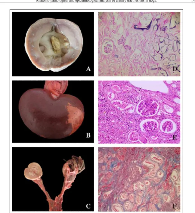
Figure 1 - Urolith obstructing the renal pelvis (A); Renal infarct (B); Exemplary of D. renalein right kidney and
hydronephrosis in contralateral kidney (C); Calcification of tubules and glomeruli (10x) H&E (D);
adenoma (1), bladder papilloma (1), bladder
papillary infiltrative transitional cell carcinoma (2),
and squamous bladder cells carcinoma (1). Only one case of circulatory disorder was recorded which was associated with congestion.
DISCUSSION
The kidney is particularly susceptible
to toxicity due to the high blood flow to it in
relation to its mass and the unique property of the renal tubular epithelium in the concentration of urine and its components, including drugs and chemicals (NEWMAN, 2013). Structural and
functional differences in renal blood flow (RBF)
and glomerular filtration rate (GFR) significantly
contribute to the increased susceptibility of elderly animals to the nephrotoxic response when compared to younger animals (KHAN, 2015). In this study, approximately 70% of the subjects were aged animals and were affected by a variety of diseases,
such as glomerulosclerosis and fibroplasia that had
led to chronic renal failure. However, age of animals was not a determining factor in the development of acute renal failure but instead represented a ‘triggering’ event.
According to KHAN et al. (2015), acute renal failure is characterized by rapid failure of
glomerular filtration rate, which can develop in a few
hours or over a number of weeks, with a consequent
Renal Lesions N/% Sex Breed Age
Tubulointerstitial injuries 313*/45.7
Acute tubular necrosis 144/46 50 F; 69 M; 25 UN 84 PB; 41 MB; 19 UN 19 P; 73 AD; 37 ED; 15 UN Tubular dilation 18/5.8 10 F; 7 M; 1 UN 15 PB; 1 MB; 2 UN 1 P; 14 AD; 1 ED; 2 UN Tubulointerstitial nephritis 142/45.4 53 F; 65 M; 24 UN 69 PB; 59 MB; 14 UN 23 P; 68 AD; 29 ED; 22 UN Granulomatous nephritis 7/2.2 3 F; 2 M; 2 UN 2 PB; 3 MB; 2 UN 4 AD; 1 ED; 2 UN
Presenceof bile pigment 2/0.6 2 F 1 PB; 1 MB 1 AD; 1 UN
Glomerular lesions 144*/21
Membrano-proliferative glomerulonephritis 20/13.9 12 F; 3 M; 5 UN 14 PB; 1 MB; 5 UN 1P; 10 AD; 6 ED; 3 UN Membranous glomerulonephritis 17/11.8 6 F; 9 M; 2 UN 10 PB; 6 MB; 1 UN 13 AD; 3 ED; 1 UN
Proliferative glomerulonephritis 3/2.1 2 F; 1 M 3 PB 1 ED; 2 UN
Glomerulosclerosis 102/70.8 38 F; 56 M; 8 UN 59 PB; 32 MB; 11 UN 6 P; 56 AD; 31 ED; 9 UN
Glomerular amyloidosis 2/1.4 1 M; 1 UN 1 PB; 1 MB 1 AD; 1 UN
Renal fibrosis 129*/18.8
Fibroplasia 129/100 48 F; 70 M; 11 UN 72 PB; 40 MB; 17 UN 7 P; 70 AD; 39 ED; 12 UN
Circulatory disorders 42*/6.1
Infarct 9/21.4 2 F; 5 M; 2 UN 8 PB; 1 MB 1 P; 5 AD; 3 ED
Congestion/Hyperemia 19/45.2 10 F; 5 M; 4 UN 7 PB; 7 MB; 4 UN 5 P; 13 AD; 1 ED
Hemorrhage 13/31 2 F; 5 M; 6 UN 6 PB; 6 MB; 1 UN 3 P; 8 AD; 2 UN
Edema 1/2.4 1 F 1 PB 1 AD
Lesions of the renal pelvis 41*/6
Pyelonephritis 14/34.1 4 F; 10 M 8 PB; 5 MB; 1 UN 1 P; 5 AD; 8 ED
Hydronephrosis 11/26.8 3 F; 7 M; 1 UN 6 PB; 1 MB; 4 UN 1 P; 4 AD; 3 ED; 3 UN
Parasitism by Dioctophyma renale 16/39.1 5 F; 9 M; 2 UN 4 PB; 11 MB; 1 UN 10 AD; 3 ED; 3 UN
Neoplasm 8*/1.2
Primary 7/87.5 4 F; 2 M; 1 UN 6 PB; 1 UN 2 AD; 4 ED; 1 UN
Metastases 1/12.5 1 F 1 PB 1 ED
Developmental abnormalities 8*/1.2
Renal dysplasia 6/75 1 F; 4 M;1 UN 3 PB; 2 MB; 1 UN 2 P; 4 AD
Renal cysts 1/12.5 1 M 1 MB 1 AD
Agenesis 1/12.5 1 M 1 MB 1 AD
Total 685
retention of nitrogenous products (prerenal azotemia) causing damage to the parenchyma and eventually the entire organ. With the progression of this process, occurs the formation of cylinders causing obstruction
of urinary flow (post-renal azotemia). KHAN et al. (2015) classified acute renal damage that was similar
to that described in this study as acute tubular necrosis, interstitial nephritis, and glomerulonephritis. Data
concerning the classification of acute tubular necrosis
presented in this study were not consistent with the protocols described in KHAN’s study. In this research, Leptospirosis was the main cause of interstitial nephritis. TOCHETTO et al. (2012) reported that in 53 cases of leptospirosis, interstitial nephritis was the characterizing lesion of the disease. Colonization of the kidney by bacteria begins in the interstitial capillary tubes and reachs the thin tubule, causing degeneration and concomitant tubular necrosis and
an inflammatory response.
Glomerular lesions in this study accounted for 21% (144) of renal lesions, and were the second most frequent group. Glomerular diseases are often associated with membranous proliferative changes that
included a significant increase in mesangial cells and
basement membrane mesangial substance, thickening of basement membranes and division of the basement membrane when combined with granular deposits of immunoglobulin (NEWMAN, 2013). In this study, four female animals had glomerulonephritis (3 membranoproliferative type and 1 proliferative type)
associated with pyometra. Renal changes in cases of pyometra are secondary to glomerulonephritis caused by deposition of immunocomplex containing bacterial endotoxins that alter the response of the renal tubule to antidiuretic hormone (NEWMAN, 2013).
Glomerulosclerosis (102) was the most frequently observed form of glomerular injury, occurring mainly in cases of chronic renal failure/ uremia. Most of animals in the study had concomitant glomerulonephritis and glomerulosclerosis. These are key symptoms of chronic kidney disease that is characterized by the formation of a vicious cycle that includes progressive loss of nephrons with
replacement by fibrous connective tissue. The
mechanism suggested for this, although not yet fully understood, involves systemic hypertension and
intra-renal glomerular hyperfiltration, hypertrophy
and/or tubular atrophy of cells (KHAN et al., 2015).
Cases of glomerulosclerosis identified in this study progressed to renal fibroplasia. Fibroplasia also
occurs in the repair infarcts and in other renal necrotic lesions (NEWMAN, 2013). Extensive glomerular
amyloidosis leads to a reduction in the efficiency
of the glomeruli, proteinuria, and kidney failure in dogs; there is a high incidence of thrombosis of the pulmonary arteries in affected animals (NEWMAN, 2013). Although NEWMAN (2013) noted an increase in the frequency of pulmonary thrombosis this was not observed in the two cases of glomerular amyloidosis reported in this study.
Table 2 - Classification of 60 lesions of lower urinary tract.
Lesions of lower urinary tract Nº/% Sex Breed Age
Inflammatory lesions 25*/41.7
Acute cystitis 19/76 4 F; 14 M; 1 UN 9 PB; 6 MB; 4 UN 7 AD; 9 ED; 3 UN
Chronic cystitis 5/20 2 F; 2 M; 1 UN 4 PB; 1 MB 4 AD; 1 ED
Acute urethritis 1/4 1 M 1PB 1P
Obstructive lesions 19*/31.6
Urolithiasis 18/94.8 2 F; 11 M; 5 UN 5 PB; 9 MB; 4 UN 10 AD; 5 ED; 3 UN
Hydroureter 1/5.2 1 M 1 UN 1 AD
Acquired anatomical variations 9*/15
Rupture of the bladder 8/88.9 1 F; 5 M; 2 UN 3 PB; 3 MB; 2 UN 1 P; 5 AD; 1 ED; 1 UN
Rupture of urethra 1/11.1 1 M 1 UN 1 P
Neoplasms 6*/10
Primary 6/100 4 F; 2 M 6 PB 3 AD; 3 ED
Circulatory disorders 1*/1.7
Congestion 1/100 1 M 1 PB 1 AD
Total 60
nematode were mixed breed and wandering animals, this can be explained by the indiscriminate eating habits of these animals (KOMMERS et al., 1999), in addition to direct contact with contaminated water. In this study, renal changes observed in parasitized animals were characterized by bone metaplasia in the renal capsule and the presence of bioperculated eggs in the lung parenchyma. These had a rough and thick bark-like appearance and had elicited no tissue response suggesting an erratic migration of the parasite.
Obstructive changes caused by uroliths can occur anywhere in the urinary tract, including the pelvic urinary bladder (INKELMANN et al., 2012b). In our study, male dogs were the most likely to be affected because they have a longer and thinner urethra (NEWMAN et al., 2013). One of the most common causes of the development of calculi is an imbalanced diet (MONFERDINI & OLIVEIRA, 2009). In the present research, most dogs affected by
uroliths were identified in the period between
1978-1989, when it was more common to feed dogs with home cooked food that did not have a proper balance of calcium and phosphate.
Primary urinary tract neoplasms occur infrequently in dogs. Primary renal tumors are rare and comprise about 1% of all tumors in dogs (NEWMAN et al., 2013). Animals in this study affected by
primary metastatic renal tumors were more than five years of age, a finding consistent with the findings
of KOBAYASHI et al. (2008). For renal neoplasms, primary renal carcinoma was the most frequent diagnose. Only one mammary adenocarcinoma case with renal metastasis was observed in this study. In other reports metastatic tumors were the most common (INKELMANN et al., 2011).
Abnormalities in urinary tract development are rare in dogs (HÜNNING et al., 2009; NEWMAN et al., 2013). In this paper, these cases represented 1.2% of renal lesions. We found two cases of renal dysplasia that were characterized as progressive juvenile nephropathy; one Shar Pei and one Shih Tzu, one year and a half, and eight months old respectively. Renal dysplasia is a congenital disease characterized by disruption of the renal parenchyma due to abnormalities in nephrogenesis, with development of structures inappropriate to the animal’s stage of development (PICUTT &LEWIS, 1987). The case of renal agenesis described here resulted in the death of the animal, as it was associated with urolithiasis of the contralateral kidney.
Among the LUT injuries described in this
paper, inflammatory causes were the most common.
the animals in this study, cystitis was more frequently observed in females, with no obvious association reported for age or breed.
Obstructive lesions of LUT, the second most common cause of urinary tract changes described in this study, were associated with uroliths and caused hydroureter and/or rupture. Tumors of LUT were all primary and occurred in the bladder in contrast to the results of INKELMANN et al. (2012b), who report a
higher number of metastatic tumors. With significant
increases in the dog population, increases in life expectancy, and poor feeding practices by many owners, we suggested that the incidence of lesions of the urinary tract is likely to increase and accurate characterization of the changes and will be important in the diagnoses of this condition.
CONCLUSION
Most diagnoses in this study were of primary lesions of the kidney, demonstrating the importance of a thorough clinical pathology analysis of the animal, since kidney failure, either acute or
chronic, is a significant cause of death in dogs. The
most frequent injury of the LUT was the acute or chronic cystitis, indicating that it deserves special attention from veterinary practitioners, as may occur the evolution of this injury and lead to worsening of the disease. The knowledge of renal alterations could be a key factor in preventive actions in order to avoid an undesirable prognosis for the animal.
Besides, the correct classification of this data will
contribute to the best comprehension of these alterations and its implications.
REFERENCES
FIGHERA, R.A. et al. Causes of death and reasons for euthanasia in dogs from the midland region of the Midwest of Rio Grande do Sul State, Brazil (1965-2004). Pesquisa Veterinária Brasileira, v.28, n.4, p.223-230, 2008. Available from: <http://www.scielo. br/pdf/pvb/v28n4/v28n4a05.pdf>. Accessed: May 4, 2015. doi: 10.1590/S0100-736X2008000400005.
HÜNNING, P.S. et al. Renal dysplasia in a dog. Acta ScientiaeVeterinariae, v.37, n.1, p.73-77, 2009. Available from: < h t t p : / / w w w. u f r g s . b r / a c t a v e t / 3 7 - 1 / a r t 8 1 5 . p d f > . Accessed: May 3, 2015.
INKELMANN, M.A. et al. Lesions of the urinary system in 1.063 dogs. Pesquisa Veterinária Brasileira, v.32, n.8, p.761-771, 2012a. Available from: <http://www.pvb.com.br/pdf_artigos/27-08-2012_11-07Vet%201225_2793%20PA.pdf>. Accessed: Jan. 15, 2015. doi: 10.1590/S0100-736X2012000800015.
INKELMANN, M.A.et al. Urolithiasis in em 76 dogs.
Pesquisa Veterinária Brasileira, v.32, n.3, p.247-253, 2012b. Available from: <http://www.scielo.br/scielo.php?pid=S0100-736X2012000300012&script=sci_arttext>. Accessed: Jun. 13, 2015. doi: 10.1590/S0100-736X2012000300012.
KHAN, T.M.; KHAN, K.N.M. Acute kidney injury and chronic kidney disease. Veterinary Pathology, v.52, n.3, p. 441-444, 2015. Available from: <http://vet.sagepub.com/content/52/3/441?etoc>. Accessed: Jun. 26, 2015. doi: 10.1177/0300985814568358.
KOBAYASHI, N. et al. Renal colleting duct carcinoma in a dog.
Veterinary Pathology, v.45, p.489-494, 2008. Available from: <http://www.ncbi.nlm.nih.gov/pubmed/18587095>. Accessed: May 14, 2015. doi: 10.1354/vp.45-4-489.
KOMMERS, G.D. et al. Dioctophymosis in dogs: 16 cases.
Ciência Rural, v.29, n.3, p.517-522, 1999. Available from: <http:// www.scielo.br/pdf/cr/v29n3/a23v29n3.pdf>. Accessed: Jan. 15, 2015. doi: 10.1590/S0103-84781999000300023.
LULICH, J.P. et al. Distúrbios do trato urinário inferior dos caninos. In: ETTINGER, S.J.; FELDMAN, E.C. Tratado de medicina interna veterinária. 5.ed. São Paulo: Manole, 2008. V.2, p.1841-1867.
MAXIE, M.G.; NEWMAN, S.J. The urinary system. In: MAXIE M.G. et al. (Eds.). Pathology of domestic animals. 5.ed. Philadelphia: Saunders Elsevier, 2007. V.2. Cap.4, p.425-522.
MONFERDINI, R.P.; OLIVEIRA, J. Manejo nutricional para cães
e gatos com urolitíase: revisão bibliográfica. Acta Veterinária Brasilica, v.3, n.1, p.1-4, 2009. Available from: <http://periodicos. ufersa.edu.br/revistas/index.php/acta/article/view/1104>. Accessed: Jun. 14, 2015.
NEWMAN, S.J. O sistema urinário. In: ZACHARY, J.F.; MCGAVIN, M.D. Bases da patologia veterinária. São Paulo: Elsevier, 2013. Cap.11, p.592-661.
PICUTT, C.A.; LEWIS, R.M. Microspic features of canine renal dysplasia. Veterinary Pathology, v.24, p.156-163, 1987. Available from: <http://vet.sagepub.com/content/24/2/156.short>. Accessed: Jun. 14, 2015. doi: 10.1177/030098588702400209.
RHA, J.Y. et al. Familial glomerulonephropathy in a litter of beagles.
Journal of the American Veterinary Medical Association, v.216, p.46-50, 2000. Available from: <http://www.researchgate. net/publication/12676797_Familial_glomerulonephropathy_ in_a_litter_of_Beagles>. Accessed: Jun. 26, 2015. doi: 10.2460/ javma.2000.216.46.
![BICORNUATE [BICORNIS, UNICOLLIS] UTERUS, A CONGENITAL MALFORMATION ASSOCIATED WITH PATHOLOGICAL LESIONS: A CLINICOPATHOLOGICAL STUDY OF 4 RARE CASES](data:image/gif;base64,R0lGODlhAQABAIAAAP///wAAACH5BAEAAAAALAAAAAABAAEAAAICRAEAOw==)