ARTICLE
Right-to-left shunt and the hypercoagulable
state: does paradoxical embolism play a role
in patients with antiphospholipid syndrome
and stroke?
Shunt direita-esquerda e hipercoagulabilidade: embolia paradoxal pode ser responsável
pelo AVC na síndrome antifosfolipídica?
Laura Nicoleti Zamproni, Viviane Flumignan Zétola, Marcos Christiano Lange
Patients with cryptogenic arterial ischemic stroke have a high prevalence of a patent foramen ovale (PFO)1. However,
the simple existence of a right-to-left shunt (RLS) does not appear suicient to explain the increased risk of ischemic stroke, and other signiicant factors must be required2. A
hy-percoagulable state could be one of these factors, because it increases thrombogenic mechanisms, such as venous throm-bosis, which is related to paradoxical embolism (PE)3.
Antiphospholipid syndrome (APS) is one of the most frequently acquired thrombophilias4. he presence of
antiphospholipid antibodies (aPL) is associated with isch-emic stroke4-7. Nevertheless, the mechanism underlying
stroke in these patients remains unclear, and there is a lack of data in the literature on the role that PE may play in the pathophysiology of arterial ischemic stroke in APS patients. herefore, the association of stroke with the combined pres-ence of APS and an RLS is of interest.
he aim of this study was to verify whether APS patients who have experienced stroke present with a higher frequency of RLS than patients with APS without a history of stroke.
Neurology Division, Hospital de Clínicas, Federal University of Paraná, Curitiba PR, Brazil.
Correspondence: Laura Nicoleti Zamproni; Hospital de Clínicas, Serviço de Neurologia; Rua General Carneiro 181 / 4º andar; 80060-900 Curitiba PR - Brasil; E-mail: [email protected]
Conflict of interest: There is no conflict of interest to declare.
ABSTRACT
Objective: Patent foramen ovale is associated with paradoxical embolism (PE) and stroke. Hypercoagulable states, such as antiphospholipid syndrome (APS), can exacerbate PE by increasing clot formation. The aim of this study was to verify whether patients with APS and stroke present a right-to-left shunt (RLS) with greater frequency than patients with APS but without stroke. Methods: Fifty-three patients with APS were tested for RLS using contrast-enhanced transcranial Doppler (cTCD): 23 patients had a history of stroke (Stroke Group) and 30 had no history of stroke (No-stroke Group). Results: cTCD was positive in 15 patients (65%) from the Stroke Group and in 16 patients (53%) in the No-stroke Group (p=0.56). The proportion of patients with a small RLS (≤10 high-intensity transient sign or HITS) and a large RLS (>10 HITS) was similar between the groups without significant difference. Conclusions: Our data do not support the theory that paradoxical embolism may play an important role in stroke in APS patients.
Key words: antiphospholipid syndrome, stroke, paradoxical embolism, right-to-left shunt.
RESUMO
Objetivo: O forame oval patente está associado com embolia paradoxal e acidente vascular cerebral isquêmico (AVCi). Estados de hiperco-agulabilidade, como a síndrome antifosfolipídica (SAF), podem facilitar esse processo, aumentando a formação de coágulos. O objetivo deste estudo foi verificar se pacientes com SAF e AVCi apresentam maior frequência de shunt direita-esquerda (SDE), comparados a pacientes com SAF sem AVCi. Métodos: Cinquenta e três pacientes com SAF foram testados para SDE usando Doppler transcraniano contrastado (DTCc): 23 com AVCi (Grupo AVC) e 30 sem história de AVCi (Grupo Controle). Resultados: DTCc foi positivo em 15 pacientes (65%) do Grupo AVC e em 16 pacientes (53%) no Grupo Controle (p=0,56). A proporção de pacientes com pequeno SDE (≤10 HITS) e grande SDE (>10 HITS) foi semelhante nos dois grupos. Não houve diferença significativa entre os grupos. Conclusões: Nossos dados não sugerem que embolia para-doxal seja causa importante de AVCi em pacientes com SAF.
METHODS
his is a cross-sectional study conducted prospectively. All patients diagnosed with APS and followed at the out-patient clinic of the Neurology and Rheumatology Division of the Hospital de Clínicas, Federal University of Paraná, were recruited between March 2009 and December 2010. After receiving written, informed consent, patients were evaluated by a single neurologist from the same institution and examined for the presence of an RLS. he Local Ethics Committee approved this study.
An APS diagnosis was made when the subject fulilled the modiied Sapporo criteria8 (at least, one clinical criterion and
one laboratory criterion must have been met). he laboratory criteria were assessed using the standards and benchmarks provided by the local laboratory that processed the patient samples. he anticardiolipin IgG or IgM value was considered positive when greater than 10 GPL or MPL. hat value is ad-opted in our laboratory although the literature only considers values over 40 GPL or MPL as positive. Several articles in the literature have discussed the lack of standardization of an-ticardiolipin tests, the diference in results between centers and especially the diference between the kits utilized9. his
reference value was maintained in our laboratory, however all patients included who had anticardiolipin antibody val-ues between 10 and 40 GPL or MPL also had a positive lupus anticoagulant ratio and a clinical history that was highly sug-gestive of APS. he level of lupus anticoagulant was consid-ered positive when its ratio was greater than one, provided that the patient was not receiving heparin. If the patient was receiving oral anticoagulants, levels of international normal-ized ratio (INR) of up to 3.0 were accepted as a positive lupus anticoagulant test. If the patient had an INR that was great-er than 3.0, the positive lupus anticoagulant test was not ac-cepted, the anticoagulation treatment was adjusted and the lupus anticoagulant test was repeated.
Clinical and neurological evaluations were conducted on all cases, and no patients indicated the presence of an ex-tracranial or an inex-tracranial stenosis in their ultrasonography results. All patients had a transthoracic echocardiogram, but no patient had any signiicant alteration. All patients with a positive stroke history were previously evaluated to exclude other etiologies before they were considered as having an ar-terial ischemic stroke due to APS and, thus, were included in the current study. All patients underwent an additional brain computed axial tomography (CT). Five patients in each group had a magnetic resonance image (MRI) in the last two years, and Flair and difusion sequences of these exams were also analyzed. Ischemic stroke was considered to be present when the patient had a previous history of a sudden neuro-logic deicit accompanied by an arterial territory lesion on CT or MRI. Arterial territories were classiied into three clusters for analysis: cortical, when the stroke included a distal lesion;
subcortical for lesions greater than 1,5 mm and not involving the cortex; and microvessels, when the lesions were small-er than 1,5 mm. he patients wsmall-ere classiied into two groups based on stroke investigation: Stroke Group, composed of pa-tients with APS with a conirmed history of stroke; and No-stroke Group, composed of patients with APS with normal brain images and without a history of stroke.
he RLS investigation was performed by a single neurol-ogist (it was not involved patients clinical evaluation) with expertise in contrast-enhanced transcranial Doppler (cTCD) ultrasonography. he cTCD (DWL Doppler-Box, Singen, Germany) procedures were performed while the patients were in a supine position, and the neurologist was blinded to the patients’ medical status. Two 2-MHz-pulsed Doppler transducers were ixed using a head frame (DWL DiaMon, Singen, Germany), and the main stems of both middle cere-bral arteries (MCAs) were insonated through the temporal window at a depth of 55 to 65 mm to capture a small sample volume of 12 mm in length with M-mode.
he contrast agent (CA) was composed of a mixture of 9 mL saline and 1 mL air. Prior to the infusion, the solution was prepared by agitating the mixture 10 times between two 10-mL syringes via a three-way tap, which was connected to a 24-gauge intravenous catheter that was inserted into the su-pericial vein of the patient’s arm. he distance from the cath-eter to the syringes was less than 10 cm. he CA was injected over ive seconds into the antecubital vein.
he procedure was conducted while the patient was rest-ing (restrest-ing phase) and before he performed a Valsalva maneu-ver (VM) protocol. he VM was performed ive seconds after the CA was injected, and its efectiveness was conirmed by a 25% decrease of MCA low velocity and by a delection of at least 40 mmHg in a manometer that the patient exhaled into during the VM. Both studies (resting phase and VM phase) were repeated three times, with each test lasting one minute.
Patients were considered RLS-positive when at least one high-intensity transient sign (HITS) was detected on the spec-tral display of at least one of the monitored MCAs. Conversely, the patient was determined to have no RLS (RLS negative) when, during the 60 seconds following the injection of the CA, there was no identiied HITS in either MCA. Based on the cTCD results, the patients were classiied as RLS-negative, “small RLS” when the patient presented with an RLS that was identiied to be small (≤10 bubbles) or “large RLS” when the patient’s RLS was identiied to be large (>10 bubbles).
Exclusion criteria comprised the following items: no tem-poral window; a lack of peripheral venous access; or patients were restricted to the bed or with serious sequelae that were incompatible with the examination; or self-exclusion.
RESULTS
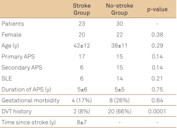
During the study period, 53 patients fulilled the criteria for APS. No patient was excluded. Of these APS patients, 23 had a history of stroke (Stroke Group), and 30 had no his-tory of stroke (No-stroke Group). In the Stroke Group, there were 20 women, and the mean±standard deviation (SD) age was 42±12 years old. Six patients had APS secondary to sys-temic lupus erythematosus (SLE), and 17 patients had pri-mary APS. Neuroimaging results revealed that only 40% of patients had multiple ischemic lesions, and 30% had lesions of diferent arterial territories (with 75% of the cases indicat-ing a stroke in the anterior circulation). he stroke was corti-cal in 53%, subcorticorti-cal in 30% and in the basal ganglia in 17% of patients. In addition to stroke, another thrombotic event was found in 13% of patients, and gestational morbidity was found in 17% of patients.
In the No-stroke Group, there were 22 women, and the mean±SD age was 38±11 years. Fourteen patients had APS secondary to SLE, one patient had APS secondary to rheu-matoid arthritis, and 15 patients had primary APS. he main clinical event present in 20 patients was deep venous throm-bosis (DVT). Four patients presented with a pulmonary em-bolism, three patients exhibited a cerebral venous throm-bosis (without brain venous ischemia), and eight presented with gestational morbidity. here was no signiicant difer-ence between Stroke Group and No-stroke Group. hese de-mographics are presented in Table 1.
cTCD was positive in 15 (65%) patients from the Stroke Group and in 16 (53%) from the No-stroke Group (p=0,56). In the Stroke Group, 8 patients showed the presence of an RLS conirmed by a resting test, and all 15 patients had conirmed RLS using the VM test. When separated into diferent grades of RLS, this group was comprised of eight patients with a small RLS and seven with a large RLS. In the No-stroke Group, small RLS and large RLS were observed in eight patients each. here was no signiicant diference between the No-stroke and Stroke Groups. hese results are shown in Table 2.
In the Stroke Group, the patients with only one ischemic territory (n=14) had a small RLS in ive cases and a large RLS in four cases; ive patients were RLS-negative. In patients with multiple ischemic territories (n=9), three patients had a small RLS, three had a large RLS, and the other three patients were RLS-negative, as shown in Table 3.
Eighteen patients had an anterior circulation stroke: nine patients had cortical lesions, six patients had subcor-tical lesions, and three patients had microvascular lesions. Approximately 50% of patients with a cortical and subcor-tical anterior circulation stroke and 100% of basal ganglia stroke cases were RLS-positive. In the five patients with a posterior circulation stroke, three had cortical lesions, and two exhibited brainstem (BS) lesions. We found that 66% of patients with a cortical posterior circulation stroke and
Stroke Group
No-stroke
Group p-value
Patients 23 30
-Female 20 22 0.38
Age (y) 42±12 38±11 0.29
Primary APS 17 15 0.14
Secondary APS 6 15 0.14
SLE 6 14 0.21
Duration of APS (y) 5±6 5±5 0.75
Gestational morbidity 4 (17%) 8 (26%) 0.64
DVT history 2 (8%) 20 (66%) 0.0001
Time since stroke (y) 8±7 - -Table 1. Demographics of APS patients, separated into Stroke and No-stroke Groups.
y: years; APS: antiphospholipid syndrome; SLE: systemic lupus erythematosus; DVT: deep venous thrombosis.
Stroke territory Patients RLS-negative
RLS-positive p-value
Anterior circulation 18
Cortical 9 4 (45%) 5 (55%) 0.5
Subcortical 6 3 (50%) 3 (50%)
Microvascular 3 0 (0%) 3 (100%)
Posterior circulation 5
Cortical 3 1 (33%) 2 (66%) 0.7
BS 2 1 (50%) 1 (50%)
Table 4. RLS and stroke characteristics in patients with APS.
RLS: right-to-left shunt; APS: antiphospholipid syndrome; BS: brainstem.
Stroke Group
No-stroke
Group p-value
Patients 23 30
RLS-positive 15 16 0.56
Positive in resting test 8 10 0.85
Only positive in VM test 7 6 0.58
Small RLS 8 8 0.74
Large RLS 7 8 0.99
Table 2. RLS in Stroke and No-stroke Groups.
RLS: right-to-left shunt; VM: Valsalva maneuver.
Single lesion
Multiple
lesions p-value
Patients 14 9
RLS-negative in VM test 5 3 0.63
Small RLS in VM test 5 3
Large RLS in VM test 4 3 Table 3. RLS in Stroke Group.
1. Cabanes L, Mas JL, Cohen A, et al. Atrial septal aneurysm and patent foramen ovale as risk factors for cryptogenic stroke in patients less than 55 years of age: a study using transesophageal echocardiography. Stroke 1993;24:1865-1873.
2. Ozdemir AO, Tamayo A, Munoz C, Dias B, Spence JD. Cryptogenic stroke and patent foramen ovale: clinical clues to paradoxical embolism. J Neurol Sci 2008;275:121-127.
3. The Antiphospholipid Antibodies in Stroke (APASS) Study Group. Anticardiolipin antibodies are an independent risk factor for first ischemic stroke. Neurology 1993;43:2069-2073.
4. Ruiz-Irastorza G, Khamashta MA. Stroke and antiphospholipid syndrome: the treatment debate. Rheumatology 2005;44:971-974.
5. Tuhrim S, Rand JH, Wu X, et al. Elevated anticardiolipin antibody titer is a stroke risk factor in a multiethnic population independent of isotype or degree of positivity. Stroke 1999;30:1561-1565.
6. Brey RL, Stallworth CL, McGlasson DL, et al. Antiphospholipid antibodies and stroke in young women. Stroke 2002;33:2396-2400.
7. Brey RL, Abbott RD, Curb JD, et al. Beta 2-glycoprotein 1-dependent anticardiolipin antibodies and risk of ischemic stroke and myocardial infarction: the honolulu heart program. Stroke 2001;32:1701-1706.
8. Miyakis S, Lockshin D, Atsumi T, et al. International consensus statement on an update of the classification criteria for definite antiphospholipid syndrome (APS). J Thromb Haemostasis 2006;4:295-306.
9. Pengo V, Biasiolo A, Bison E, et al. Antiphospholipid antibody ELISAs: References
50% of patients with a BS stroke were RLS-positive. No dif-ference was noted between the presence of an RLS and the pattern of ischemic stroke (Table 4).
DISCUSSION
In this study, there was no relationship between stroke and an RLS in patients with APS, even when multiple isch-emic territories or diferent lesion patterns were present.
PFO can be associated with cryptogenic stroke; the mecha-nism is postulated to be a PE, where clots originate in the venous circulation and travel to the arterial side through an RLS10,11.
Systemic hypercoagulable states can potentially increase clot formation12. It is reasonable to hypothesize that patients with
hypercoagulable states may be at a higher risk of developing a paradoxical embolism and stroke. To our knowledge, there is no literature to prove this association13,14. Most of the published
data on this issue are either anecdotal or case series12.
Di Tullio et al.15 reported a signiicantly higher prevalence of
protein C deiciency in 25 stroke patients with PFO compared with 195 without PFO. However, this study diagnosed the pres-ence of RLS using transthoracic contrast echocardiography, which may introduce substantial bias. Barinagarrementeria et al.16 and Chaturvedi11 obtained similar indings in a small case
series, whereas Schwarze et al.17 found an increased prevalence
of RLS and activated protein C resistance in patients with stroke compared with patients without this thrombophilic disorder.
Florez et al.13 studied patients with stroke and veriied
the coexistence of PFO and a hypercoagulable state. he frequency of a hypercoagulation abnormality was not high-er in patients with a PFO compared with patients without a PFO. Similar results were obtained by Carod-Artal et al.18.
In their study, the prevalence of thrombophilia in PFO-positive and PFO-negative patients with cryptogenic stroke and stroke patients with known etiology was examined. he authors found no diference between the PFO-positive and PFO-negative groups18. Notably, a group of control subjects
with no history of stroke was lacking in all of these studies.
Recently, the PICCS-APASS study analyzed the presence of APL antibodies and a PFO in stroke patients. his study did not report a relationship among positive APL antibodies, PFO and the recurrence of cerebral ischemic events14. Notwithstanding,
this study considered the only presence of APL antibodies and did not consider patients with diagnosed APS.
In our study, patients with conirmed APS, with and without a history of stroke, were evaluated for the pres-ence of an RLS. We also attempted to correlate the char-acteristics of arterial stroke with the presence of an RLS. he data did not support the theory that patients with APS who had experienced stroke have a greater frequency of RLS than patients with APS who did not have a history of cerebral ischemic events. No relationship was found be-tween multiple ischemic events and cortical stroke and the presence of an RLS. Interestingly, a high prevalence of RLS was found in both groups with APS (65 and 53%) versus the population prevalence estimated in the literature (30%)12.
Unfortunately, there is no data in the literature comparing the prevalence of RLS in the APS population versus healthy subjects. It is well known that APS predisposes patients to cardiac defects, such as valvar abnormalities. Although speculative, it is likely that APS patients also present with more cardiac shunts.
he limitations of this study include the small number of patients (APS is a rare disease) and the lack of transesoph-ageal echocardiography to conirm the presence of a PFO. ß2-Glycoprotein testing was unavailable for patients in this study, because it is not routine in our institution.
survey on the performance of clinical laboratories assessed by using lyophilized affinity-purified IgG with anticardiolipin and anti-beta2-Glycoprotein I activity. Thromb Res 2007;120:127-133.
10. Karttunen V, Hiltunen L, Rasi V, Vahtera E, Hillbom M. Factor V Leiden and prothrombin gene mutation may predispose to paradoxical embolism in subjects with patent foramen ovale. Blood Coagul Fibrinolysis 2003;14:261-268.
11. Chaturvedi S. Coagulation abnormalities in adults with cryptogenic stroke and patent foramen ovale. J Neurol Sci 1998;160:158-160.
12. Lange MC, Zamproni LN, Braatz V, Fernandes AF, Zetola VF, Nóvak EM. Ischemic stroke in a patient with Crohn’s disease: a confirmed paradoxical embolism mechanism. Arq Neuropsiquiatr 2010;68:651-652.
13. Florez JC, Ay H, Van Cott EM, Buonanno FS. Patent foramen ovale and hypercoagulability as combined risk factors for stroke. J Stroke Cerebrovasc Dis 2003;12:114-118.
14. Rajamani K, Chaturvedi S, Jin Z, et al. Patent foramen ovale, cardiac valve thickening, and antiphospholipid antibodies as risk factors for subsequent vascular events: the PICSS-APASS study. Stroke 2009;40;2337-2342.
15. Di Tullio MR, Santoni-Rugiu F, Sacco RL, et al. Patent foramen ovale and hypercoagulable states in ischemic stroke patients. Circulation1994;90:398.
16. Barinagarrementeria F, Santos J, Ruiz-Sandoval JL, et al. Prothrombotic states as triggering of cerebral infarction in patients with patent foramen ovale. Neurology 1998;50:155.
17. Schwarze JJ, Klingelhofer J, Kim S, et al. Coexistence of a prothrombotic state and right-to-left shunts: a potential mechanism of paradoxical embolism? Neurology 1998;50:A154.