Printed version ISSN 0001-3765 / Online version ISSN 1678-2690 http://dx.doi.org/10.1590/0001-3765201520140712
www.scielo.br/aabc
Toxicological Evaluation of Anti-Scrapie Trimethoxychalcones and Oxadiazoles
ClAudiA P. FiguEirEdO1
, NATAliA C. FErrEirA1
, giSEllE F. PASSOS1
, rObSON dA COSTA1
, FErNANdA S. NEvES1
, ClAriCE S.C. MAChAdO1
, AlESSANdrA MASCArEllO2
, lOuiSE d. ChiArAdiA-dElATOrrE2
, PATríCiA d. NEuENFEldT2
, riCArdO J. NuNES2
and YrAiMA COrdEirO1
1
Faculdade de Farmácia, Universidade Federal do Rio de Janeiro, Av. Carlos Chagas Filho, 373, 21941-902 Rio de Janeiro, RJ, Brasil 2
Departamento de Química, Universidade Federal de Santa Catarina, Campus Universitário Trindade, 88040-900 Florianópolis, SC, Brasil
Manuscript received on December 23, 2014; accepted on publication April 9, 2015
AbSTrACT
An altered form of the cellular prion protein, the PrPSc
or PrPRes
, is implicated in the occurrence of the still untreatable transmissible spongiform encephalopathies. We have previously synthesized and characterized aromatic compounds that inhibit protease-resistant prion protein (PrPRes) accumulation in scrapie-infected cells. These compounds belong to different chemical classes, including acylhydrazones, chalcones and oxadiazoles. Some of the active compounds were non-toxic to neuroblastoma cells in culture and seem to possess drugable properties, since they are in agreement with the Lipinski´s rule of 5 and present
desirable pharmacokinetic profiles as predicted in silico. Before the evaluation of the in vivo efficacy of the aromatic compounds in scrapie-infected mice, safety assessment in healthy mice is needed. Here we
used Swiss mice to evaluate the acute toxicity profile of the six most promising anti-prionic compounds,
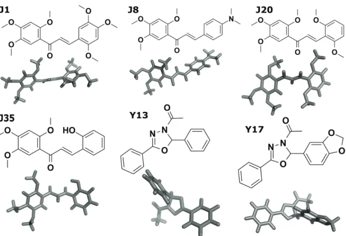
the 2,4,5-trimethoxychalcones (J1, J8, J20 and J35) and the 1,3,4-oxadiazoles (Y13 and Y17). One single oral administration (300 mg/kg) of J1, J8, J20, J35, Y13 and Y17 or repeated intraperitoneal administration (10 mg/kg, 3 times a week, for 4 weeks) of J1, J8 and J35, did not elicit toxicity in mice. We strongly believe that the investigated trimethoxychalcones and oxadiazoles are interesting compounds to be further analyzed in vivo against prion diseases.
Key words: prion, acute toxicity, organic compound, drug safety, scrapie, pharmacokinetics.
Correspondence to: Yraima Cordeiro E-mail: [email protected]
iNTrOduCTiON
Prion diseases or transmissible spongiform
enceph-alopathies (TSEs) are severe neurodegenerative
disorders that still lack efficient treatment. An ab -normal form of the cellular prion protein (PrP), the
scrapie PrP (PrPSc), is involved in the establishment
of TSEs (Prusiner 1998). PrPSc is partially
protease-resistant, poorly soluble in aqueous solvents and
accumulates in its aggregated form in the brain of
affected individuals. Most experimental
approach-es against TSEs focused either on inhibiting the
conversion of PrPC to PrPSc or on controlling the
clearance of PrPSc from infected cells and tissues
(Kocisko et al. 2003, Kocisko and Caughey 2006,
Rainov et al. 2007, Forloni et al. 2013, Cordeiro
or-ganic compounds have been screened for these ef-fects, such as quinolines, acridines, oxadiazoles, chalcones, 2-aminothiazoles, arylamides, among others (Doh-ura et al 2000, Korth et al. 2001, Koster et al. 2003, Cashman and Caughey 2004, Ghaemmaghami et al. 2010, Macedo et al. 2010, Sim 2012, Li et al. 2013, Ferreira et al. 2014). The majority of tests have used cultured mammalian cells infected with the scrapie form of PrP obtained from different sources (murine, hamster) (Kocisko et al. 2003, Kocisko and Caughey 2006, Sim 2012, Ferreira et al. 2014). Their ability to reduce PrPSc (or PrPRes) levels in vitro is usually evaluated using anti-PrP antibodies in proteinase-K treated cells. Although several of these compounds show low IC50 values (nM range), only a few were able to cross the blood brain barrier (BBB) and/or were
evaluated for their pharmacokinetic profile (Koster
et al. 2003, Silber et al. 2013). Flupirtine, quina-crine, chlorpromazine, pentosan polysulfate (PPS) and doxycycline have even been tested in humans, and even though clinical improvement and margin-ally increase in the survival time (for PPS) were observed in some cases, none of them were able to reverse or even to halt the clinical course of the dis-ease (Furukawa et al. 2002, Cashman and Caughey 2004, Otto et al. 2004, Martínez-Lage et al. 2005, Rainov et al. 2007, Tsuboi et al. 2009, Collinge et al. 2009, Forloni et al. 2013, Haïk et al. 2014). The investigation of drugs/phytotherapics already commercialized with other clinical indications has helped to overcome the toxicological limitations of studying new compounds and administering them to animal models and humans. However, newly
de-veloped molecules might possess better efficacy
and safety compared to old ones and could repre-sent an important step in the development of anti-scrapie therapeutic strategies. Nevertheless, initial toxicological analysis and evaluation of the
phar-macokinetic (PK) profile of these new substances
are indeed needed.
We have recently identified a new panel of aromatic hetero and homocyclic compounds with anti-scrapie activity by screening in scrapie-infected neuroblastoma (ScN2a) cells (Ferreira et al. 2014, Cordeiro and Ferreira 2015). Among ~ 50 active
compounds, we identified four trimethoxychalcones
(named J1, J8, J20, J35) and two oxadiazoles (Y13 and Y17) with IC50 ~1 µM (Js) and ~10 µM (Ys), respectively, obtained after incubation with ScN2a cells for 4 days. In silico PK and physico-chemical predictions indicated that these compounds have promising drug characteristics, such as reduced mutagenicity, high permeation to the central nervous system and high oral bioavailability (Ferreira et al. 2014). One study has shown that the chalcones J8, J20 and J35 at 100 µM, reduced the viability of human K562 acute myeloid leukemia cells from 30-50 % (Costa et al. 2014). J1 inhibited the activity of cruzain from T. cruzi with an IC50 of 100 µM (Borchhardt et al. 2010). However, even for the compounds that had different biological/ pharmacological activities, they did not match the potential anti-scrapie activity, as the IC50 values
obtained in ScN2a cells were significantly lower
than those reported (Borchhardt et al. 2010, Costa et al. 2014). Therefore, we do not expect that the compounds are at a risk of presenting unwanted pharmacological side effects during their preclinical and clinical trials.
Here, we assessed the safety of compounds J1, J8, J20, J35, Y13 and Y17 (Fig. 1) by evaluating their toxic effects after acute oral administration or following repeated intraperitoneal administration in mice. The development of behavioural alterations (Hippocratic test), food and water intake, were evaluated. In addition, the effect of the compounds on lethality was investigated during the
first 24 h and for 14 or 28 days in order to assess
the possible alterations related to the course of
treatment. Animals were sacrificed at the end of the
alterations in several organs and/or glands was performed.
None of the investigated compounds caused death of the mice at the administered dose and no macroscopic alterations were seen in the selected organs or glands following oral administration. Our results indicate that LD50 is higher than 300 mg/kg for all studied compounds, and that oral administration of 2,4,5-trimethoxychalcones and 1,3,4-oxadiazoles is safe in doses of up to 300 mg/kg. In addition, they can also be considered safe for systemic administration, since only one mouse treated with J8 and one with J35 (both intraperitoneal administration) showed minor lung bleeding, kidney discoloration and increased relative weight of the heart, when compared with
the control group. This toxicological evaluation provides preliminary information for in vivo studies in scrapie-infected murine models.
MATEriAlS ANd METhOdS
Compounds
ethanone, and Y17 (1-[(2R)-2-(1,3-benzodioxol-5-yl)-5-phenyl-1,3,4-oxadiazol-3(2H)-yl]ethanone) were synthesized and characterized as previously described (Borchhardt et al. 2010, Nunes et al. 2012, Costa et al. 2014).
AnimAls
Acute oral toxicity experiments were conducted on 36 male and 38 female adult Swiss mice (6-8 weeks old, 22-27 g), and repeated intraperitoneal doses were conducted on 24 female adult Swiss mice (8-12 weeks old, 30-35 g). The animals were purchased from Central Biotery of Fundação Oswaldo Cruz (FIOCRUZ – Rio de Janeiro/ RJ). Animals were housed in the vivarium at the School of Pharmacy, CCS, UFRJ. Animals were housed in clear microisolator cages (40x32x17 cm) (ALESCO, Campinas, Brazil), with controlled temperature (22 ± 2 °C), humidity between 60-80 % on a 12-h light/dark cycle, with food and water
ad libitum. All experiments were conducted in
accordance with the ethical principles set forth by the Brazilian School of Animal Experimentation (COBEA 1991) and with the approval of the Ethics Committee for Animal Experimentation (CEUA) of the Federal University of Rio de Janeiro (Protocol FARMACIA07-04/16). Prior to experimentation, animals were individually identified in order to evaluate behaviour and body weight during the period of study, as well as to evaluate organ weight at the end of the experiments.
In silicoprediCtionof toxiCologiCAl properties
Physicochemical and toxicity properties of compounds J1, J8, J20, J35, Y13 and Y17 were predicted with software modules from ACD/ Labs Percepta Platform, version 12.01, Advanced Chemistry Development, Inc., Toronto, On, Canada (www.acdlabs.com 2014) (ttp://www. acdlabs.com/products/percepta/predictors.php) from the uploaded three-dimensional structure of the compounds. Software components provided by ACD/Labs (from PhysChem Modules and Toxicity
Modules) were used to predict aqueous solubility, toxicity category and LD50 values for the selected compounds. We used the LD50 module that predicts LD50 values (mg/kg) for rats and mice according to various routes of administration, and the Toxicity
Categories module that classifies compounds into
OECD categories for acute oral toxicity. OECD
assigns chemicals to one of the five Oral Acute
Toxicity Hazard Categories according to the LD50 values after oral administration to mice, which are: V, 2000 to 5000 mg/kg (may be harmful if swallowed); IV, 300 to 2000 mg/kg (harmful if swallowed); III, 50 to 300 mg/kg (toxic if swallowed); II, 5 to 50 mg/kg (fatal if swallowed); I, < 5 mg/kg (fatal if swallowed). Predictions are provided as a list of probabilities that the compound’s LD50 (rat, oral route) would exceed the cut-off values separating different categories. On the basis of these values, the software selects the most probable OECD Hazard categories for that compound. For both solubility and LD50 calculation a reliability index (RI) value is provided that represents a quantitative evaluation of prediction confidence. In general, RI values below 0.3 indicate that the respective prediction is not reliable.
prepArAtionofthe J1, J8, J20, J35,
Y13 And Y17 Compounds
Stocks solutions were prepared according to the proposed doses (based on in silico LD50 prediction) and considering the estimated water solubility of the compounds. Stock solutions of compounds were prepared by solubilising the compound in 0.9 % NaCl supplemented with Tween 80 at 8 % (for i.p. administration) or 10 % (for oral administration).
orAl ACute toxiCitY
vehicle (0.9 % NaCl containing 10 % Tween 80, 10 mL/kg, p.o.). After oral administration of vehicle or the compounds, animals were arranged in groups and kept in clear microisolator cages for 14 days. Parameters indicative of toxicity, such as abdominal constriction, ptosis, piloerection, tremors, paralysis, tremor, reduction of body tonus, convulsions
(seizures), corneal reflex, touch response, tail grip,
and mortality were evaluated 0.25, 0.5, 1, 2, 4, 8 and 24 h after treatment (Brandão 2004, OECD 2001). The dose selection was based on OECD/ OCDE Guideline 423, which suggests that when there is no information on a substance to be tested, for animal welfare reasons, it is recommended to use the starting dose of 300 mg/kg body weight (OECD 2001).
The possible influence of acute administration of
compounds on food and water intake, weight gain, as well as on some vital organs was assessed. Thus, animals were kept in microisolator cages and on the 1st
, 4th, 7th, 9th, 11th and 14th day after administration of J1, J8, J20, J35, Y13 and Y17 (300 mg/kg) or vehicle, body weight, food and water intake were evaluated. On the 14th day of observation, animals
were anesthetized and sacrificed by cervical displacement and subjected to laparotomy, which allowed macroscopic observation (general aspect, colour and weight) of organs and/or glands, such as: heart, lung, liver, kidney, spleen, stomach, testis, ovary and uterine tubes, salivary gland, adrenal gland, thymus, mesenteric lymph node and encephalon.
sYstemiC toxiCitY
To analyze the possible systemic toxicity of the trimethoxychalcones, mice were grouped in microisolator cages (n = 6), and received J1, J8 or J35 (10 mg/kg), administered via intraperitoneal (i.p.) route three times a week for four weeks. The control group received vehicle (0.9 % NaCl containing 8 % Tween 80, 10 mL/kg, i.p.) at the same intervals. Parameters indicative of toxicity, as well as body weight and relative weight of some
organs were assessed as described for the oral acute toxicity experiments. Mice were evaluated every
other day and sacrificed after 28 days. Macroscopic
observation of organs was performed as described for the oral acute toxicity experiments.
stAtistiCAl AnAlYses
Results are expressed as mean ± standard error
mean or as mean with 95 % of confidence interval.
Statistical analysis of results was performed using one- or two-way analysis of variance (ANOVA) followed by post-hoc Newman-Keuls test when appropriate. For absolute body weight comparison, two-way ANOVA with repeated measures was performed. Differences from the control group
were considered to be significant when P < 0.05.
rESulTS
One of the limitations to explore new compounds as possible drug candidates is the lack of pharmacokinetic and toxicological information, which limits their application in clinical studies. We have previously described novel chalcones and oxadiazoles with potent anti-scrapie activity, as seen by the clearance of protease-resistant PrP (PrPRes) from prion scrapie infected cells (Ferreira et al. 2014). Here we describe a preliminary acute toxicity evaluation of the compounds that had low IC50 values and suitable PK properties predicted in silico.
reliable because RI values were very low (0.13 and 0.32), probably due to the lack of experimental values for compounds with similar structures. However, they were partially solubilized in Tween 80 from 8 to 10 %. We used two software components related to acute toxicity provided by ACD/Labs Acute Toxicity predictor. Compounds from the J series (2,4,5-trimethoxychalcones) were predicted as being less toxic than compounds from the Y series (1,3,4-oxadiazoles), according to the
classification into OECD toxicity categories and
the calculated LD50 values (Table I).
The compounds had anti-scrapie activity in the dot-blot assay at IC50 values ranging from 1 to 10
µM (from ~0.4 to 4.0 mg/L) (Ferreira et al. 2014); these values, along with the predicted LD50 and solubility values reported here, guided us to select the best dosing regimen for the toxicology studies. Our toxicity studies were performed in accordance
with the specifications of the Agência Nacional de Vigilância Sanitária/Brazilian Sanitary Agency (ANVISA) and The Organisation for Economic Co-operation and Development (OECD). For acute toxicity tests, ANVISA recommends the use of a dose which corresponds to at least tenfold the clinical exposure dose (ANVISA 2010). Since our compounds were effective from 1 to 10 µM, the maximum dose would be 4.0 mg/kg and the acute toxicity test should be performed using a dose of at least 40 mg/kg. However, to work with a wide safety margin and based on OECD Guideline 423 (OECD 2001) we used a single dose of 300 mg/ kg. Regarding the multiple dose toxicity study, ANVISA recommends a dose lower than 1,000 mg/ kg/day.
For 14 days we investigated the acute toxicity of J1, J8, J20, J35, Y13, and Y17 (diluted in 10 % Tween 80), administered orally at a single dose of
TAblE i
Prediction of aqueous solubility and acute toxicity of compounds.
Compound Probable solubility in watera
(mg/ml)
Toxicity Categoryb
ld50 = < 5000 (Js) or < 2000 mg/kg
(Ys)
ld50> 300 (Js) or> 50 mg/kg (Ys)
Prediction of ld50 values (mg/kg)c
J1
> 0.01 (RI 0.68)
P = 0.93
4 or 5 82 % 94 %
i.p.: 830
oral: 1300
J8
> 0.010 (RI 0.57)
P = 0.8
4 or 5 82 % 95 %
i.p.: 650
oral: 1100
J20
> 0.01 (RI 0.56)
P = 0.8
4 or 5 79 % 95 %
i.p.: 680
oral: 1100
J35
> 0.01 (RI 0.59)
P = 0.97
4 or 5 78 % 96 %
i.p.: 580
oral: 1300
Y13
> 0.01 (RI 0.13)
P = 0.85
3 or 4 85 % 93 %
i.p.: 960
oral: 560
Y17
> 0.01 (RI 0.32)
P = 0.97
3 or 4 8 % 92 %
i.p.: 640
oral: 310
a
Qualitative aqueous solubility predicted with PhysChem Module of ACD/Labs. bIn accordance to OECD classification of compounds into one of five OECD categories for acute oral toxicity (Acute Toxicity Prediction Module). c
300 mg/kg to 74 Swiss mice (36 male and 38 female, 10 to 12 animals per group). Acute treatment with all compounds (at 300 mg/kg) or vehicle, administered orally, caused 10 % of lethality in mice, which was
not significantly different from the control group
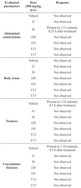
(Table II). In addition, acute treatment with the compounds or vehicle did not induce behavioral alterations indicative of acute toxicity in either male or female mice, such as piloerection, tremors, reduction of body tonus, abdominal constrictions, convulsions (seizures), touch response, tail grip
or corneal reflex, when compared with the control
group (vehicle) (Table III, IV and V).
Food and water intake were not modified
significantly upon acute oral treatment with the
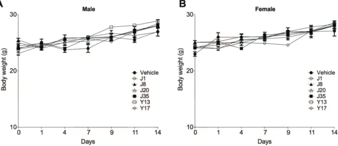
compounds or vehicle compared with the control group (Table VI). Corroborating these findings, the treatment with J1, J8, J20, J35, Y13 and Y17 (300 mg/kg) or vehicle, administered orally
to mice, did not significantly change absolute body weight of animals when compared with the control group (Fig. 2). Three animals treated
with Y17 had diarrhea in the first hour following
administration. In addition, mice which received acute treatment with the compounds at 300 mg/ kg, administered orally, showed no important macroscopic alterations considering aspect and color of the selected organs and glands (heart, lung, liver, kidney, spleen, stomach, testis, ovary and uterine tubes, salivary gland, adrenal gland,
TAblE ii
Effect of acute treatment with J1, J8, J20, J35, Y13 and Y17 (300 mg/kg) or vehicle, administered orally, on the
mice mortality percentage.
Compound p.o. (300 mg/kg)
Sex n Mortality
(%)
vehicle Male 5 10
Female 5
J1 Male 7 0
Female 5
J8 Male 4 0
Female 7
J20 Male 4 0
Female 6
J35 Male 7 10
Female 3
Y13 Male 4 0
Female 7
Y17 Male 5 0
Female 5
n: number of animals.
TAblE iii
Effect of acute treatment with J1, J8, J20, J35, Y13 and Y17 (300 mg/kg) or vehicle, observed during 0.25, 0.5, 1, 2, 4, 8 and 24 h after oral administration, on some behavioural
parameters indicative of acute toxicity in mice.
Evaluated parameters
dose (300 mg/kg,
p.o.)
response
Touch response
Vehicle Not observed
J1 Present in 1/12 animals, 2 h after treatment
J8 Not observed
J20 Not observed
J35 Not observed
Y13 Not observed
Y17 Not observed
Tail grip
Vehicle Not observed
J1 Not observed
J8 Not observed
J20 Not observed
J35 Not observed
Y13 Not observed
Y17 Not observed
Corneal
reflex
Vehicle Not observed
J1 Not observed
J8 Not observed
J20 Not observed
J35 Not observed
Y13 Not observed
TAblE v
Effect of acute treatment with J1, J8, J20, J35, Y13 and Y17 (300 mg/kg) or vehicle, observed during 0.25, 0.5, 1, 2, 4, 8 and 24 h after oral administration, on some behavioural
parameters indicative of acute toxicity in mice.
Evaluated parameters
dose (300 mg/kg, p.o.)
response
Straub’s Signal
Vehicle Not observed
J1 Not observed
J8 Not observed
J20 Not observed
J35 Not observed
Y13 Not observed
Y17 Not observed
Ptosis
(lacrimation)
Vehicle Not observed
J1 Not observed
J8 Not observed
J20 Not observed
J35 Not observed
Y13 Not observed
Y17 Not observed
Piloerection
Vehicle Not observed
J1 Not observed
J8 Not observed
J20 Not observed
J35 Not observed
Y13 Not observed
Y17 Not observed
Values are expressed as the number of animals that presented behavioural alterations in comparison with the total number of evaluated animals.
TAblE vi
Effect of acute treatment with J1, J8, J20, J35, Y13 and Y17 (300 mg/kg) or vehicle, after oral administration in males and females, on changes in the average of food and
water intake evaluated at the end of treatment.
group (300 mg/kg, p.o.)
n Food (g) Water (ml)
vehicle 9 5.87 + 0.86 5.79 + 0.28
J1 12 6.04 + 0.54 5.98 + 0.36
J8 11 6.12 + 0.47 6.10 + 0.39
J20 10 7.45 + 1.02 6.96 + 0.39
J35 10 7.63 + 0.97 7.01 + 0.53
Y13 11 7.90 + 1.07 7.67 + 1.59
Y17 10 6.23 + 0.55 6.56 + 0.46
Values represent the variation of mean solid (g/animal/day) and liquid (mL/animal/day) intake of 9-12 animals ± standard error mean. Two-way ANOVA with repeated measures shows no difference between the groups.
TAblE iv
Effect of acute treatment with J1, J8, J20, J35, Y13 and Y17 (300 mg/kg) or vehicle, observed during 0.25, 0.5,
1, 2, 4, 8 and 24 h after oral administration, on some behavioural parameters indicative of acute toxicity in mice.
Evaluated parameters
dose (300 mg/kg,
p.o.)
response
Abdominal constrictions
Vehicle Not observed
J1 Not observed
J8 Present in 1/11 animals, 0.25 h after treatment
J20 Not observed
J35 Not observed
Y13 Not observed
Y17 Not observed
body tonus
Vehicle Not observed
J1 Not observed
J8 Not observed
J20 Not observed
J35 Not observed
Y13 Not observed
Y17 Not observed
Tremors
Vehicle Present in 1/10 animals, 0.5 h after treatment
J1 Not observed
J8 Not observed
J20 Not observed
J35 Not observed
Y13 Not observed
Y17 Not observed
Convulsions/ Seizures
Vehicle Present in 1/10 animals, 0.5 h after treatment
J1 Not observed
J8 Not observed
J20 Not observed
J35 Not observed
Y13 Not observed
Y17 Not observed
thymus, mesenteric lymph node and encephalon), when compared with the control group (vehicle), all of which were evaluated at the 14th day of study. Furthermore, mice acutely treated orally with J1, J8, J20, J35, Y13 and Y17 (300 mg/kg), did not
show signifi cant differences in either relative or
absolute weight of heart, lung, kidney, spleen, stomach, salivary gland, adrenal gland, thymus and encephalon when compared with the control group, all of which were evaluated at the 14th day of study (Table VII). Only treatment with J1 resulted
in signifi cant increase in the relative weight of the
testis (Table VII).
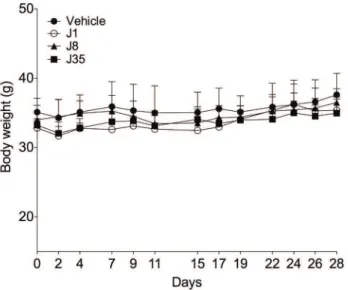
Next, we evaluated the systemic effects of intraperitoneal administration of selected compounds from the J series (J1, J8 and J35). Only three compounds were assayed in this set of experiments because limited amounts of the other compounds were available to complete the assay. In this set of experiments, mice were treated with multiple doses (10 mg/kg, 3 times a week, i.p.), as this will be the probable dosing regimen for future in vivo assays with scrapie-infected mice. Moreover, at this selected dose, the compound concentration
will be above the estimated IC50 values (Ferreira et al. 2014). When evaluated for 28 days, none of the tested compounds induced mouse mortality
(data not shown) and no signifi cant alterations in
body weight were observed in mice treated with J1, J8 or J35 (10 mg/kg, 3 times a week, i.p., for 28 days) when compared with control mice (Fig. 3). Macroscopic evaluation of the selected organs and glands (heart, lung, liver, kidney, spleen, stomach, adrenal gland, and encephalon) demonstrated no alterations of aspect and color in mice treated with J1 (10 mg/kg, 3 times a week, i.p.), evaluated at the 28th day of study, when compared with the control group (vehicle). Similar results were found for J8 and J35; however, one mouse treated with J8 and one with J35 (10 mg/kg, 3 times a week, i.p.) showed minor lung bleeding, and one mouse treated with J35 presented kidney discoloration. In addition, systemic treatment with J1, J8, or J35 (10 mg/kg, 3 times a week, i.p.) for 28 days was
not capable of inducing signifi cant alterations in
TAblE vii
Effect of acute treatment with J1, J8, J20, J35, Y13 and Y17, after oral administration, on relative weight of some organs and glands of male and female mice.
Structure vehicle J1 J8 J20 J35 Y13 Y17
Spleen 0.33
(0.27 – 0.40)
0.41 (0.34 – 0.47)
0.33 (0.31 – 0.36)
0.31 (0.27 - 0.35)
0.42 (0.33 - 0.51)
0.27 (0.24 - 0.34)
0.27 (0.23 - 0.31)
Kidney 0.75
(0.72 – 0.78)
0.59 (0.55 – 0.62)
0.76 (0.69 – 0.83)
0.70 (0.64 - 0.75)
0.75 (0.70 - 0.80)
0.67 (0.52 - 0.82)
0.76 (0.60 - 0.91)
heart 0.50
(0.46 – 0.54)
0.46 (0.42 – 0.49)
0.48 (0.44 – 0.52)
0.43 (0.40 - 0.46)
0.51 (0.47 - 0.54)
0.53 (0.41 - 0.64)
0.46 (0.36 - 0.56)
lung 0.87
(0.66 – 1.07)
0.79 (0.71 – 0.87)
0.75 (0.64 – 0.86)
0.65 (0.53 - 0.76)
0.80 (0.65 - 0.94)
0.69 (0.60 - 0. 70)
0.63 (0.59 - 0.68)
liver 5.40
(4.90 – 5.90)
4.30 (4.00 – 4.60)
5.10 (4.80 – 5.30)
4.80 (4.50 - 5.10)
4.90 (4.40 - 5.20)
4.70 (4.20 - 5.20)
4.90 (4.60 - 5.20) Mesenteric
lymph node
0.17 (0.10 – 0.24)
0.09 (0.04 – 0.15)
0.15 (0.10 – 0.20)
0.11 (0.07 - 0.15)
0.08 (0.04 - 0.13)
0.05 (0.009-0.09)
0.12 (0.01 - 0.27)
Adrenal gland 0.02 (0.01 – 0.02)
0.02 (0.01 – 0.02)
0.02 (0.01 – 0.02)
0.01 (0.008-0. 02) 0.01 (0.009-0.02) 0.01 (0.01-0.016) 0.02 (0.01 - 0.04)
Thymus 0.18
(0.13 – 0.23)
0.20 (0.17 – 0.23)
0.20 (0.16 – 0.23)
0.17 (0.11 - 0.24)
0.15 (0.10 - 0.20)
0.13 (0.07 - 0.17)
0.21 (0.15 - 0.27)
Stomach 0.72
(0.64 – 0.81)
0.86 (0.79 – 0.93)
0.86 (0.69 - 0.91)
0.84 (0.72 - 0.96)
0.68 (0.47 - 0.89)
0.72 (0.65 - 0.78)
0.74 (0.49 - 0.99)
Encephalon 1.50 (1.40 – 1.60)
1.60 (1.50 –1.70)
1.50 (1.40 – 1.60)
1.30 (1.20 - 1.40)
1.60 (1.50 - 1.70)
1.60 (1.50 - 1.70)
1.80 (0.60 – 2.90)
Testis 0.32
(0.30 – 0.33)
1.00* (0.90 – 1.20)
0.27 (0.21 – 0.33)
0.26 (0.20 - 0.32)
0.30 (0.26 - 0.34)
0.28 (0.23 - 0.33)
0.28 (0.19 - 0.37)
Salivary gland 0.30 (0.25 – 0.36)
0.23 (0.14 – 0.32)
0.31 (0.27 – 0.35)
0.28 (0.24 - 0.33)
0.46 (0.33 - 0.95)
0.33 (0.31 - 0.35)
0.29 (0.20 - 0.37)
Values represent the mean of organs weight of 9-12 animals with 95 % of confidence interval.
Values were obtained from the relation (percentage) of organ weight to body mass of animals (using the following formula: organ weight/body weight x 100). Two-way ANOVA except for testis where one-way ANOVA was used. * p≤ 0.05 when compared with the vehicle group.
TAblE viii
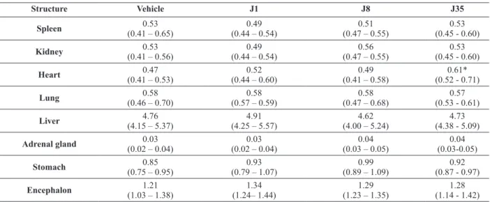
Effect of repeated i.p. administration of J1, J8 and J35 on relative weight of some organs of mice.
Structure vehicle J1 J8 J35
Spleen 0.53
(0.41 – 0.65)
0.49 (0.44 – 0.54)
0.51 (0.47 – 0.55)
0.53 (0.45 - 0.60)
Kidney 0.53
(0.41 – 0.56)
0.49 (0.44 – 0.54)
0.56 (0.47 – 0.55)
0.53 (0.45 - 0.60)
heart 0.47
(0.41 – 0.53)
0.52 (0.44 – 0.60)
0.49 (0.41 – 0.58)
0.61* (0.52 - 0.71)
lung 0.58
(0.46 – 0.70)
0.58 (0.57 – 0.59)
0.58 (0.47 – 0.68)
0.57 (0.53 - 0.61)
liver 4.76
(4.15 – 5.37)
4.91 (4.25 – 5.57)
4.62 (4.00 – 5.24)
4.73 (4.38 - 5.09)
Adrenal gland 0.03
(0.02 – 0.04)
0.03 (0.02 – 0.04)
0.04 (0.03 – 0.05)
0.04 (0.03-0.05)
Stomach 0.85
(0.75 – 0.95)
0.93 (0.79 – 1.07)
0.99 (0.89 – 1.09)
0.92 (0.87 - 0.97)
Encephalon 1.21
(1.03 – 1.38)
1.34 (1.24– 1.44)
1.29 (1.23 – 1.35)
1.28 (1.14 - 1.42)
Values represent the mean of organs weight of 5-6 animals with 95 % of confidence interval.
J35 caused significant increase in the relative weight of heart of mice following i.p. treatment (10 mg/kg, 3 times a week, during 4 weeks, i.p.) (Table VIII).
In conclusion, analyses of results demonstrated that acute oral administration of J1, J8, J20, J35, Y13 and Y17, and intraperitoneal repeated doses of J1, J8 and J35 showed good tolerability and had no acute toxic effects in mice, when evaluated on several behavioral parameters. The six compounds showed low acute toxicity and did not cause alterations in body mass or food and water intake in mice. In addition, the acute oral administration of these compounds did not lead to important macroscopic alteration concerning the aspect, color and size of organs and/or glands, such as heart, lung, liver, kidney, spleen, stomach, ovary and uterine tubes, salivary gland, adrenal gland, thymus, mesenteric lymph node and encephalon, when compared with organs of control mice. J1, J8 and J35 also indicated being safe when administered systemically, with only one mouse treated with J8 and one with J35 showing minor lung bleeding,
kidney discoloration and increased relative weight of the heart when compared with the control group.
diSCuSSiON
Chalcones and oxadiazoles are chemical classes of compounds with several biological activities, but to date only our previous report revealed their putative anti-scrapie activity (Ferreira et al. 2014). Particularly, 1,3,4-oxadiazoles with different substituents have been assayed for
anti-inflammatory, anti-tumor, analgesic, anti-viral and
antimicrobial activities (Omar et al. 1996, Sun et al. 2013), being considered privileged structures in medicinal chemistry (Rane et al. 2013). Acute toxicity studies of substituted 1,3,4-oxadiazoles
that have anti-inflammatory and analgesic activities
showed LD50 values of ~1000 mg/kg after i.p. administration to mice (Omar et al. 1996). As we did not reach such dose in our study, we can only suppose that Y13 and Y17 will also have high LD50 values. However, we are dealing with different oxadiazole derivatives, what can completely alter
the PK profile. Recently, anti-bacterial, anti-fungal
and anti-tubercular activities were described for 4-nitropyrrole-based 1,3,4-oxadiazoles. Their IC50 values for cytotoxicity in mammalian VERO cells ranged from 100-700 µM (Rane et al. 2013), much higher than the minimum inhibitory concentrations (MIC). Our previous study also showed that Y13 and Y17 are non-toxic to cultured mammalian cells at and above the effective anti-scrapie concentration (Ferreira et al. 2014).
Besides having several known pharmacological activities, the synthesis of chalcones is rapid, low cost and normally with high yields, which renders this class of compounds promising for drug development. Other 3,4,5-trimethoxychalcone analogues presented potent activity against H.
pylori (Lai et al. 2010) and inhibited both nitric
oxide production and tumor cell proliferation (Rao et al. 2009). The 3,4,5-trimethoxyphenyl ring is increasingly associated with anti-tumor activity of several chalcone derivatives. Although differing in the methoxy position, the 2,4,5-trimethoxychalcones investigated here (J8, J20 and J35) also had anti-proliferative activity against K652 leukemia cells (Costa et al. 2014). However, J8 and J20 reduced cell viability in only 30-35 %, and J35 to 50 % at 100 µM. J1 had no
significant activity (Costa el al. 2014). Cytotoxicity
against non-tumoral cell lines was investigated by us, and what we found is that the chalcones were not toxic to N2a cells in concentrations of up to 25 µM (Ferreira et al. 2014). Nonetheless, it remains
to be confirmed whether these compounds are safe
to non-tumoral cells at higher concentrations. Regarding the ability to cross the BBB, there is lack of in vivo PK studies with the chalcones and oxadiazoles investigated here. A trimethoxychalcone derivative was detected in implanted glioblastoma tumors and in the brain of
i.p. treated mice, showing that at least this specific
chalcone crosses the BBB (Boumendjel et al. 2009). Previously, we used a computational approach to predict some pharmacokinetic parameters that helped us select the most promising compounds to assay in vivo. Our in silico analysis indicated that
the fraction of J1, J35, Y13 and Y17 that reaches circulation after oral administration is theoretically expected to be higher than 70 %, while for J8 and J20 it stands between 30 and 70 %. These compounds
were also classified as sufficiently CNS-permeable
to be active in the CNS. These ADMET predictions do not substitute in vivo evaluation, but give us preliminary information on the potential drugability of these compounds (Cordeiro and Ferreira 2015).
All compounds were predicted to have low solubility in water (Table I), what renders them prone to cross the BBB. However, we still have not evaluated (in vitro or in silico) binding to plasma proteins, a feature that compromises permeation to biological membranes, even for lipophilic compounds. Prediction of toxicity indicated that all compounds from the J series are less toxic than compounds from the Y series (Table I). We were not able to calculate LD50 values for the compounds, as oral and i.p. administration of both Ys and Js as single or multiple doses did not cause death of the treated mice. Consequently, although the in silico
toxicity prediction supported the in vivo results for the chalcones, we could not correlate the LD50 prediction for compounds of the Y series (Table I) to the obtained in vivo results.
Sub-acute and chronic toxicity studies are needed to provide data on genotoxicity, mutagenicity, and on long-term effects in organs and glands and overall animal behavior. However, the acute toxicity results presented here show that at least the compounds are safe regarding oral and i.p. administration at the investigated doses. Previous data showing the pharmaceutical importance of the 1,3,4-oxadiazole and trimethoxychalcone scaffolds (Sun et al. 2013, Rane et al. 2013, Omar et al. 1996, Costa el al. 2014, Ferreira et al. 2014), together with our in vivo acute toxicity results strongly indicate that J1, J8, J20, J35, Y13 and Y17 are interesting compounds for further investigation as anti-scrapie drugs or for other activities. Further
structural modifications might also generate novel
ACKNOWlEdgMENTS
We thank Dr. Maria Letícia de Castro Barbosa and Dr. Julia R. Clarke from the Faculty of Pharmacy, UFRJ for critical reading and revision of the manuscript and Wesley J. A. da Conceição for helping to elaborate Fig. 1. We also thank Dr. Patricia N. Fernandes for excellent technical support. This work was supported by grants from
Conselho Nacional de Desenvolvimento Científico
e Tecnológico (CNPq); from the Instituto Nacional de Ciência e Tecnologia de Biologia Estrutural e Bioimagem (INBEB); Fundação de Amparo à Pesquisa do Estado do Rio de Janeiro (FAPERJ); Coordenação de Aperfeiçoamento de Pessoal de Nível Superior (CAPES)- PROCAD/ Casadinho: Fortalecimento e Consolidação da Área de Farmacologia e Toxicologia Pré-Clínica do Programa de Pós-Graduação em Ciências Farmacêuticas da Universidade Federal do Rio de Janeiro (UFRJ/UFSC).
rESuMO
Uma forma alterada da proteína prion celular, a PrPSc ou PrPRes
, está envolvida na ocorrência das encefalopatias espongiformes transmissíveis. Nós sintetizamos e caracterizamos previamente compostos aromáticos que inibem o acúmulo da proteína prion resistente a proteases (PrPRes) em células infectadas com prion scrapie. Estes compostos pertencem a diferentes classes químicas, incluindo acilhidrazonas, chalconas e oxadiazóis. Alguns dos compostos ativos não apresentaram toxicidade para células de neuroblastoma em cultura e parecem apresentar propriedades de fármacos, uma vez que todos obedecem aos 5 quesitos da regra de
Lipinski e apresentam perfil farmacocinético desejável,
como predito in silico. Antes de avaliar a eficácia in vivo dos compostos aromáticos em camundongos infectados com prion scrapie, é necessário estimar sua segurança em camundongos saudáveis. Neste trabalho nós utilizamos camundongos Suíços para avaliar o
perfil de toxicidade aguda dos seis compostos mais
promissores, as 2,4,5-trimetoxichalconas (J1, J8, J20 e J35) e os 1,3,4-oxadiazóis (Y13 e Y17). Administração oral em dose única (300 mg/kg) de J1, J8, J20, J35, Y13
e Y17 ou administração repetida via intraperitoneal (10 mg/kg, 3 vezes por semana, por 4 semanas) de J1, J8 e J35, não geraram toxicidade nos camundongos. Nós acreditamos fortemente que as trimetoxichalconas e os oxadiazóis investigados são compostos interessantes para serem futuramente avaliados in vivo contra as doenças causadas por prions.
Palavras-chave: prion, toxicidade aguda, composto orgânico, segurança de fármacos, scrapie, farmacocinética.
rEFErENCES
ACD/LABS PERCEPTA PLATFORM. 2014. Version 12.01, Advanced Chemistry Development, Inc. (ACD/Labs), Toronto, On, Canada. www.acdlabs.com.
ANVISA. 2010. Guia para a condução de estudos não clínicos de segurança necessários ao desenvolvimento de medicamentos. Brasília, Brasil, p. 6-8.
BORCHHARDT DM, MASCARELLO A, CHIARADIA LD, NUNES
RJ, OLIVA G, YUNES RA ANDANDRICOPULO AD. 2010. Biochemical evaluation of a series of synthetic chalcone and hydrazide derivatives as novel inhibitors of cruzain from Trypanosoma cruzi. J Braz Chem Soc 21: 142-150.
BOUMENDJEL A, MCLEER-FLORIN A, CHAMPELOVIER P,
ALLEGRO D, MUHAMMAD D, SOUARD F, DEROUAZI M,
PEYROT V, TOUSSAINT B AND BOUTONNAT J. 2009. A
novel chalcone derivative which acts as a microtubule depolymerising agent and an inhibitor of P-gp and BCRP in in-vitro and in-vivo glioblastoma models. BMC Cancer 9: 242.
BRANDÃO DC. 2004. Resolução-REno 90, 16 de março. In:
Registro de Fitoterápicos, PHC Pharma Consulting.
CASHMAN NR AND CAUGHEY B. 2004. Prion diseases-close to
effective therapy? Nat Rev Drug Discov 3: 874-884. COBEA – COLÉGIO BRASILEIRO DE EXPERIMENTAÇÃO
ANIMAL. 1991. Os princípios éticos da experimentação
animal. São Paulo, Brasil.
COLLINGE J ET AL. 2009. Safety and efficacy of quinacrine
in human prion disease (PRION-1 study): a patient-preference trial. Lancet Neurol 8: 334-344.
CORDEIRO Y AND FERREIRA NC. 2015. New approaches for the
selection and evaluation of anti-prion organic compounds. Mini Rev Med Chem 15: 84-92.
COSTA A ET AL. 2014. Apoptotic effect of synthetic
2′,4′,5′-trimethoxychalcones in human K562 and Jurkat leukemia cells. Med Chem Res 23: 4301-4319.
FERREIRA NC ET AL. 2014. Anti-prion activity of a panel of aromatic chemical compounds: in vitro and in silico approaches. PLoS One 9(1): e84531.
FORLONI G, ARTUSO V, ROITER I, MORBIN M AND TAGLIAVINI
F. 2013. Therapy in prion diseases. Curr Top Med Chem 13: 2465-2476.
FURUKAWA H, TAKAHASHI M, NAKAJIMA M AND YAMADA
T. 2002. Prospects of the therapeutic approaches to Creutzfeldt-Jacob disease: a clinical trial of antimalarial quinacrine. Nihon Rinsho 60: 1649-1657.
GHAEMMAGHAMI S, MAY BC, RENSLO AR AND PRUSINER SB.
2010. Discovery of 2-aminothiazoles as potent antiprion compounds. J Virol 84: 3408-3412.
HAÏK S ET AL. 2014. Doxycycline in Creutzfeldt-Jakob disease:
a phase 2, randomised, double-blind, placebo-controlled trial. Lancet Neurol 13: 150-158.
KOCISKO DA, BARON GS, RUBENSTEIN R, CHEN J, KUIZON
S AND CAUGHEY B. 2003. New inhibitors of
scrapie-associated prion protein formation in a library of 2000 drugs and natural products. J Virol 77: 10288-10294.
KOCISKO DA AND CAUGHEY B. 2006. Searching for
anti-prion compounds: cell-based high-throughput in vitro assays and animal testing strategies. Methods Enzymol 412: 223-234.
KORTH C, MAY BC, COHEN FE AND PRUSINER SB. 2001. Acridine and phenothiazine derivatives as pharmacotherapeutics for prion disease. Proc Natl Acad Sci USA 98: 9836-9841.
KOSTER T, SINGH K, ZIMMERMANN M AND GRUYS E. 2003.
Emerging therapeutic agents for transmissible spongiform encephalopathies: a review. J Vet Pharmacol Ther 26: 315-326.
LAI CH, RAO YK, FANG SH, SING YT AND TZENG YM. 2010. Identification of 3’,4’,5’-trimethoxychalcone analogues as potent inhibitors of Helicobacter pylori-induced inflammation in human gastric epithelial cells. Bioorg Med Chem Lett 20: 5462-5465.
LI Z, RAO S, GEVER JR, WIDJAJA K, PRUSINER SB AND
SILBER BM. 2013. Optimization of Arylamides as Novel,
Potent and Brain-penetrant Antiprion Lead Compounds. ACS Med Chem Lett 4: 647-650.
MACEDO B, KASCHULA CH, HUNTER R, CHAVES JA, VAN
DER MERWE JD, SILVA JL, EGAN TJ AND CORDEIRO Y.
2010. Synthesis and anti-prion activity evaluation of aminoquinoline analogues. Eur J Med Chem 45: 5468-5473.
MARTÍNEZ-LAGE JF, RÁBANO A, BERMEJO J, MARTÍNEZ PÉREZ
M, GUERRERO MC, CONTRERAS MA AND LUNAR A.
2005. Creutzfeldt-Jakob disease acquired via a dural graft: failure of therapy with quinacrine and chlorpromazine. Surg Neurol 64: 542-54.
NUNES RJ ET AL. 2012. Compostos acil-hidrazonas e oxadiazóis,
composições farmacêuticas compreendendo os mesmos e seus usos. Brazilian patent: PCT/BR2012/000480. OECD. 2001. Guideline for the testing of chemicals, Section
4: Health Effects. Acute oral toxicity – Acute toxic class method, Test no 423, 14 p.
OMAR F, MAHFOUZ N AND RAHMAN M. 1996. Design, synthesis and antiinflammatory activity of some 1,3,4-oxadiazole derivatives. Eur J Med Chem 31: 819-825.
OTTO M ET AL. 2004. Efficacy of flupirtine on cognitive
function in patients with CJD: A double-blind study. Neurology 62: 714-718.
PRUSINER SB. 1998. Prions. Proc Natl Acad Sci USA 95:
13363-13383.
RAINOV NG, TSUBOI Y, KROLAK-SALMON P, VIGHETTO A AND
DOH-URA K. 2007. Experimental treatments for human transmissible spongiform encephalopathies: is there a role for pentosan polysulfate? Expert Opin Biol Ther 7: 713-726.
RANE RA, BANGALORE P, BORHADE SD AND KHANDARE PK. 2013. Synthesis and evaluation of novel 4-nitropyrrole-based 1,3,4-oxadiazole derivatives as antimicrobial and anti-tubercular agents. Eur J Med Chem 70: 49-58. RAO YK, FANG SH AND TZENG YM. 2009. Synthesis and
biological evaluation of 3’,4’,5’-trimethoxychalcone analogues as inhibitors of nitric oxide production and tumor cell proliferation. Bioorg Med Chem 17: 7909-7914.
SILBER BM ET AL. 2013. Pharmacokinetics and metabolism of
2-aminothiazoles with antiprion activity in mice. Pharm Res 30: 932-950.
SIM VL. 2012. Prion disease: chemotherapeutic strategies. Infect Disord Drug Targets 12: 144-160.
SUN J, MAKAWANA JA AND ZHU HL. 2013. 1,3,4-oxadiazole derivatives as potential biological agents. Mini Rev Med Chem 13: 1725-1743.
TSUBOI Y, DOH-URA K AND YAMADA T. 2009. Continuous