Spatiotemporal expression of
MYD88
gene in pigs from birth to adulthood
LiNa Gan
1, WeiYun Qin
1, Sen Wu
1, ShengLong Wu
1,2and WenBin Bao
1,21
Key Laboratory for Animal Genetics, Breeding, Reproduction and Molecular Design of Jiangsu Province,
College of Animal Science and Technology, Yangzhou University, Yangzhou, China.
2
Joint International Research Laboratory of Agriculture & Agri-Product Safety, Yangzhou University,
Yangzhou, China.
Abstract
MYD88 plays an important role in the immune response against infections. To analyzeMYD88 gene expression
dur-ing different stages of pig development, we used real-time PCR.MYD88 was seen expressed in all tissues
exam-ined.MYD88 expression in spleen, lungs, and thymus reached its highest value from 7 to 14 days of age and
decreased thereafter. Expression in lymph nodes was high until 28 days of age and then it declined after weaning, with stable low levels in adult pigs.MYD88 expression was high before 35 days of age in the small intestine (duode-num, jeju(duode-num, and ileum), where it reached its highest value from 7 to 14 days of age.MYD88 expression in the small intestine declined post-weaning and remained relatively low during adulthood. The results of this study suggest that weaning stress and development of the immune system might be positively correlated withMYD88 expression regu-lation. Moreover, this study provided evidence that the high expression ofMYD88 may diminish weaning stress and increase disease resistance in Meishan pigs.
Keywords:MYD88,pig, developmental expression, immune response. Received: January 28, 2017; Accepted: August 30, 2017.
Introduction
Mammalian species have an innate and an acquired immune system. The innate immune system is the first line of defense against pathogens (Zhouet al., 2015). The toll-like receptor (TLR) family is a group of pattern recognition receptors (PRRs) that play important roles in sensing pathogen-associated molecular patterns (PAMPs) in innate immunity (Wack and Gallorini, 2008). The TLR family consists of 13 members with different downstream effects. The myeloid differentiation factor 88 (MYD88) is an adap-tor protein for TLR signaling, with the exception of TLR3 (Shchebliakovet al., 2010; Serezaniet al., 2011). MYD88 is a soluble cytoplasmic protein that belongs to both the Toll/IL-1R (TIR) family and the death domain family. The protein contains three functional areas: an N-terminal DD, a middle region, and a C-terminal TIR domain (Naro and Sette, 2016). The MYD88 TIR domain combines the TLRs and the TIR domain of IL-1R, which activates Interleukin-1 Receptor-Associated-Kinase-1/4 (IRAK-1/4) and tumor necrosis factor receptor-associated factor-6 (TRAF-6), re-sulting in the activation of nuclear factor kappa B (NF-kB)
and the release of pro-inflammatory factors and cellular mediators. The activation of the TLR/MyD88/NF-kB sig-nal pathway leads to lymphocyte activation and increased synthesis of pro-inflammatory proteins (Janssens and Beyaert, 2003; Arancibiaet al., 2007; Gayet al., 2014; Liu
et al., 2014; Suet al., 2015), thereby leading to an acquired immune response.
PorcineMYD88, which is located on chromosome 13 and has a coding sequence region consisting of 882 nucleo-tides, encodes 293 amino acids with 87% to 88% homo-logies between porcine and human proteins. MYD88 is widely expressed in several tissues, especially in immune and intestinal tissues (Tohnoet al., 2007; Liet al., 2009). The expression pattern of the gene is related to the immune response triggered by bacterial infections. Porcine MYD88 is a key protein in the TLRs/IL-1R signaling pathway that sends inflammatory signals and enhances the intensity of the inflammatory response. Additionally, MYD88 triggers the release of intestinal inflammatory mediators (Sunet al., 2015).
The signal transduction pathway mediated by MYD88 is involved in the occurrence and development of several diseases. At present, most studies focus on its im-mune regulation (Singh et al., 2015) and pathological mechanisms (Parpaleixet al., 2016). However, no study has assessed the expression pattern of MYD88. In this study, we used real-time PCR technology to quatify
Send correspondence to WenBin Bao. Key Laboratory for Animal Genetics, Breeding, Reproduction and Molecular Design of Jiangsu Province, College of Animal Science and Technology, Yangzhou University, Yangzhou, Jiangsu 225009, China. E-mail: [email protected].
MYD88 expression at eight post-natal developmental stages in twelve tissues of Meishan pigs. Our goal was to provide a theoretical basis for further research on the genet-ics ofMYD88in pig breeding for disease resistance.
Materials and Methods
Ethics statement
The animal study proposal was approved by the Insti-tutional Animal Care and Use Committee (IACUC) of the Yangzhou University Animal Experiments Ethics Commit-tee (permit number: SYXK [Su] IACUC 2012-0029). All experimental procedures involving piglets were performed in accordance with the Regulations for the Administration of Affairs Concerning Experimental Animals and approved by the State Council of People’s Republic of China.
Materials
Meishan pigs obtained from the Meishan Pigs Con-servation Breeding Company (Jiangsu China) were used in this study. All the experimental pigs were maintained under standard piggery conditions. The animals hadad libitum
access to a commercial-type compound feed with 21.7% crude protein and no antimicrobial additives or organic ac-ids. We selected one animal from five litters at different developmental stages (newborn, 7-day old, 14-day old, 21-day old, 28-day old, 35-day old, 3-month old, and 6-month old). A total of 40 animals (five animals per group) were used, and the animals at the same developmental stage had similar characteristics (e.g., size and weight). Animals were electrically stunned (300 V for 5 s) and bled by heart puncture under the left armpit. Tissue samples from heart, liver, spleen, lung, kidney, stomach, muscle, thymus, lym-ph, duodenum, jejunum, and ileum were collected, frozen in liquid nitrogen and stored at -80 °C. All the experiments were conducted in the Yangzhou Key Laboratory of Ani-mal Genetics and Breeding of Yangzhou University (Jiangsu, China).
RNA extraction and reverse transcription
Total RNA was extracted from the tissues (50–100 mg) using Trizol reagent (TaKaRa Biotechnology Dalian Co., Ltd, China). Precipitated RNA was suspended in 20 mL RNase-free H2O, diluted to 2 ng/mL, and stored at -80
°C. RNA quality was assessed by formaldehyde denaturing
gel electrophoresis. The concentrations and purity of RNA were determined spectrophotometrically (Nanodrop ND-1000, NanoDrop Technologies Co., Ltd, USA).
Total RNA was reverse transcribed into cDNA using a HiScript Q RT SuperMix for qPCR (+gDNA wiper) kit (Vazyme Biotech Co.,Ltd, China), which includes a genomic DNA removal module. The reaction mixture (10 mL) for cDNA synthesis consisted of 2mL 5qRT SuperMix II, 500 ng total RNA, and RNase-free H2O. The reaction
was carried out at 25 °C for 10 min, 50 °C for 5 min, 85 °C for 5 min and the products were then stored at 4 °C.
Real-time PCR primer design and quantitative fluorescence PCR
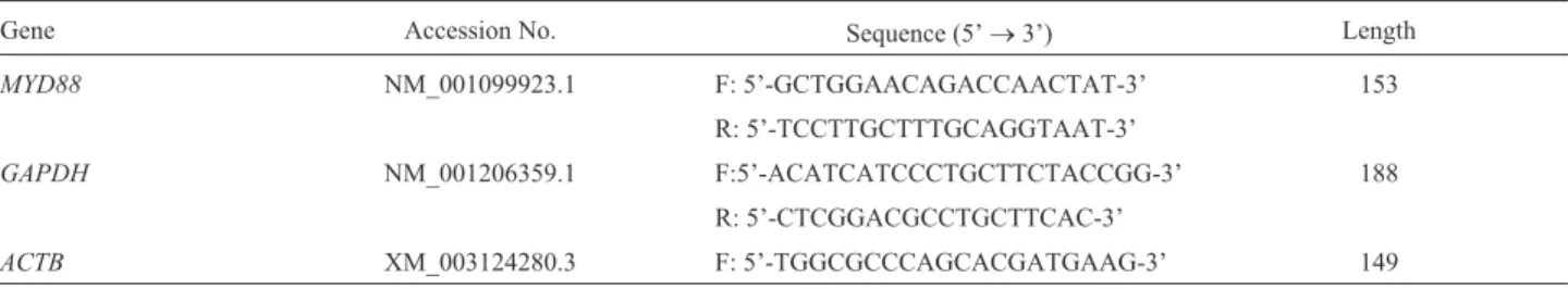
Using Primer Express 2.0, we designedMYD88 prim-ers based on the gene sequence deposited in GenBank. Primers were synthesized by Takara Biotechnology Dalian Co., Ltd. (China).GAPDHandACTBwere used as internal control genes to normalize the threshold cycle (Ct) values of other transcripts. The primer sequences used for amplifi-cations ofMYD88, GAPDH,andACTBare listed in Table 1.
Real-time PCR amplification was performed using a PCR kit (Vazyme Biotech Co., Ltd, China) in a 25-mL reac-tion mixture containing 2mL of cDNA (500 ng), 0.5mL of the forward and reverse primer (10mM each), 0.5mL of 50x ROX Reference Dye II, 10mL of 2x SYBR Green Real-time PCR Master Mix, and ddH2O. PCR conditions were
set at 95 °C for 5 min, followed by 40 cycles of 95 °C for 5 s and 60 °C for 34 s. Dissociation curve analysis was done af-ter amplification. A peak melting temperature (Tm) of 85± 0.8 °C in the dissociation curve was used to determine the specificity of the amplification product. The Tm value for each sample was calculated from the average of triplicate technical samples. The 2-DDCtmethod was used to calculate relative gene expression (Oparinaet al., 2012).
Statistical analysis
Statistical analyses were carried out using SPSS 17.0 software (SPSS Inc, USA). The multivariate general linear model (GLM) was used to determine differences in tran-script levels among different developmental stages. Data are reported as means± SD. Significance was set at P< 0.05.
Table 1- Real-time PCR primer sequences.
Gene Accession No. Sequence (5’®3’) Length
MYD88 NM_001099923.1 F: 5’-GCTGGAACAGACCAACTAT-3’ 153
R: 5’-TCCTTGCTTTGCAGGTAAT-3’
GAPDH NM_001206359.1 F:5’-ACATCATCCCTGCTTCTACCGG-3’ 188
R: 5’-CTCGGACGCCTGCTTCAC-3’
Results
Purity and integrity of total RNA
We assessed RNA quality using denaturing gel elec-trophoresis and RNA quantity by measuring nucleic acid and protein concentrations. On the gel, we observed three bands, corresponding to 28S, 18S, and 5S. Based on gel electrophoresis results, there was no DNA contamination or significant degradation (data not shown). The A260/A280
ratios of the samples ranged from 1.9 to 2.0.
Quantitative fluorescence PCR amplification and melting curves
The real-time PCR amplification and melting curves for MYD88 were consistent between amplification reac-tions. A single specific peak forMYD88 with no primer dimers or non-specific reaction products was obtained. The standard curves forMYD88, ACTB,andGAPDHrevealed that the amplification efficiencies of the target and refer-ence genes were almost the same; therefore, the 2-DDCt method was used (Figure S1).
MYD88gene expression in different tissues and
development stages
Using the established SYBR green real-time quantita-tive PCR method, we measured the relaquantita-tive expression lev-els ofMYD88in different tissues and at different develop-ment stages.MYD88expression levels were normalized to
ACTBandGAPDHexpression levels, and the expression of
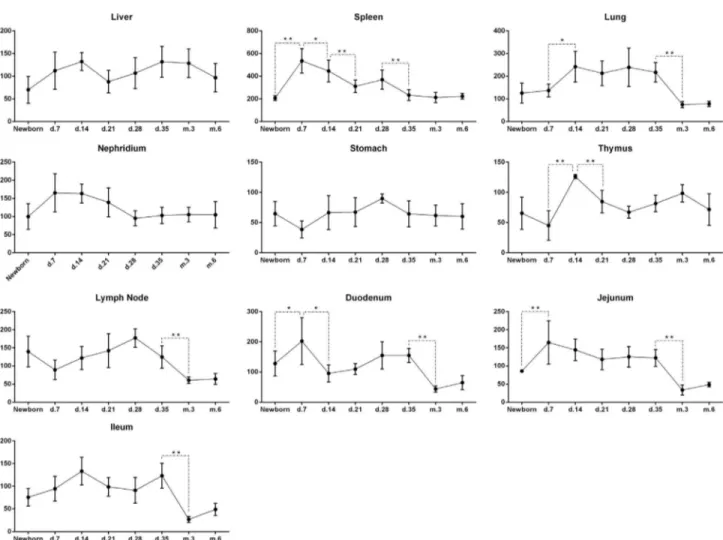
MYD88in the muscle of the 3-month old group was used as reference (calibrator) value (1.0). At all development stages, the average expression levels of MYD88 in the spleen was the highest (Figure 1), while they were ex-tremely low in heart and muscle. Expression was high in immunity-related tissues (liver, lung, kidney, thymus, and
lymph nodes) and also digestive tract tissues (stomach, du-odenum, jejunum, and ileum). Before weaning, MYD88
expression was lower in the stomach than in the duodenum, jejunum or ileum.
Analysis ofMYD88expression at different development stages in the same tissue
We analyzedMYD88 expression in 10 tissues (ex-cluding heart and muscle, which had extremely low
MYD88expression levels during all development stages). Different tissues had different expression patterns during development (Figure 2). The expression ofMYD88in the spleen, lung, and thymus was highest in 7-day old and 14-day old pigs and decreased in adulthood. Expression of this gene in lymph nodes was maintained at a high level, but declined from 28 days of age onwards. There was no signif-icant difference inMYD88expression in the liver, kidney, and stomach during development. Expression ofMYD88in intestinal tissues (duodenum, jejunum, and ileum) was high before 35 days (i.e., at weaning), with a peak from 7 to 14 days and decreasing post weaning. During adulthood the level was maintained low.
Discussion
MYD88, an important transduction protein in myeloid cell differentiation, plays a vital role in the TLR signaling pathway. The TLR signaling pathway transfers signals via MYD88-dependent and MYD88-independent pathways. The MYD88-dependent pathway is the most im-portant signal transduction pathway of TLRs (Serezaniet al., 2011). Studies have shown that MYD88 is indispens-able for the downstream signal transduction of several TLRs. Specifically, the protein interacts with IRAK-4 (IL-1R-associated kinase-4), which subsequently activates other IRAK family members such as IRAK-1 and IRAK-2.
This promotes the transfer of NF-kB into the nucleus and the activation of pro-inflammatory cytokine genes (Kawa-goeet al., 2008; Takeuchi and Akira, 2010). Kaderet al.
(2016) investigated the role of MYD88 using MYD88 -knockout mice and found that MYD88 plays a protective role in ehrlichiosis via the suppression of IL-10 and IL-17. Frantzet al.(2012) usingMYD88knocked out mice entero-cytes, found an increase in the number of intestinal mu-cus-associated bacteria and a decrease in the expression of polymeric immunoglobulin receptor (epithelial transport receptor of IgA) and mucin 2 (the main protein of intestinal mucus). In addition, the composition of the intestinal flora was significantly different between the enterocyte of
MYD88-knockout mice and the wild-type mice, decreasing resistance to acute colitis in knockout mice. These results showed that the MYD88 signal might be essential for the homeostasis of the intestinal tract. Therefore,MYD88plays an important role in the immune response and defense sys-tem.
In this study, the analysis of theMYD88expression profile revealed thatMYD88was expressed in all 12 tissues from Meishan pigs. The expression patterns changed over time, in agreement with Qiang (2008).MYD88expression
was highest in spleen and higher in other immune tissues (liver, lung, kidney, thymus, and lymph nodes) and diges-tive tissues (stomach, duodenum, jejunum, and ileum) than in heart and muscle.MYD88is mainly expressed in T cells, B cells, NK cell lines, dendritic cell lines, thymus cell lines, and other immune cells, indicating that it may play an im-portant role in immune responses (Hardimanet al., 1996). Nishiyaet al.(2007) observed that MYD88 is located in the cytoplasm and is not secreted. The authors hypothesized that the body wide expression ofMYD88may be associated with the transduction of TLR signaling pathways. This may be the reason whyMYD88showed a high expression level in most tissue samples from Meishan pigs, and hence we in-fer that the highest expression level ofMYD88found in spleen may be related with the important function of spleen in innate and adaptive immunity.
Most studies on MYD88 transcription were per-formed in humans and mice; few were done in livestock. Furthermore, there are no reports on the expression of
MYD88 in pigs in different developmental stages. This study analyzed the expression ofMYD88in different tis-sues and at different developmental stages using real-time PCR. Piglets gain immune protection in two ways: passive
immunization through sow milk and active immunization via their developing immune system (Salmonet al., 2009; Levastet al., 2014). Newborn piglets obtain specific anti-bodies from the sow colostrum, which can protect against infections (Ogawaet al., 2016). However, maternal anti-bodies acquired from sow milk cannot be maintained over time (Levastet al., 2014). After weaning, piglets experi-ence psychological and physiological stress from being separated from the sow and leaving the familiar environ-ment. The piglets may have problems adapting to the new environment, leading to conditional diarrhea.
The development of the digestive system is stimu-lated by early supplement feeding of 5 to 7-day old piglets (Wanget al., 2002). In this study, the expression ofMYD88
increased in spleen, duodenum, and jejunum from birth to 7 days of age, which coincided with the feeding period. Ex-pression ofMYD88was significantly increased in the lung and thymus from 7 to 14 days of age. The small intestine (duodenum, jejunum, and ileum) has the highest absorptive ability (Caspary, 1992). Direct stimulation of the intestine may explain the increased expression inMYD88in the duo-denum and jejunum after feeding. As the piglets aged, the expression level ofMYD88in duodenum, jejunum and il-eum showed a decreasing trend. Recent studies have shown that different parts of the intestine contain different micro-flora (Kelly et al., 2017). Furthermore, the bacteria and function of the intestinal microflora may affect the level of inflammatory cytokines secreted by immune cells (Schir-meret al., 2016). Therefore, the distribution of the micro-bial flora may affect the expression ofMYD88and account for the discrepancies inMYD88expression patterns among the three intestinal sections.
Both the spleen and thymus are important immune or-gans. Expression ofMYD88was significantly higher in the spleen than in the thymus, suggesting that the spleen may react more quickly in response to external stimuli com-pared to the thymus. The lung is a connective site between the organism and the environment and it is potentially sus-ceptible to harmful substances and pathogenic bacteria. Therefore, MYD88 expression significantly increased in the lung. The expression ofMYD88was maintained at a rel-atively high level during early post-natal development, but decreased significantly in the heart, lung, lymph, duode-num, jejuduode-num, and ileum from 35 days to 3 months of age and stabilized after sexual maturity. These results suggest that the high expression ofMYD88during weaning proba-bly enhanced the non-specific immune response and in-flammatory response in piglets. As the immune system in Meishan pigs matures, the MYD88expression gradually decreases to a stable level.
This study explored the differences of MYD88 ex-pression in Meishan pigs at different development stages. We speculate that weaning stress and immune system de-velopment are the main reasons for the differences found. The high expression ofMYD88may be beneficial for
im-proving disease resistance in Meishan pigs. Future studies should examine the molecular mechanisms ofMYD88 reg-ulation and develop breeding strategies for disease resistant Chinese pigs.
Acknowledgments
This work was financially supported by the National Natural Science Foundation of China [grant numbers 31472066, 31572360], the Science and Technology Sup-porting Project of Suzhou City [grant numbers SNG201628] and the Priority Academic Program Develop-ment of Jiangsu Higher Education Institutions (PAPD).
References
Arancibia SA, Beltrán CJ, Aguirre IM, Silva P, Peralta AL, Malinarich F and Hermoso MA (2007) Toll-like receptors are key participants in innate immune responses. Biol Res 40:97-112.
Caspary WF (1992) Physiology and pathophysiology of intestinal absorption. Am J Clin Nutr 55:299S-308S.
Frantz AL, Rogier EW, Weber CR, Shen L, Cohen DA, Fenton LA, Bruno ME C and Kaetzel CS (2012) Targeted deletion of MYD88 in intestinal epithelial cells results in compro-mised antibacterial immunity associated with downregu-lation of polymeric immunoglobulin receptor, mucin-2, and antibacterial peptides. Mucosal Immunol 5:501-512. Gay NJ, Symmons MF, Gangloff M and Bryant CE (2014)
As-sembly and localization of Toll-like receptor signalling complexes. Nat Rev Immunol 14:546-558.
Hardiman G, Rock FL, Balasubramanian S, Kastelein RA and Bazan JF (1996) Molecular characterization and modular analysis of human MYD88. Oncogene 13:2467-2475. Janssens S and Beyaert R (2003) Functional diversity and
regula-tion of different interleukin-1 receptor-associated kinase (IRAK) family members. Mol Cell 11:293-302.
Kader M, Alaoui-El-Azher M, Kode B, Kode B, McArthur M, Shinde A, Wells A and Ismail N (2016) MYD88 suppresses IL-10 and IL-17 production in response to obligate intra-cellular Ehrlichia infection. J Immunol 196:206.1.
Kawagoe T, Sato S, Matsushita K, Kato H, Matsui K, Kumagai Y, Kawai T, Takeuchi O and Akira S (2008) Sequential control of Toll-like receptor-dependent responses by IRAK1 and IRAK2. Nat Immunol 9:684-691.
Kelly J, Daly K, Moran AW, Ryan S, Bravo D, Shirazi-Beechey SP (2017) Composition and diversity of mucosa-associated microbiota along the entire length of the pig gastrointestinal tract; dietary influences. Environ Microbiol 19:1425-1438. Levast B, Berri M, Wilson HL, Meurens F and Salmon H (2014)
Development of gut immunoglobulin A production in piglet in response to innate and environmental factors. Dev Comp Immunol 44:235-244.
Li X, Liu H, Shulin Y, Tang Z, Ma Y, Chu M and Li K (2009) Characterization analysis and polymorphism detection of the porcine MYD88 gene. Genet Mol Biol 32:295-300. Liu P, Qiu M and He L (2014) Expression and cellular distribution
Naro C and Sette C (2016) Dissecting a hub for immune response: Modeling the structure of MYD88. Structure 24:349-351. Nishiya T, Kajita E, Horinouchi T, Nishimoto A and Miwa S
(2007) Distinct roles of TIR and non–TIR regions in the subcellular localization and signaling properties of MYD88. FEBS Lett 581:3223-3229.
Ogawa S, Tsukahara T, Imaoka T, Nakanishi N, Ushida K, Inoue R (2016) The effect of colostrum ingestion during the first 24 hours of life on early postnatal development of piglet im-mune systems. Anim Sci J 87:1511-1515.
Oparina NY, Sadritdinova AF, Snezhkina AV, Dmitriev AA, Krasnov GS, Senchenko VN, Melnikova NV, Belenikin MS, Lakunina VA, Veselovsky VA,et al.(2012) Increase in NETO2 gene expression is a potential molecular genetic marker in renal and lung cancers. Russ J Genet 48:506-512. Parpaleix A, Amsellem V, Houssaini A, Abid S, Breau M, Marcos
E, Sawaki D, Delcroix M, Quarck R, Maillard A, et al. (2016) Role of interleukin-1 receptor 1/MYD88 signalling in the development and progression of pulmonary hyperten-sion. Eur Respir J 48:470-483.
Qiang L (2008) Molecular cloning, tissues expression profile on porcine TLR7 and MYD88 and expression of MYD88 in pichia pastoris. Doctoral thesis, Sichuan Agricultural Uni-versity, Sichuan.
Salmon H, Berri M, Gerdts V and Meurens F (2009) Humoral and cellular factors of maternal immunity in swine. Dev Comp Immunol 33:384-393.
Schirmer M, Smeekens SP, Vlamakis H, Jaeger M, Oosting M, Franzosa EA, Horst R, Jansen T, Jacobs L and Bonder MJ (2016) Linking the human gut microbiome to inflammatory cytokine production capacity. Cell 167:1125-1136. Serezani CH, Lewis C, Jancar S and Peters-Golden M (2011)
Leukotriene B 4 amplifies NF-kB activation in mouse macrophages by reducing SOCS1 inhibition of MYD88 ex-pression. J Clin Invest 121:671-682.
Shchebliakov DV, Logunov DY, Tukhvatulin AI, Shmarov MM, Naroditsky BS and Ginzburg AL (2010) Toll-like receptors (TLRs): The role in tumor progression. Acta Nat 2:3. Singh MV, Cicha MZ, Chapleau MW and Abboud FM (2015)
In-teractions of MYD88 and TRIF-pathways of innate imune
responses regulate Angiotensin II hypertension. Circulation 132:A19953-A19953.
Su B, Luo T, Zhu J, Fu J, Zhao X, Chen L, Zhang H, Ren Y, Yu L, Yang X,et al.(2015) Interleukin–1b/Iinterleukin–1 recep-tor–associated kinase 1 inflammatory signaling contributes to persistent Gankyrin activation during hepatocarcinoge-nesis. Hepatology 61:585-597.
Sun L, Xia RW, Yin XM, Yu LH, Zhu GQ, Wu SL and Bao WB (2015) Analysis of differential expression of TLR4 and TLR4 signaling pathway genes under lipopolysaccharide-induced pig intestinal epithelial cells. Chin J Anim Vet Sci 2015:1095-1101. [in Chinese]
Takeuchi O and Akira S (2010) Pattern recognition receptors and inflammation. Cell 140:805.
Tohno M, Shimazu T, Aso H, Kawai Y, Saito T and Kitazawa H (2007) Molecular cloning and functional characterization of porcine MYD88 essential for TLR signaling. Cell Mol Immunol 4:369-376.
Wack A and Gallorini S (2008) Bacterial polysaccharides with zwitterionic charge motifs: Toll-like receptor 2 agonists, T cell antigens, or both?. Immunopharmacol Immunotoxicol 30:761-770.
Wang WQ, Fan B, Wang LL, Cai TH and Huang K (2002) Effect of early supplementary feeding and ablactation on piglet weight. Guizhou Agric Sci 30:44-46. [in Chinese]
Zhou M, Duan Q, Li Y, Yang Y, Hardwidge PR and Zhu G (2015) Membrane cholesterol plays an important role in entero-pathogen adhesion and the activation of innate immunity via flagellin–TLR5 signaling. Arch Microbiol 197:797-803.
Supplementary material
The following online material is available for this article: Figure S1 - Amplification plot and melting curve analysis for
MYD88.
Associate Editor: Alexandre Rodrigues Caetano