1. IntroductIon
Drug photosensitivity can be defined as an abnormal cuta-neous reaction to light (usually ultraviolet radiation (UVR), es-pecially UVA) occurring in the setting of drug exposure.1,2
One can, in theory, categorize drug photosensitivity by its pathomechanisms as immune-mediated (mostly photoallergy) or non-immune mediated (phototoxicity). Most immune-me-diated reactions present as an eczematous eruption whereas classical phototoxicity is described as an acute sunburn. Both are predominant in sun-exposed skin, although the eczematous
pattern tends to be more widespread. There are, however, ins-tances in which both types of reactions are intertwined and can-not be separated, both in their clinical features and underlying pathomechanisms. Furthermore, there are other examples of drug-induced photosensitivity, often overlooked and under diagnosed. That is the case of drug-induced lupus erythemato-sus (LE)3 or drug-induced non-melanoma skin cancer (NMSC). In the latter case, there has been increasing evidence for the role of widely prescribed photoactive drugs in the development of actinic keratosis and NMSC.4-7
More than 300 drugs, topical or systemic, can cause
Fotossensibilidade Induzida por Fármacos
Miguel Gouveia1, Ana Gameiro1, Inês Coutinho2, Margarida Gonçalo3
1Médico interno do Internato Complementar de Dermatovenereologia, Serviço de Dermatologia, Centro Hospitalar e Universitário
de Coimbra, Coimbra, Portugal/Resident, Dermatology and Venereology, Centro Hospitalar e Universitário de Coimbra, Portugal
2Assistente Hospitalar de Dermatovenereologia, Serviço de Dermatologia, Centro Hospitalar e Universitário de Coimbra, Coimbra,
Portugal/Consultant of Dermatology, Dermatology Department, Centro Hospitalar e Universitário de Coimbra, Portugal
3Assistente Hospitalar Graduado Sénior de Dermatologia, Serviço de Dermatologia, Centro Hospitalar e Universitário de Coimbra,
Coimbra, Portugal e Professor Auxiliar da Faculdade de Medicina da Universidade de Coimbra, Coimbra, Portugal/Senior Graduate Assistant in Dermatology, Dermatology Department, Centro Hospitalar e Universitário de Coimbra, Portugal and Professor of Dermatology, Dermatology Department, Centro Hospitalar Universitário de Coimbra, Portugal
rESuMo – A fotossensibilidade induzida por fármacos traduz uma resposta cutânea anormal à luz em indivíduos expostos a um fár-maco, os quais, na sua ausência, tolerariam o mesmo grau de fotoexposição. Pode ocorrer como uma reação aguda ou retardada, assumindo características clínicas polimórficas que variam desde a queimadura solar exagerada até aspectos de fotoonicólise, pseudo--porfíria, pigmentação, eczema agudo e lúpus eritematoso cutâneo. Os autores descrevem os fármacos mais frequentemente envol-vidos neste tipo de reações e as formas clínicas mais frequentes e discutem o possível contributo destes fármacos na potenciação da fotocarcinogénese. O reconhecimento d estes padrões clínicos de fotossensibilidade e dos fármacos responsáveis é fundamental para a evicção do fármaco causal e consequente melhoria ou resolução da reação de fotossensibilidade.
PALAVrAS-cHAVE – Dermatite Fotoalérgica; Dermatite Fototóxica; Fotossensibilidade/induzida quimicamente; Pele/efeito de medicamentos.
Drug-Induced Photosensitivity
ABSTRACT – Drug-induced photosensitivity is an abnormal skin reaction to light in individuals exposed to a drug, whom, in the absence of the culprit drug, would tolerate the same amount of light exposure. It can present as an acute or delayed clinical picture, with poly-morphic features ranging from an exaggerate sunburn to acute eczema, photoonycholysis, pseudo-porphyria, pigmentation or cuta-neous lupus erythematosus. The authors describe the drugs involved in these photosensitivity reactions and their main clinical pictures and discuss the possibility that these drugs can enhance photocarcinogenesis. Clinical recognition of these clinical patterns is of outmost importance in order to avoid the culprit drug and consequently improve or resolve photosensitivity.
KEY-WORDS – Dermatitis, Photoallergic; Dermatitis, Phototoxic; Patch Tests; Photosensitivity Disorders/chemically induced; Skin/drug effects.
correspondência: Miguel Gouveia
Centro Hospitalar e Universitário de Coimbra - Serviço de Dermatologia Praceta Mota Pinto - 3000-075 Coimbra, Portugal
E-mail: miguelpgouveia@gmail.com
recebido/Received 11 Janeiro/11 January 2016 Aceite/Accepted
photosensitivity and this list continues to grow, as new drugs enter the market and patients’ complexity increases. This pa-radigm is evident in HIV infection, where photosensitivity inhe-rent to the disease blends with the added risk of photoactive drugs (as efavirenz and tenofovir).8
2. BASIc concEPtS In drug PHotoSEnSItIVIty Most drug photosensitivity reactions occur within the spec-trum of UVA wavelength, although some can extend to UVB or visible light, generally from natural sun exposure. Artificial light, as used in UV lamps in aesthetic, therapeutic or occupa-tional setting may also be involved. For drug-induced photo-sensitivity, however, the depth of penetration achieved by UVA is paramount in the elicitation of the reaction.9
Classically, drug photosensitivity is divided in photoaller-gy and phototoxicity (Table 1). Photoallerphotoaller-gy is an immune--mediated reaction involving T-cell-dependent mechanisms and can result in photoallergic contact dermatitis or systemic photoallergy. In typical photoallergic reactions, the energy from the photon converts the drug into an unstable photopro-duct, able to combine with an endogenous peptide forming a hapten or an antigen. Dendritic cells uptake this antigen and pair it with HLA molecules, carry it to the skin-draining lymph nodes, where, in the presence of cytokines and co-stimulatory molecules, they can stimulate and eventually sensitize naïve T cells. The resulting drug-specific T-cells will be mostly respon-sible for the effector response.10
These reactions develop only in a limited number of in-dividuals and need previous sensitization. After a certain threshold, they are not dose-dependent and can develop even with low UV exposure. They resemble mostly eczema, with a
predominant localization on sun-exposed areas but they can spread to non-exposed sites (Fig. 1). There can be cross-reac-tivity with structurally similar drugs. Histology reveals dermal table 1 -
Distinction between phototoxicity and photoallergy
PHototoXIcIty PHotoALLErgy
Frequency Moderate to High Low
Latency period / sensitization None Yes
UV doses / photosensitizer High Low
Cross-reactions No Yes
Basic morphology of lesions Sunburn; Monomorphic Eczema, erythema-multiforme-like
Limits Sharp Diffuse
Covered areas Not involved Possibly involved
Resolution Fast May recur; Persistent reactors
Residual hyperpigmentation Yes Usually not
Histology Sunburn cells Eczematous dermatitis
Pathomechanism DNA damage/cell deathROS* production Inflammation
Type IV hypersensitivity to photoproducts
*ROS – reactive oxygen species
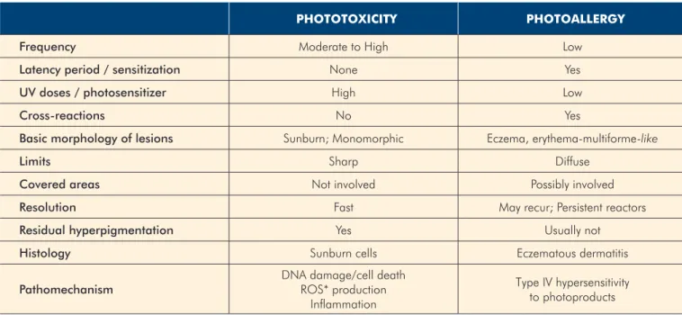
Figure 1 - Photoallergy reaction to St. John’s Wort infusion. Note the eczema spreading to non-photoexposed areas.
and epidermal T-cell infiltration and spongiosis with formation of vesicles.11
Phototoxicity, by definition, does not involve specific im-mune mechanisms and is caused by the presence of an ab-normal chromophore in exaggerated amounts in the skin. This chromophore can be either the drug itself, a drug metabolite or an endogenous chromophore induced by the drug. When excited by an UV photon, the chromophore’s energy increa-ses, entering a singlet state (a short-lived excited state) or a triplet state (a more stable, biologically active and long-lived state). These molecules then react with neighbouring molecu-les in a photodynamic reaction, leading to disruption of lipi-dic cell membranes and changes in the aromatic aminoacids of pyrimidine bases of DNA and RNA. Free radicals are also formed resulting in the damage of cellular organelles and ul-timately, cytotoxicity. Inflammatory cytokines such as IL-1, IL-6, TNF-alpha and other inflammatory mediators such as prosta-glandins and leukotrienes also contribute to this inflammatory response.1,9,12
Typically, these are the most frequent reactions, developing in any individual as long as there is enough photosensitizer and sun exposure. They can occur on a first contact, with no particular aggravation on further contacts. Clinically they re-semble acute sunburn with well-demarcated erythema exclusi-vely on sun-exposed areas, resolving with hyperpigmentation. There is no cross-reactivity with other drugs and histology shows apoptotic keratinocytes (sunburn cells).11
However, even though the mechanisms of photoaller-gy and phototoxicity are well established there are overlap-ping mechanisms as well as clinical manifestations. In fact, many drugs can induce photoallergic and phototoxic reac-tions. For example, the phototoxic furocoumarins, contained in plant extracts that are used in “folk medicine” or during photochemotherapy, can induce photoallergy.13 The same is true for promethazine and lomefloxacin, which have a well--established phototoxic potential but can also elicit photoal-lergic reactions.14-16
Less commonly, other mechanisms of photosensitivity can be considered, some immune-mediated like drug-induced LE while others are more phototoxic in nature, namely pseudo-porphyria, photoaging and photocarcinogenesis.
3. cLInIcAL FEAturES oF drug PHotoSEnSItI-VIty
Photosensitivity can result from systemic uptake or topical application of drugs. Cutaneous lesions can vary from urtica-ria through eczema, subacute LE, vitiligo-like depigmentation, dyschromia, NMSC to acute sunburn1,17 (Table 2). The timefra-me between drug introduction and beginning of skin findings can range from a few minutes (in vemurafenib-induced pho-tosensitivity, for example) to years (in NMSC from voricona-zole). Most photoallergic and phototoxic reactions, however, occur 1 to 2 days after introduction of the drug and sun expo-sure. For pseudoporphyria, drug-induced LE and photoony-colysis, it may take several days to weeks.
3.1 Systemic Photosensitvity
From a clinical standpoint, it is useful to divide patterns of drug-induced photosensitivity in acute, subacute and delayed reactions, since, as we have established previously, they can overlap in their pathomechanisms.
3.1.1 Acute Reactions
In acute photoallergy from systemic drugs lesions are mainly confluent or non-confluent eczematous patches in photo-exposed areas but can sometimes resemble erythema multiforme.18,19 They usually involve, in a symmetrical distribu-tion, the face and forehead, V-shaped area of the neck and upper chest, dorsum of the hands and forearms. The shaded areas of the face (upper eyelids, upper lip, deep furrows), re-troauricular and submentonian regions are usually spared. In more extensive sun exposure, large body folds, like the axillae, groins, finger webs and covered areas (clothes, watch string, shoes) are also usually spared. This is especially important table 2 -
Clinical patterns of photosensitivity
PrEdoMInAnt In PHototoXIcIty PrEdoMInAnt In PHotoALLErgy
Exaggerated “sunburn”
Urticaria in sun-exposed areas Pseudoporphyria
Photoonycholysis
Acute and subacute eczema Hyperpigmentation
Hypopigmentation (vitiligo-like)
Cheilitis Telangiectasia
Purpura Erythema multiforme-like lesions
Pellagra-like Subacute or chronic lupus erythematosus
when distinguishing systemic photoallergy from airborne der-matitis, in which the shaded areas typically spared in exposure to photoactive drugs can be involved in airborne drug expo-sure, for example in nurses or other care-givers who crash tablets.
A slightly different distribution of lesions can occur, for example, in car drivers, who only expose the left side of body, giving rise to an asymmetrical pattern of sun-exposure. Fur-thermore, lesions can be found only on the lower lip, because of its higher exposure and thinner stratum corneum.20
Most systemic phototoxic reactions occur between 12-24 hours after sun-exposure and resemble acute sunburn, with sharply delimitated erythema that can progress to vesicles and bullae and later to desquamation in large epidermal sheets. Residual hyperpigmentation is frequent.
Some phototoxic drugs, however, can induce immediate prickling and burning with transient erythema1 (amiodarone, for example – Fig. 2). Immediate burning is also seen with more than 50% of patients under vemurafenib treatment for metastatic melanoma.21 This can be prevented by sun avoi-dance and sun protection extending to the long UVA.22, 23
3.1.2 Subacute Reactions
These reactions usually take several days to weeks to de-velop, and mainly evoke phototoxic mechanisms as in pseu-doporphyria, photoonycolysis, dyschromia, telangiectasia and purpura, whereas annular lesions may suggest drug-induced subacute cutaneous LE.
Drug-induced pseudoporphyria is clinically and histo-logical indistinguishable from classical porphyria cutanea tarda, presenting with chronic skin fragility and flaccid bullae on non-inflamed, sun-exposed skin, occasionally progressing with the formation of millia. It usually develops within weeks to months after drug exposure. This pattern was described ini-tially with nalidixic acid, furosemide and naproxen1 but it has been recognized more recently with celecoxib,24,25 ciprofloxa-cin,26 voriconazole,27,28 torsemide,29 metformin,30 finasteride31 and imatinib.32-34
Pseudoporphyria occurs in individuals with no inborn error in porphyrin metabolism and as such, no elevation in endo-genous porphyrins is detected, apart from occasional transient increase of uroporphyrins with voriconazole.35
Drug-induced cutaneous lupus erythematosus seems to be the consequence of the exaggerated expression of the Ro/ SSA antigen on the surface of keratinocytes in the presence of the drug, however, the precise mechanisms underlying this reaction are not known.36 Annular lesions are clinically and on histopathology similar to idiopathic form of subacute cuta-neous LE and are located in photoexposed areas and also in usually UV-shaded areas.37,38
Drug-induced subacute cutaneous LE usually develops weeks or months after drug exposure and is associated with a long list of drugs,39 namely thiazide diuretics, calcium channel blockers, ACE inhibitors,36 but particularly with terbinafine3,39 – the drug with the highest odds ratio for this event.39
Photoonycholysis refers to the half-moon distal onycholy-sis of one or several nails, described usually 2-3 weeks after exposure to tetracyclines (doxycycline)40 (Fig. 3), psoralens and fluoroquinolones.41 Although there is no definite explanation for this peculiar presentation of photosensitivity, most authors point out that the nail plate is relatively unprotected from sun-light (since it displays less melanin) and suffers from augmen-tation of sun exposure through the nail plate, acting like a lens through which concentrated UVR can enhance inflammation and result in nail detachment.40-42
Dyschromia can result from residual hyperpigmentation following an acute phototoxicity (Fig.4) and from photoaging (enhancement of solar lentigines) induced by some drugs (vo-riconazole, vandetanib43,44). The accumulation of photoactive Figure 2 - Amiodarone-induced erythema. Note the sparing of the
wrinkles.
drugs or their metabolites in the dermis may also lead to dys-chromia, as is the case for amiodarone,45 minocycline46,47 and phenotiazines (especially thioridazine).1 A golden-brown or slate grey, bluish colour on sun-exposed areas can even per-sist longer after stopping these drugs.
Other clinical manifestations of subacute photosensitivity include telangiectasia in sun-exposed areas reported for cal-cium channel blockers,1 petechial purpura with sharp limits on the transition to the shaded areas for ciprofloxacin,48 pella-gra as a consequence of niacin consumption during prolon-ged therapy with isoniazid and pellagroid reactions reported for anti-cancer agents such as 6-mercaptopurine and 5-fluo-rouracil.49
3.1.3 Delayed Reactions
Chronic exposure to photoactive drugs can lead to accele-rated photoaging, actinic keratosis and skin cancers. Vorico-nazole can result in dyschromia, lentigines, actinic keratosis and squamous cell carcinomas, even in children,7,50 and there is consensual agreement on dose-dependent increased risk for skin cancers after long-time PUVA phototherapy.51 Naproxen, chlorpromazine and fluoroquinolones, especially lomefloxa-cin, can augment DNA aggression induced in vitro by UV and result in epidermal neoplasia in animals.52 In humans, po-tentially photosensitizing drugs, such as diuretics and cardio-vascular drugs, are being associated with a rise in cutaneous precancerous lesions.4,6 For vemurafenib there is a known risk for developing actinic keratoses, keratoacanthoma-like NMSC and even new melanomas but, probably, this is independent of photosensitivity and mostly dependent on the activation of alternate signalling pathways after BRAF inhibition.21,22
3.2 topical Photossensitivity
Topical photosensitizers are responsible mostly for acute reactions. Generally, in phototoxic contact dermatitis lesions develop minutes to days after sun-exposure, and in photoal-lergic there is a delay of usually 24 to 48 hours after ultraviolet exposure.53 Immediate urticarial reactions, like photocontact urticaria, have also been described with chlorpromazine54 and 5-aminolevulinic acid used in photodynamic therapy.55
Clinically, photoallergic contact dermatitis presents as an eczematous response, whereas phototoxic appears as erythe-ma, oedema and bullous lesions. In both types of photo-contact dermatitis, lesions are localized in sun-exposed skin where the drug has been applied. Nevertheless, distant lesions can be the result of accidental contact (such as kissing lesions in the inner thighs or inadvertent spread by hands or contami-nated objects56,57). Connubial dermatitis has been described for ketoprofen and benzydamine.20,58-61 Also, when the drug is used topically in the mouth lesions can manifest as lip and chin dermatitis.20,62 If the contact photoallergen is significantly absorbed through the skin, it can mimic the distribution of sys-temic photosensitivity.
Next to the topical NSAIDs, UV filters are the main topical photosensitizers,15,63 and given their importance, a brief over-view of photoallergy to sunscreens evolution is noteworthy.
PABA (p-aminobenzoic acid) was initially the main respon-sible for photoallergy contact dermatitis although nowadays has been largely replaced and is only seldom used.64 Oxy-benzone (benzophenone 3) was introduced in the 1970 and 1980’s but despite being replaced in many sunscreens cur-rently, is still one of the leading causes of positive photopatch tests.63 The reasons for the high level of positivity to benzophe-none 3 are possibly the wide presence in cosmetic products and the cross-reactivity with other agents containing benzo-phenone nucleus, such as ketoprofen and fenofibrate.53,65
Cinnamates and salicylates have also been responsible for photoallergic reactions although apparently in a lesser de-gree.66 Octocrylene, as a result of its wider use and in higher concentrations, is being responsible for a raising number of cases, notably in children and in adults previously photosensi-tized to ketoprofen.65,67,68
Concerning the newer UV filters, Mexoryl SX (terephthalyli-dene dicamphor sulfonic acid), Tinosorb S® (bis-ethylhexylo-xyphenol methoxyphenyl triazine) and Tinosorb M® (methylene bis-benzotriazolyl tetramethylbutylphenol) photoallergys are rare, but Tinosorb M® is frequently responsible for contact der-matitis due to decyl glucoside.65,69-71
As new UV absorbers are introduced, the incidence of photoallergic contact dermatitis and causative allergens is li-kely to evolve.
4. MAIn drugS cAuSIng PHotoSEnSItIVIty The list of photosensitizers is large and ever-growing and comprises drugs that can be used topically or systemically (Table 3). Sometimes, a drug can induce photosensitivity by both ways, as piroxicam, for example. Other drugs, like ke-toprofen, frequently induce a photoallergic contact dermatitis with topical use but its concentration on the skin from systemic exposure is usually insufficient for inducing photosensitization.
Topical drugs NSAIDs, namely ketoprofen, etofenama-te, benzydamine and phenothiazine derivatives are the main agents responsible for photoallergic contact dermatitis15,63,72-74 and, by far, responsible for most positive patch-tests in sou-thern Europe15,72-74 as recently shown also in a multicentre eu-ropean photopatch test study (The Europen Multicentre Study Figure 4 - Residual hyper and hypopigmented lesions in fenofibrate
Photopatch Test EMCPPTS – 2012).63 Although not considered a drug, UV filters are also an important source of photoaller-gic contact dermatitis as we have discussed previously.
Main systemic photosensitizers include antimicrobials, es-pecially tetracyclines, fluoroquinolones, sulphonamides and antifungals, NSAIDs and cardiovascular drugs.
Antimicrobials, including tetracyclines and quinolo-nes are among frequent photosensitizers. Doxycycline and less frequently minocycline are phototoxic, can induce pho-toonycholysis (Fig. 3), pseudoporphyria and for the latter there is also the risk of dyschromia, already covered in this arti-cle.1,40,47
Quinolones, especially fluoroquinolones, can induce pho-totoxic reactions and pseudoporphyria.26 This was evident for the first quinolone antibiotic – nalidixic acid,47 but phototoxicity can also occur in up to 15% of patients treated with fleroxacin, lomefloxacin, sparfloxacin and pefloxacin, and less frequently for ciprofloxacin, norfloxacin, ofloxacin and enoxacin1. Admi-nistering the drug at night, to reduce drug concentrations in
circulation during midday can diminish its phototoxic potential. Photoallergy with lomefloxacin14,16 (Fig. 5) and enoxacin47 as well as cross-reactions with other fluorquinolones (ciprofloxa-cin and flerofloxa(ciprofloxa-cin)75,76 have been described. Fluoroquino-lones can also photosensitize DNA and have photomutagenic and photocarcinogenic properties.52
Sulphonamides and sulpha-drugs, like thiazide diu-retics, sulfonylureas and celecoxib, as well as dapsone (diaminodiphenylsulfone), have been reported to cause pho-tosensitivity.47,77 Apparently, this side effect is not so frequent with cotrimoxazole.1,47
Systemic phenothiazines (chlorpromazine and thiorida-zine) are not only phototoxic but can also induce lichenoid lesions with residual hyperpigmentation.1 Promethazine, still used as a topical antipruritic, can induce both a phototoxic and photoallergic contact dermatitis.15,78 Other topical pheno-thiazines, like chlorproethazine used as a muscle relaxant and isothipendyl chlorhydrate, used as an antipruritic agent caused photoallergy and positive patch tests to chlorpromazine.79,80 table 3 -
Main drugs causing photosensitivity
SyStEMIc PHotoSEnSItIVty toPIcAL PHotoSEnSItIVIty
Antimicrobials Antidepressants NSAIDs
Tetracyclinesa (doxycline, minocycline) Clomipramine, imipramine, sertraline
Ketoprofenc
Piroxicamc, etofenamatec
Piroxicamc, etofenamatec benzydamine
Diclofenac
Sulphonamides (sulfamethoxazole) Cardiovascular drugs Phenothiazines Fluoroquinolones (lomefloxacin,
ciprofloxacin)a Amiodaronea, quinidine
Chlorpromazine
Promethazine, chlorhydrate chlorproethazine Voriconazolea,b Furosemide, torasemide and thiazide
diuretics Plants (used as drugs)
Terbinafine, griseofulvina Anti-cancer agents Ruta graveola (common rue)
Efavirenz, tenofovir, faldeprevir Paclitaxel, docetaxel Photodynamic therapy agents NSAIDs Methotrexate, 5-fluoracil 5-aminolevulinic acid
Arylpropionic acids: Tiaprofenic acida, suprofen
Naproxen, ibuprofen, ibuproxam, carprofena
Dacarbazine
Miscellaneous Psoralensb, Fenofibrate, simvastatin
Piroxicamc,d Sulfonylureas, sitagliptin, metformin
Flutamide, finasteride, pirfenidone Celecoxib, diclofenacd
Azapropazone, phenylbutazone,
indomethacin Retinoids
Phenothiazines Plants (used as drugs)a
Chlorpromazined, thioridazine Hypericum perforatum (St. John’s wort)
Targeted therapies
Kava extracts Vemurafenibb, imatinib, vandetanib
Amiodarone, as previously discussed, can induce erythe-ma (Fig. 2) followed by a bluish-grey hyperpigmentation in sun-exposed areas.45
Antifungals comprise many agents with photosensitizing properties, namely griseofulvin, terbinafine and voriconazo-le. The first two can be found to aggravate lupus erythemato-sus, even inducing subacute lupus erythematosus in patients who develop anti-Ro/SS-A antibodies.3,39
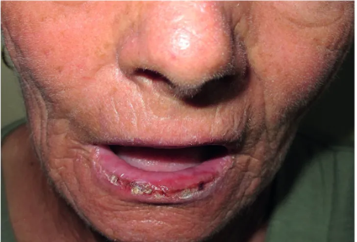
Photosensitivity to voriconazole is seemingly not extensive to other azole antifungals. This drug is used mainly in invasi-ve aspergillosis or refractory candidiasis, generally in patients with previous immunosuppression, either from underlying di-seases or from therapeutic immunosuppressants, therefore in individuals with a considerably risk for photosensitivity50,81 Photosensitivity from voriconazole can manifest as a sun-burn reaction with cheilitis and erosions of the lower lip28,50,82 (Fig.6), as pseudoporhyria,27 but also with photoaging with solar lentigines and actinic keratosis progressing into multi-focal invasive squamous cell carcinoma,5,7,83,84 even with de--novo melanoma.85
Antiviral agents, especially those used in the treatment of HIV and HCV infection, have been described as photosensiti-zing. Efavirenz, for example, induced papulosquamous annu-lar lesions on photoexposed areas, only within a few days to weeks after initiation of treatment.86,87 Tenofovir, a newer anti--retroviral drug has also been reported as inducing systemic photoallergy, with positive photopatch tests.88 This is especially important when you consider HIV infection in itself as a pho-tosensitizing condition and a known risk-factor for a variety of photosensitive disorders.
Nonsteroidal anti-inflammatory drugs (NSAIDs) are a
frequent cause of photosensitivity. This was initially seen with benoxaprofen, calling attention for this adverse event not only with this agent but to many others within this class (carprofen, naproxen, suprofen, ketoprofen, ibuprofen and tiaprofenic acid). Other NSAIDs, namely diclofenac, piroxicam, celeco-xib, benzydamine and etofenamate were also documented as photosensitizers. For tiaprofenic acid, for example, in vitro and in vivo phototoxic potencial was reported,89,90 but in other pu-blications also photoallergic reactions were described,91-93 un-derlining that both patterns of photosensitivity can be elicited by the same agent.
Most topically applied NSAIDs are absorbed through the skin and may cause distant lesions resembling a systemic pho-tosensitivity. Benzydamine, used for oral or genital hygiene, causes photosensitivity at distant sites,15 as well as cheilitis and chin dermatitis or hand dermatitis caused by the application of the drug.15,20
Ketoprofen may cause severe photoallergic reactions, with oedema, bullae and even erythema multiforme-like le-sions.60,81,94-96 These extend well beyond the area of applica-tion, and can recur on sun-exposure even without further drug application,60,94 as the drug or its metabolite can persist in the skin for several days.96 Cross-reactions with benzophenone and octocrylene in sunscreens60 or benzophenones in magazi-ne ink97 have also been described. Cross-reactivity also occurs between arylpropionic acid derivatives that share the benzo-phenone structure, namely, tiaprofenic acid and suprofen, but is not extensive to naproxen or ibuprofen.
The analogues of ketoprofen, piketoprofen and dexketo-profen have a similar behaviour concerning photosensitivi-ty.61,98 New topical formulations of ketoprofen in plaster aim to reduce UV exposure of the drug, but do not completely hinder this particular side-effect.
Piroxicam is a known photosensitizer since the 1980’s, usually reacting on a first exposure because of its close rela-tion to thiomersal99 and its main sensitizing moiety of the mo-lecule, thiosalicylic acid.100 Photoallergy can occur both from topical and systemic use of the drug but, as this NSAID has been replaced by newer drugs, this side effect is becoming less Figure 5 - Acute systemic photoallergy reaction induced by
lome-floxacin.
frequent. However, a few cases were still found in the recent European multicentre photopatch test study.63 Systemic pho-tosensitivity develops within 24-48 hours as an acute eczema involving the whole face or as scattered erythematous papu-les and vesicpapu-les on the face and dorsum of the hands, often pompholyx-type.101-103 Patients displaying this photoallergy do not react against tenoxicam, meloxicam or lornoxicam neither on photopatch nor on drug rechallenge, as these oxicams do not share the thiosalicylate moiety.104 On the contrary, cross--reaction between piroxicam and other oxicams occurs regu-larly in fixed drug eruption.105
More recently, the new kinase inhibitors and new anti--cancer drugs deserve a place among the drugs capable of eliciting photosensitivity. Vandetanib,106 imatinib and in par-ticular vemurafenib are known to cause phototoxic reactions. Regarding the latter, more than 50% of patients develop bur-ning and oedematous erythema on sun-exposure22,23 and also actinic keratosis and squamous cell carcinoma, as early as within 8 weeks of starting therapy.21
Finally, “folk” medicines, mostly based on plant extracts, some of them rich in furocoumarins, can obviously result in systemic or topical photosensitivity, such as home-made in-fusions of St. John’s wort (Hypericum perforatum L.)107 (Fig. 1) and topically applied infusions of Ruta graveolens.108
5. dIAgnoStIc ProcEdurES In drug PHoto-SEnSItIVIty
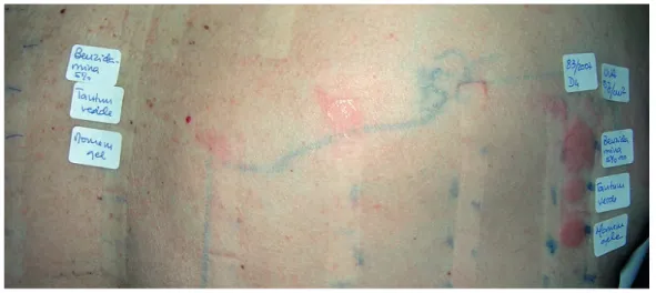
A photosensitive eruption demands a careful and syste-matic review of all the drugs taken by the patient. Photopatch tests are indicated mainly for photoallergic contact dermati-tis but can also be used to assess systemic drug photoaller-gy.17,109,110 The recommended European baseline photopatch test series includes ketoprofen, etofenamate, piroxicam and benzydamine, with the extended series covering also pike-toprofen, dexkepike-toprofen, ibuprofen, diclofenac, fenofibrate and chlorpromazine,111 but any suspected drug can be tested according to the general standardized procedures of photo-patch testing.
Briefly, allergens are applied in duplicate on the back, followed by irradiation of only one of the sets at day 1 or day 2 with 5 J/cm2 of UVA, whereas the other set is shielded from the light. Readings should be performed immediately after ir-radiation and also 48 and/or 72 h afterwards.112
Photopatch tests results have to be carefully interpreted: positive reactions in both sets mean contact allergy that can be photoaggravated if the reaction is 1+ more on the irradiated side. A photopatch test is positive when erythema and papules covering the whole test area is observed only in the irradiated set112 (Fig. 7). If the reaction is mainly erythema and oedema, without pruritus and exclusively limited to the test chamber area, beginning shortly after irradiation, reaching its highest intensity by 24h and regressing in 48 to 72h, then it is proba-bly a phototoxic event. If there is pruritic exanthema with ve-sicles, diffuse limits extending beyond the chamber area, with increasing intensity until 48-72h after irradiation, this is more suggestive of a photoallergic reaction.113
In systemic photosensitivity, oral photoprovocation with skin irradiation after drug exposure or with the calculation of the minimal erythema dose (MED) when exposed to the drug and after drug withdrawal may help to identify the culprit.
In phototoxic reactions, both photopatch and photopro-vocation tests are positive in the majority of tested patients. Therefore they are not particularly useful for confirming the aetiology of a phototoxic reaction but can disclose a hidden photoallergy.
6. gEnErAL PrIncIPLES oF trEAtMEnt oF drug PHotoSEnSItIVIty
Drug suspension and sun avoidance are recommended to resolve drug photosensitivity. If the drug is essential and life--saving, when there is no alternative drug or if the alternative drug is inadequate, sun avoidance, physical protection and a broad-spectrum sunscreen that covers the spectrum of UVA may be adequate to improve photosensitivity. For phototoxic reactions, this protective effect of sunscreen is particularly use-ful, as shown for voriconazole, vemurafenib and amiodarone. Figure 7 - Positive photopatch test to benzydamine. Note that the erythema is only seen in the irradiated site.
Moreover, broad-spectrum sunscreen can be implemented as a preventive measure when initiating a known photosensi-tizer, however one must acknowledge that chemical UV filters represent an important cause of contact photosensitivity, parti-cularly in patients with previous dermatoses.
In cases of acute photoallergy, suspension of the culprit drug and sun avoidance won’t resolve the skin lesions within a short time and active treatment may be necessary. Topical corticosteroids may be prescribed for a few days and severe reactions may need an additional short course of oral corti-costeroids with fast dose tapering.
Acute phototoxicity, presenting mainly as acute sunburn, may benefit greatly from emollients and photoprotection even for some time after the reaction, and the efficacy of corticoste-roids is highly questioned in this setting.
7. concLuSIon
Phototoxic, photoallergic and overlapping photosensitive reactions are still a frequent problem. They can exhibit clinical polymorphism, different time courses and late consequences. Culprit drugs often depend on geographic areas and pres-cription habits, changing also over time.
The dermatologist must be alert not only for the multiple clinical patterns that can result from photosensitivity but also for the many drugs that can cause it. A thorough review of all systemic and topical agents, including “folk” medicine, should be conducted and complementary tests such as photo-patch tests, phototests and photoprovocation may contribute to a final etiologic diagnosis. These proceedings may finally allow adequate patient advice concerning further eviction of the photosensitizer and related chemicals and greatly improve the patient’s quality of life.
rEFErEncES
1. Ferguson J. Drug and chemical photosensitivity. In: Hawk J, editor. Photodermatology. New York: Oxford University Press; 1999. p. 155-69.
2. Gonçalo M. Phototoxic and photoallergic reactions. In: Johansen J, Frosch P, Lepoittevin J-P, editors. Contact Der-matitis. 5th ed. Berlin: Springer-Verlag; 2011. p. 361-76. 3. Farhi D, Viguier M, Cosnes A, Reygagne P, Dubertret L,
Revuz J, et al. Terbinafine-induced subacute cutaneous lupus erythematosus. Dermatology. 2006; 212:59-65.
4. Placzek M, Eberlein-Konig B, Przybilla B. Association be-tween actinic keratoses and potentially photosensitizing drugs. N Engl J Med. 1999; 341:1474-5.
5. McCarthy KL, Playford EG, Looke DF, Whitby M. Severe photosensitivity causing multifocal squamous cell carcino-mas secondary to prolonged voriconazole therapy. Clin Infect Dis. 2007;44:e55-6.
6. Jensen AO, Thomsen HF, Engebjerg MC, Olesen AB, So-rensen HT, Karagas MR. Use of photosensitising diuretics and risk of skin cancer: a population-based case-control study. Br J Cancer. 2008; 99:1522-8.
7. Cowen EW, Nguyen JC, Miller DD, McShane D, Arron ST, Prose NS, et al. Chronic phototoxicity and aggressi-ve squamous cell carcinoma of the skin in children and adults during treatment with voriconazole. J Am Acad Dermatol. 2010; 62:31-7.
8. Bilu D, Mamelak AJ, Nguyen RH, Queiroz PC, Kowalski J, Morison WL, et al. Clinical and epidemiologic characteri-zation of photosensitivity in HIV-positive individuals. Pho-todermatol Photoimmunol Photomed. 2004; 20:175-83. 9. Hawk J. Photodermatology. New York: Oxford University
Press; 1999.
10. Rustemeyer T, van Hoogstraaaten I, van Blomberg B, Gibbs S, Scheper R. Mechanisms of irritant and allergic contact dermatitis. In: Johansen JD, Frosch P, Lepoittevin J-P, editors. Contact Dermatitis. 5th ed. Berlin: Springer--Verlag; 2011. p. 43-90.
11. Gonçalo M, Gimenez-Arnau A. Drug Photosensitivity. In: Katsambas AD, Lotti TM, Dessinoti C, D'Erme AM, editors. European Handbook of Dermatological Treatments. 3rd ed. Berlin: Springer-Verlag; 2015. p. 233-51.
12. Kutlubay Z, Sevim A, Engin B, Tuzun Y. Photodermatoses, including phototoxic and photoallergic reactions (internal and external). Clin Dermatol. 2014; 32:73-9.
13. Karimian-Teherani D, Kinaciyan T, Tanew A. Photoallergic contact dermatitis to Heracleum giganteum. Photoderma-tol Photoimmunol Photomed. 2008; 24:99-101.
14. Oliveira HS, Goncalo M, Figueiredo AC. Photosensitivity to lomefloxacin. A clinical and photobiological study. Pho-todermatol Photoimmunol Photomed. 2000; 16:116-20. 15. Cardoso JC, Canelas MM, Goncalo M, Figueiredo A. Pho-topatch testing with an extended series of photoallergens: a 5-year study. Contact Dermatitis. 2009; 60:325-9. 16. Kurumaji Y, Shono M. Scarified photopatch testing in
lo-mefloxacin photosensitivity. Contact Dermatitis. 1992; 26:5-10.
17. Gonçalo M. Explorations dans les photo-allergies médi-camenteuses. In: GERDA, editor. Progrès en Dermato-al-lergologie. Nancy: John Libbey Eurotext; 1998. p. 67-74. 18. Cohen PR. Photodistributed erythema multiforme: paclita-xel-related, photosensitive conditions in patients with can-cer. J Drugs Dermatol. 2009; 8:61-4.
19. Rodriguez-Pazos L, Gomez-Bernal S, Rodriguez-Grana-dos MT, Toribio J. Photodistributed erythema multiforme. Actas Dermosifiliogr. 2013; 104:645-53.
20. Canelas MM, Cardoso JC, Goncalo M, Figueiredo A. Conflitos de interesse: Os autores declaram não possuir
con-flitos de interesse. Suporte financeiro: O presente trabalho não foi suportado por nenhum subsídio ou bolsa. Direito à privaci-dade e consentimento escrito: Os autores declaram que pedi-ram consentimento ao doente para usar as imagens no artigo.
Conflicts of interest: The authors have no conflicts of interest to declare. Financing Support: This work has not received any contribution, grant or scholarship. Privacy policy and informed consent: The authors declare that the patient gave written infor-med consent for the use of its photos in this article.
Photoallergic contact dermatitis from benzydamine pre-senting mainly as lip dermatitis. Contact Dermatitis. 2010; 63:85-8.
21. Rinderknecht JD, Goldinger SM, Rozati S, Kamarashev J, Kerl K, French LE, et al. RASopathic skin eruptions during vemurafenib therapy. PLoS One. 2013; 8:e58721. 22. Gelot P, Dutartre H, Khammari A, Boisrobert A, Schmitt C,
Deybach JC, et al. Vemurafenib: an unusual UVA-indu-ced photosensitivity. Exp Dermatol. 2013; 22:297-8. 23. Dummer R, Rinderknecht J, Goldinger SM. Ultraviolet A
and photosensitivity during vemurafenib therapy. N Engl J Med. 2012; 366:480-1.
24. Cummins R, Wagner-Weiner L, Paller A. Pseudoporphyria induced by celecoxib in a patient with juvenile rheumatoid arthritis. J Rheumatol. 2000; 27:2938-40.
25. Schmutz JL, Barbaud A, Trechot P. Pseudoporphyrie et Coxib. Ann Dermatol Venereol. 2006; 133:213.
26. Schmutz JL, Barbaud A, Trechot P. Ciprofloxacine et pseu-doporphyrie. Ann Dermatol Venereol. 2008; 135:804. 27. Tolland JP, McKeown PP, Corbett JR. Voriconazole-induced
pseudoporphyria. Photodermatol Photoimmunol Photo-med. 2007; 23:29-31.
28. Auffret N, Janssen F, Chevalier P, Guillemain R, Amrein C, Le Beller C. Photosensibilisation au voriconazole: 7 cas. Ann Dermatol Venereol. 2006; 133:330-2.
29. Perez-Bustillo A, Sanchez-Sambucety P, Suarez-Amor O, Rodriguez-Prieto MA. Torsemide-induced pseudopor-phyria. Arch Dermatol. 2008; 144:812-3.
30. Lenfestey A, Friedmann D, Burke WA. Metformin-induced pseudoporphyria. J Drugs Dermatol. 2012; 11:1272. 31. Santo Domingo D, Stevenson ML, Auerbach J, Lerman
J. Finasteride-induced pseudoporphyria. Arch Dermatol. 2011; 147:747-8.
32. Timmer-de Mik L, Kardaun SH, Kramer MH, Hayes DP, Bousema MT. Imatinib-induced pseudoporphyria. Clin Exp Dermatol. 2009; 34:705-7.
33. Berghoff AT, English JC. Imatinib mesylate-induced pseu-doporphyria. J Am Acad Dermatol. 2010; 63:e14-6. 34. Perez NO, Esturo SV, Viladomiu Edel A, Moreno AJ, Valls
AT. Pseudoporphyria induced by imatinib mesylate. Int J Dermatol. 2014; 53:e143-4.
35. Hickman G, Duval A, Picard C, Petit A. Porphyrie cutanee tardive revelee par le voriconazole. Ann Dermatol Vene-reol. 2010; 137:36-9.
36. Sontheimer RD, Henderson CL, Grau RH. Drug-induced subacute cutaneous lupus erythematosus: a paradigm for bedside-to-bench patient-oriented translational clinical investigation. Arch Dermatol Res. 2009; 301:65-70. 37. Biazar C, Sigges J, Patsinakidis N, Ruland V, Amler S,
Bonsmann G, et al. Cutaneous lupus erythematosus: first multicenter database analysis of 1002 patients from the European Society of Cutaneous Lupus Erythematosus (EUSCLE). Autoimmun Rev. 2013; 12:444-54.
38. Lamond NW, Younis T, Purdy K, Dorreen MS. Drug-induced subacute cutaneous lupus erythematosus associated with nab-paclitaxel therapy. Curr Oncol. 2013; 20:e484-7.
39. Gronhagen CM, Fored CM, Linder M, Granath F, Nyberg F. Subacute cutaneous lupus erythematosus and its asso-ciation with drugs: a population-based matched case--control study of 234 patients in Sweden. Br J Dermatol. 2012; 167:296-305.
40. Passier A, Smits-van Herwaarden A, van Puijenbroek E. Photo-onycholysis associated with the use of doxycycline. BMJ. 2004; 329:265.
41. Baran R, Juhlin L. Photoonycholysis. Photodermatol Pho-toimmunol Photomed. 2002; 18:202-7.
42. Gregoriou S, Karagiorga T, Stratigos A, Volonakis K, Kontochristopoulos G, Rigopoulos D. Photo-onycholysis caused by olanzapine and aripiprazole. J Clin Psycho-pharmacol. 2008; 28:219-20.
43. Malani AN, Aronoff DM. Voriconazole-induced photosen-sitivity. Clin Med Res. 2008; 6:83-5.
44. Giacchero D, Ramacciotti C, Arnault JP, Brassard M, Bau-din E, Maksimovic L, et al. A new spectrum of skin toxic effects associated with the multikinase inhibitor vandeta-nib. Arch Dermatol. 2012; 148:1418-20.
45. Ammoury A, Michaud S, Paul C, Prost-Squarcioni C, Al-varez F, Lamant L, et al. Photodistribution of blue-gray hyperpigmentation after amiodarone treatment: molecu-lar characterization of amiodarone in the skin. Arch Der-matol. 2008; 144:92-6.
46. Wetter DA. Minocycline hyperpigmentation. Mayo Clin Proc. 2012; 87:e33.
47. Vassileva SG, Mateev G, Parish LC. Antimicrobial pho-tosensitive reactions. Arch Intern Med. 1998; 158:1993-2000.
48. Urbina F, Barrios M, Sudy E. Photolocalized purpura du-ring ciprofloxacin therapy. Photodermatol Photoimmunol Photomed. 2006; 22:111-2.
49. Stevens HP, Ostlere LS, Begent RH, Dooley JS, Rustin MH. Pellagra secondary to 5-fluorouracil. Br J Dermatol. 1993; 128:578-80.
50. Frick MA, Soler-Palacin P, Martin Nalda A, Guarner ME, Nadal CF. Photosensitivity in immunocompromised pa-tients receiving long-term therapy with oral voriconazole. Pediatr Infect Dis J. 2010; 29:480-1.
51. Stern RS. The risk of squamous cell and basal cell can-cer associated with psoralen and ultraviolet A thera-py: a 30-year prospective study. J Am Acad Dermatol. 2012;66:553-62.
52. Klecak G, Urbach F, Urwyler H. Fluoroquinolone antibac-terials enhance UVA-induced skin tumors. J Photochem Photobiol B. 1997; 37:174-81.
53. Deleo VA. Photocontact dermatitis. Dermatol Ther. 2004; 17:279-88.
54. Lovell CR, Cronin E, Rhodes EL. Photocontact urtica-ria from chlorpromazine. Contact Dermatitis. 1986; 14:290-1.
55. Kerr AC, Ferguson J, Ibbotson SH. Acute phototoxicity with urticarial features during topical 5-aminolaevulinic acid photodynamic therapy. Clin Exp Dermatol. 2007; 32:201-2.
56. Hindsen M, Isaksson M, Persson L, Zimersson E, Bruze M. Photoallergic contact dermatitis from ketoprofen induced by drug-contaminated personal objects. J Am Acad Der-matol. 2004; 50:215-9.
57. Lasa Elgezua O, Gorrotxategi PE, Gardeazabal Garcia J, Raton Nieto JA, Perez JL. Photoallergic hand eczema due to benzydamine. Eur J Dermatol. 2004; 14:69-70. 58. Matthieu L, Meuleman L, Van Hecke E, Blondeel A,
De-zfoulian B, Constandt L, et al. Contact and photocontact allergy to ketoprofen. The Belgian experience. Contact Dermatitis. 2004; 50:238-41.
59. Hindsen M, Zimerson E, Bruze M. Photoallergic contact dermatitis from ketoprofen in southern Sweden. Contact Dermatitis. 2006; 54:150-7.
60. Devleeschouwer V, Roelandts R, Garmyn M, Goossens A. Allergic and photoallergic contact dermatitis from keto-profen: results of (photo) patch testing and follow-up of 42 patients. Contact Dermatitis. 2008; 58:159-66. 61. Fernandez-Jorge B, Goday Bujan JJ, Paradela S, Mazaira
M, Fonseca E. Consort photocontact dermatitis from pike-toprofen. Contact Dermatitis. 2008; 58:113-5.
62. Conti R, Bassi A, Difonzo EM, Moretti S, Francalanci S. A case of photoallergic contact dermatitis caused by unu-sual exposure to ketoprofen. Dermatitis. 2012; 23:295-6. 63. A European multicentre photopatch test study. Br J
Der-matol. 2012; 166:1002-9.
64. Waters AJ, Sandhu DR, Lowe G, Ferguson J. Photocontact allergy to PABA in sunscreens: the need for continued vi-gilance. Contact Dermatitis. 2009; 60:172-3.
65. Heurung AR, Raju SI, Warshaw EM. Adverse reactions to sunscreen agents: epidemiology, responsible irritants and allergens, clinical characteristics, and management. Der-matitis. 2014; 25:289-326.
66. Wong T, Orton D. Sunscreen allergy and its investigation. Clin Dermatol. 2011;29:306-10.
67. de Groot AC, Roberts DW. Contact and photocontact al-lergy to octocrylene: a review. Contact Dermatitis. 2014; 70:193-204.
68. Karlsson I, Vanden Broecke K, Martensson J, Goossens A, Borje A. Clinical and experimental studies of octocrylene's allergenic potency. Contact Dermatitis. 2011; 64:343-52. 69. Gonzalez ME, Soter NA, Cohen DE. Positive patch- and
photopatch-test reactions to methylene bis-benzotriazolyl tetramethylbutylphenol in patients with both atopic der-matitis and chronic actinic derder-matitis. Derder-matitis. 2011; 22:106-11.
70. de Groot AC, van Zuuren EJ, Hissink D. Contact allergy to Tinosorb(R) M: recommendations for diagnostic improve-ment. Contact Dermatitis. 2014; 70:251-4.
71. Pereira N, Coutinho I, Andrade P, Goncalo M. The UV filter tinosorb M, containing decyl glucoside, is a frequent cause of allergic contact dermatitis. Dermatitis. 2013; 24:41-3.
72. de la Cuadra-Oyanguren J, Perez-Ferriols A, Lecha--Carrelero M, Gimenez-Arnau AM, Fernandez-Redondo V, Ortiz de Frutos FJ, et al. Resultados y evaluacion del
fotoparche en Espana: hacia una nueva bateria estandar de fotoalergenos. Actas Dermosifiliogr. 2007; 98:96-101. 73. Leonard F, Adamski H, Bonnevalle A, Bottlaender A,
Bourrain JL, Goujon-Henry C, et al. Etude prospective multicentrique 1991-2001 de la batterie standard des photopatch-tests de la Societe Francaise de Photoderma-tologie. Ann Dermatol Venereol. 2005; 132:313-20. 74. Pigatto PD, Guzzi G, Schena D, Guarrera M, Foti C,
Fran-calanci S, et al. Photopatch tests: an Italian multicentre study from 2004 to 2006. Contact Dermatitis. 2008; 59:103-8.
75. Kimura M, Kawada A. Photosensitivity induced by lome-floxacin with cross-photosensitivity to ciprolome-floxacin and fleroxacin. Contact Dermatitis. 1998; 38:180.
76. Correia O, Delgado L, Barros MA. Bullous photodermato-sis after lomefloxacin. Arch Dermatol. 1994; 130:808-9. 77. Yazici AC, Baz K, Ikizoglu G, Kokturk A, Uzumlu H,
Tata-roglu C. Celecoxib-induced photoallergic drug eruption. Int J Dermatol. 2004; 43:459-61.
78. Katsarou A, Makris M, Zarafonitis G, Lagogianni E, Gre-goriou S, Kalogeromitros D. Photoallergic contact derma-titis: the 15-year experience of a tertiary reference center in a sunny Mediterranean city. Int J Immunopathol Phar-macol. 2008; 21:725-7.
79. Barbaud A, Collet E, Martin S, Granel F, Trechot P, Lambert D, et al. Contact sensitization to chlorproethazine can in-duce persistent light reaction and cross-photoreactions to other phenothiazines. Contact Dermatitis. 2001; 44:373. 80. Bibas N, Sartor V, Bulai Livideanu C, Bagheri H, Nougue J,
Giordano-Labadie F, et al. Contact photoallergy to isothi-pendyl chlorhydrate. Dermatology. 2012; 224:289-91. 81. Beani JC. Les photosensibilisations graves. Ann Dermatol
Venereol. 2009; 136:76-83.
82. Hansford JR, Cole C, Blyth CC, Gottardo NG. Idiosyncra-tic nature of voriconazole photosensitivity in children un-dergoing cancer therapy. J Antimicrob Chemother. 2012; 67:1807-9.
83. Williams K, Mansh M, Chin-Hong P, Singer J, Arron ST. Voriconazole-associated cutaneous malignancy: a litera-ture review on photocarcinogenesis in organ transplant recipients. Clin Infect Dis. 2014; 58:997-1002.
84. Morice C, Acher A, Soufir N, Michel M, Comoz F, Leroy D, et al. Multifocal aggressive squamous cell carcinomas in-duced by prolonged voriconazole therapy: a case report. Case Rep Med. 2010; 2010:351084.
85. Miller DD, Cowen EW, Nguyen JC, McCalmont TH, Fox LP. Melanoma associated with long-term voriconazole thera-py: a new manifestation of chronic photosensitivity. Arch Dermatol. 2010; 146:300-4.
86. Yoshimoto E, Konishi M, Takahashi K, Murakawa K, Maeda K, Mikasa K, et al. The first case of efavirenz-duced photosensitivity in a Japanese patient with HIV in-fection. Intern Med. 2004; 43:630-1.
87. Isaacs T, Ngwanya MR, Dlamini S, Lehloenya RJ. An-nular erythema and photosensitivity as manifestations of efavirenz-induced cutaneous reactions: a review of
five consecutive cases. J Antimicrob Chemother. 2013; 68:2871-4.
88. Verma R, Vasudevan B, Shankar S, Pragasam V, Suwal B, Venugopal R. First reported case of tenofovir-indu-ced photoallergic reaction. Indian J Pharmacol. 2012; 44:651-3.
89. Figueiredo A, Ribeiro CA, Goncalo M, Baptista AP, Teixei-ra F. Experimental studies on the mechanisms of tiapro-fenic acid photosensitization. J Photochem Photobiol B. 1993; 18:161-8.
90. Neumann NJ, Holzle E, Plewig G, Schwarz T, Panizzon RG, Breit R, et al. Photopatch testing: the 12-year expe-rience of the German, Austrian, and Swiss photopatch test group. J Am Acad Dermatol. 2000; 42:183-92.
91. Foti C, Bonamonte D, Conserva A, Stingeni L, Lisi P, Lio-netti N, et al. Allergic and photoallergic contact dermatitis from ketoprofen: evaluation of cross-reactivities by a com-bination of photopatch testing and computerized confor-mational analysis. Curr Pharm Des. 2008; 14:2833-9. 92. Pigatto P, Bigardi A, Legori A, Valsecchi R, Picardo M.
Cross-reactions in patch testing and photopatch testing with ketoprofen, thiaprophenic acid, and cinnamic al-dehyde. Am J Contact Dermat. 1996; 7:220-3.
93. Le Coz CJ, Bottlaender A, Scrivener JN, Santinelli F, Cribier BJ, Heid E, et al. Photocontact dermatitis from ketoprofen and tiaprofenic acid: cross-reactivity study in 12 consecu-tive patients. Contact Dermatitis. 1998; 38:245-52. 94. Veyrac G, Paulin M, Milpied B, Bourin M, Jolliet P. Bilan
de l'enquete nationale sur les effets indesirables cutanes du ketoprofene gel enregistres entre le 01/09/1996 et le 31/08/2000. Therapie. 2002; 57:55-64.
95. Izu K, Hino R, Isoda H, Nakashima D, Kabashima K, Tokura Y. Photocontact dermatitis to ketoprofen presen-ting with erythema multiforme. Eur J Dermatol. 2008; 18:710-3.
96. Sugiura M, Hayakawa R, Kato Y, Sugiura K, Ueda H. 4 cases of photocontact dermatitis due to ketoprofen. Con-tact Dermatitis. 2000; 43:16-9.
97. Infante Hernando L, Serra-Baldrich E, Dordal T, Puig Sanz L. Photoallergic contact dermatitis caused by ben-zophenones in magazine inks. Contact Dermatitis. 2013; 69:124-6.
98. Marmgren V, Hindsen M, Zimerson E, Bruze M. Successful photopatch testing with ketoprofen using one-hour occlu-sion. Acta Derm Venereol. 2011; 91:131-6.
99. de Castro JL, Freitas JP, Brandao FM, Themido R. Sensiti-vity to thimerosal and photosensitiSensiti-vity to piroxicam. Con-tact Dermatitis. 1991; 24:187-92.
100. Goncalo M, Figueiredo A, Goncalo S. Hypersensitivity to thimerosal: the sensitizing moiety. Contact Dermatitis. 1996; 34:201-3.
101. Varela P, Amorim I, Massa A, Sanches M, Silva E. Piro-xicam-beta-cyclodextrin and photosensitivity reactions. Contact Dermatitis. 1998; 38:229.
102. Youn JI, Lee HG, Yeo UC, Lee YS. Piroxicam photosensi-tivity associated with vesicular hand dermatitis. Clin Exp Dermatol. 1993; 18:52-4.
103. Serra D, Gonçalo M, Figueiredo A. Two decades of cuta-neous adverse drug reactions from piroxicam. Contact Dermatitis. 2008; 58:35.
104. Goncalo M, Figueiredo A, Tavares P, Ribeiro CA, Teixeira F, Baptista AP. Photosensitivity to piroxicam: absence of cross-reaction with tenoxicam. Contact Dermatitis. 1992; 27:287-90.
105. Gonçalo M, Oliveira HS, Fernandes B, Robalo-Cordeiro M, Figueiredo A. Topical provocation in fixed drug erup-tion from nonsteroidal anti-inflammatory drugs. Exog Dermatol. 2002; 1:81-6.
106. Chang CH, Chang JW, Hui CY, Yang CH. Severe pho-tosensitivity reaction to vandetanib. J Clin Oncol. 2009; 27:e114-5.
107. Lovell CR. Phytophotodermatitis. Boca Raton, Florida: CRC Press; 2000.
108. Arias-Santiago SA, Fernandez-Pugnaire MA, Almazan--Fernandez FM, Serrano-Falcon C, Serrano-Ortega S. Phytophotodermatitis due to Ruta graveolens prescribed for fibromyalgia. Rheumatol. 2009; 48:1401.
109. Barbaud A, Goncalo M, Bruynzeel D, Bircher A. Guide-lines for performing skin tests with drugs in the investi-gation of cutaneous adverse drug reactions. Contact Dermatitis. 2001; 45:321-8.
110. Gonçalo M. Photopatch testing. In: Johansen JD, Frosch P, Lepittevin J-P, editors. Contact dermatitis. 5th ed. Berlin: Springer-Verlag; 2011. p. 519-32.
111. Goncalo M, Ferguson J, Bonevalle A, Bruynzeel DP, Gime-nez-Arnau A, Goossens A, et al. Photopatch testing: re-commendations for a European photopatch test baseline series. Contact Dermatitis. 2013; 68:239-43.
112. Bruynzeel DP, Ferguson J, Andersen K, Goncalo M, En-glish J, Goossens A, et al. Photopatch testing: a consensus methodology for Europe. J Eur Acad Dermatol Venereol. 2004; 18:679-82.
113. Neumann NJ, Holzle E, Lehmann P, Benedikter S, Taper-noux B, Plewig G. Pattern analysis of photopatch test reac-tions. Photodermatol Photoimmunol Photomed. 1994; 10:65-73.
1. Which of the following features is not charac-teristic of a photoallergic reaction?
a) Immune-mediated reaction involving T-cell depen-dent mechanism
b) Resolves with hyperpigmentation c) Onset even with low UV exposure
d) Clinically resembles eczema that is predominant on sun-exposed areas but can spread to non-ex-posed sites
2. Which of the following features is not charac-teristic of a phototoxic reaction?
a) More frequent than photoallergy
b) Can develop on any individual as long as there is enough photosensitizer and sun exposure c) Cross reactions with other drugs may occur d) Clinically resembles acute sunburn
3. Which drug is most likely to induce subacute cutaneous lupus erythematosus?
a) Terbinafine b) Hydroclorothiazide c) Amlodipine d) Perindopril
4. considering photocarcinogenesis, select the the wrong sentence
a) Voriconazole can result in squamous cell carcino-ma even in children
b) PUVA is not associated with increased risk for skin cancer
c) Vemurafenib can result in de novo melanomas d) Diuretics are being associated with a rise in
cuta-neous precancerous lesions
5. nSAIds are important photosensitizers. Select the correct sentence
a) Systemic ketoprofen commonly induces photosen-sitization
b) The new topical formulation of ketoprofen in plas-ter is effective in preventing photosensitization c) Piroxicam-induced photoallergy can occur from
both topical or systemic use
d) Piroxicam cross-reacts with tenoxicam, meloxicam or lornoxicam
6. Minocycline can be responsible for which reac-tions?
a) Photoonycholysis b) Pseudoporhyria c) Dyschromia d) All of the above
7. considering photopatch tests, select the wrong sentence
a) Allergens are applied in duplicate on the back, followed by irradiation of only one of the sets at day 1 or day 2 with 5 J/cm2 of UVA, whereas the other set is shielded from the light.
b) Positive reaction in both irradiated and non-irra-diated set of allergens means contact allergy c) A photopatch test is positive when erythema and
papules covering the whole test area is observed only in the irradiated region
d) Early reaction, with erythema, oedema and pru-ritus reaching its highest intensity by 24h and regressing in 48 to 72 h suggests photoallergy
ASSESS WHAT YOU HAVE LEARNED
Key: 1-b),
2-c), 3-a), 4-b), 5-2-c), 6-d), 7-d)