SHORT REPORTS
Sclerosing Encapsulating Peritonitis:
A Case Successfully Treated with
Immunosuppression
Sclerosing encapsulating peritonitis (SEP) associ-ated with peritoneal dialysis (PD) has been reported frequently since its initial description by Gandhi in 1980 (1). The etiology remains elusive: peritonitis might be the most important risk factor, but acetate-based dialysis solutions, chlorhexidine as a disinfec-tant, and β−blocking drugs have also been implicated as causative agents.
Sclerosing encapsulating peritonitis, a particular presentation of sclerosing peritonitis (SP) should be distinguished from simple peritoneal sclerosis (SS). The latter is almost always present, in variable degrees, in PD patients for more than a few months and leads to a deterioration of membrane permeability. Bioincom-patibility of solutions and time on PD are its presumed etiological factors. Usually, SEP presents as small bowel obstruction, malnutrition, sometimes with blood-stained dialysate, and is characterized by cocooning of the small bowel by a very thick fibrous sheath, often with sparse calcified plates lining the peritoneum.
The mean SP incidence in recently published stud-ies ranged between 3.5 (2) and 4.2/1000 patients/year (3). Sclerosing encapsulating peritonitis is a severe complication, although rare, with a mortality rate exceeding 60%, due to sepsis and malnutrition. The treatment is still debated: surgical management is difficult and frequently leads to fatal bowel perfora-tion. Immunosuppressive agents have been associated with some success, supporting the hypothesis that SEP could be immunologically mediated.
We report a clinical case of a continuous ambula-tory peritoneal dialysis (CAPD) patient, with clinical and radiological signs of SEP, successfully treated with prednisone and azathioprine, parenteral nutrition, and transfer to hemodialysis (HD). She recovered from the intestinal obstruction and remains asymptomatic 18 months later.
CLINICAL REPORT
The patient is a female Caucasian, born in Novem-ber of 1975, with juvenile nephronophthisis. She began
HD in May 1984. In March 1985 she had her first cadaveric kidney transplant (CKT), with immediate function but it failed 4 months later because of non-compliance. A definitive vascular access was not pos-sible and she was transferred to CAPD with a lactate-based solution. She had two peritonitis episodes by unknown agents before her second CKT in October 1986. This graft had excellent function, but was com-plicated with ureteral necrosis, which was surgically resolved, and one corticosensitive acute rejection. She recovered and her Tenckhoff catheter was removed 5 months later. In June 1993 a late rejection, again due to noncompliance, subsided. A second Tenckhoff catheter was introduced and she returned to CAPD, with some residual renal function, in November 1994 using four bags per day of a lactate-based solution, glucose 1.36%, calcium 1.25 mmol/L Baxter system.
In February 1995 she had a new Staphylococcus epidermidis uncomplicated peritonitis. She had a third CKT in May 1995, complicated by immediate vascu-lar thrombosis and graft infarction; she remained on CAPD. In July 1995, she had another uncomplicated S. epidermidis peritonitis. Following this incident, she needed one 3.86% glucose dialysis per day to main-tain adequate ultrafiltration (UF). She became hyper-sensitized, with a panel-reactive antigen of 84%. In January 1996, a peritoneal equilibration test (Twar-dowski method) showed a low-average transport with a 4-hour dialysate-to-plasma ratio (D/P) creatinine of 0.54, similar to that of 1 year before (0.58).
During 1996, there were three new peritonitis sodes (one Streptococcus viridans and two S. epi-dermidis) which promptly resolved. In December 1996, acute rejection episodes on stopping steroids led to transplantectomy (second graft). Hemoperito-neum without peritonitis persisted for 3 days. Since then, she has needed one 2.27% and one 3.86% bag per day. On 6 February 1997, she was admitted with a new peritonitis episode, which was treated with van-comycin and ceftazidime. A methicillin-sensitive S. epidermidis was identified and treatment was changed to cefazolin. Severe abdominal pain and fever persisted, and the peritoneal catheter was substituted 6 days later. On 18 February 1997, with persisting fever and abdominal complaints, Candida parap-silosis was detected in the culture of the catheter tip and in a new sample of peritoneal effluent.
by on September 21, 2011
www.pdiconnect.com
nal ultrasonography (US) showed no collections and was interpreted as normal. The patient was treated with intraperitoneal fluconazole for 2 days, then changed to intravenous amphotericin B. She was transferred to HD, with intradialytic nutritional supplementation. The Tenckhoff catheter stayed in place for 5 more days, maintaining peritoneal washings. Her fever disappeared but her abdominal pain increased severely, relieved only by opioids, and obstructive symptoms, nausea, vomiting, and malnu-trition with an 8-kg weight loss developed. A new US was interpreted as normal.
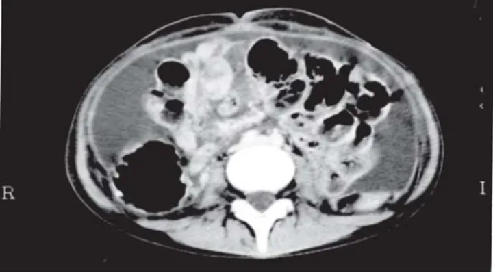
On 10 March 1997, an abdominal computed tomog-raphy (CT) showed intestinal adhesions and the bowel abnormally restricted to the central part of the abdo-men, peritoneal thickening, and loculation of the ab-dominal cavity (Figure 1). The typical cocoon image emerged and allowed the diagnosis of SEP. Total parenteral nutrition, epidural analgesia, and immu-nosuppression with azathioprine 50 mg/day plus pred-nisone 100 mg/day were prescribed. Surgical management was discussed but not done owing to the unsatisfactory results previously reported. Three days later complaints diminished, epidural analgesia could be stopped and 10 days later she started progressive oral nutrition with intradialytic parenteral supple-mentation for 1 month more. A follow-up CT scan performed on 18 March 1997 showed some improve-ment. Azathioprine was stopped at 2.5 months of treatment and steroids were continued for 2 months more with progressive tapering without relapse of pain or occlusion. She remains on HD using a central catheter. The patient recovered her lost weight and had a serum albumin of 3.7 g/dL. A last CT performed in August 1997 showed normal distribution of the bowel; the “cocoon” had disappeared (Figure 2).
DISCUSSION
With the increasing number of PD patients and the increasing survival on PD, SP becomes an impor-tant problem. An Australian study (3) showed a near exponential increase in SP incidence with time on PD: from 1.9% for patients on dialysis less than 2 years, to 6.4%, 10.8%, and 19.4% for more than 5, 6, and 8 years, respectively, suggesting the duration of the exposure of the peritoneum to PD to be a major risk factor. However, some cases occur early in the course of PD.
Peritoneal sclerosis has two distinct forms: the SS form — thought mainly dependent on the bio-incompatibility of the solutions — with a sclerotic submesothelial layer not exceeding 40 μ, and without significant inflammatory infiltrate (4); and the SP form where there is a dramatic progression of the sclerosis after an inflammatory insult, such as peritonitis. The
thickness of the sclerotic tissue reaches much higher values (1000 – 4000 μ) than in SS, and a marked chronic inflammatory infiltrate is invariably present (4). Although bioincompatibility is a risk factor, a de-finitive etiology for SEP is still unknown. Peritonitis is the most commonly invoked, but not obligatory, pathogenic factor. The underlying process of SP may be immunological. A particular form of SP, SEP (en-capsulating form), is characterized by bowel obstruc-tion due to the extensive fibrosis covering and enclosing the gut like a rigid bag. The case we described pre-sented the clinical and radiological features of SEP. The image of a cocoon bowel observed on CT, based on a suggestive clinical presentation, was diagnostic (2,3) and we considered peritoneal biopsy too risky.
This patient was on PD for 42 months and had an incidence of peritonitis of 2.6 episodes/year, much higher than our mean incidence of 0.7 episodes/pt/ year. The impact of peritonitis on mesothelial cells, and on their intrinsic fibrinolytic activity are well known. After this damage, the multipotential stem cells of the subserosa may differentiate as mesothe-lial cells, causing a new epitheliation of the
perito-Figure 2 — Abdominal computed tomography image 5 months after Figure 1, showing normal distribution of the bowel loops, filling of all the cavity, with lumenal patency. Figure 1 — Abdominal computed tomography image at time of diagnosis of sclerosing encapsulating peritonitis, showing adherent bowel loops, with posterior tethering and lumenal narrowing, peritoneal thickening, and loculated fluid.
by on September 21, 2011
www.pdiconnect.com
neum or, to the contrary, leading to fibroneogenesis and peritoneal sclerosis. This process of regeneration versus fibrosis is affected not only by the number and duration of peritonitis episodes, but also by their char-acteristics: persisting peritonitis (2), late peritonitis of a previously damaged peritoneum (2), and the se-verity of the last peritonitis (2). More aggressive agents such as S. aureus, Pseudomonas, and fungi are more likely to damage the peritoneum. The increased fibrinous exudate due to increased coagulability and decreased fibrinolysis justifies the decision of some authors, including these authors, to maintain the cath-eter in place for 48 hours, if possible, to maintain peri-toneal washing. The fact that two of three SP cases present after transfer from PD suggests that PD helps to remove fibrin accretion (3).
Peritonitis is only one of many risk factors for SP (3). The peritonitis rate has decreased in the past 10 years and, on the contrary, SP has progressively increased: from 1.5/1000 in the past decade, to 4.2/ 1000 (3) in the first half of the present decade. In many cases, a previous peritonitis episode could not be docu-mented, which suggests other factors are implicated in SP genesis.
The number of abdominal surgeries, related or not to the catheter, might be another risk factor (2,5). The use of acetate-based PD solution in the early 1980s was undoubtedly related with SP process. Chlor-hexidine and β-blocking agents have also been respon-sible for a considerable number of SP cases. Peritoneal exposure to glucose, hypertonicity, low pH, plasticiz-ers, glucose degradation products (GDP) by heat ster-ilization, and even trauma from the tip of the catheter (5), have all been implicated as risk factors (2,3). Some antimicrobial agents administered intraperitoneally are likely to negatively affect re-epithelization of the peritoneum. Our patient received fluconazole intra-peritoneally for 3 days, as proposed by peritonitis treatment recommendations, although there is little experience with this route of administration. We do not know if it contributed to the SP; however, small bowel obstructive symptoms appeared before its use. Some authors consider UF failure as an alerting sign for PS, and suggest systematic screening for SP in pa-tients on PD for more than 4 – 5 years who present loss of UF (5). Peritoneal effluent CA125 may also be a sign. Our patient presented UF deterioration.
Imaging of the abdominal cavity became progres-sively more important during the present decade for the diagnosis of SEP. Abdominal x ray is of low utility (5). Ultrasonography can show peritoneal thickening and a preperitoneal membrane, forming the typical image of trilayer membrane or “sandwich-like” mem-brane, and a fixed and dilated bowel (5). The CT is the most useful method for this diagnosis (2,3,5). The characteristic image of a cocoon, with the small bowel
restricted to the central part of the abdomen due to the fibrotic sheath covering and closing the loops, and the resulting contracted mesentery, peritoneal thick-ening, calcifications, and the septa leading to fluid loculation are the features often seen on CT (2,3,5), and were present in our patient. The colonic transit study, although now simplified, takes 2 or 3 days to show an increased colonic transit time (5).
Therapeutic strategy is a difficult decision. One invariable attitude is to stop PD and oral nutrition and transfer the patient to total parenteral nutrition and HD. The cases diagnosed after stopping PD sug-gest a possible causal relationship between this stop and SP expression, and it may be advisable to main-tain the catheter and lavage if infection can be ruled out. Surgical adhesiolysis is invasive and difficult to perform: to find a cleavage plane for the lysis of the adhesions and remove the fibrotic sheath is difficult and sometimes impossible work, especially if fibrosis overpasses the superficial layer. Occasional intesti-nal perforation may be fatal.
Immunosuppression is another option of therapeu-tic strategy, as an alternative to surgery or sometimes trying to facilitate the ensuing surgery. The improve-ment of small bowel obstruction in some SP patients after transplantation encouraged this strategy. Some authors only have SP survivors among those who were treated with immunosuppressive drugs, mostly renal transplanted patients (6). Immunosuppressive regi-mens were variable between reports, but all of them had prednisone and another drug, usually azathio-prine. This supported our decision to use these two drugs, but the ideal prescription is not known; some have advocated intraperitoneal prednisone alone. When we started the immunosuppression, the fun-gal peritonitis seemed fully treated, with microbio-logical tests repeatedly negative, but we maintained amphotericin for 14 days.
From the experience acquired with this case, we believe that the therapeutic strategy adopted, which included prednisone and azathioprine, was decisive for the favorable outcome.
La Salete S. Martins Anabela S. Rodrigues António N. Cabrita Serafim Guimaraes
Department of Nephrology Hospital de Santo António Porto, Portugal
R E F E R E N C E S
1. Gandhi VC, Humayun HM, Ing TS, Daugirdas JT, Jablokow VR, Iwatsuki S, et al. Sclerotic thickening of
by on September 21, 2011
www.pdiconnect.com
the peritoneal membrane in maintenance peritoneal dialysis patients. Ann Intern Med 1980; 140:1201–3. 2. Hendriks P, Pannekeet M, Gulik T, Struijk D, Phoa S,
Sie L, et al. Peritoneal sclerosis in chronic peritoneal dialysis patients: analysis of clinical presentation, risk factors, and peritoneal transport kinetics. Perit Dial Int 1997; 17:136–43.
3. Rigby RJ, Hawley CM. Sclerosing peritonitis: the ex-perience in Australia. Nephrol Dial Transplant 1998; 13:154–9.
4. Garosi G and Di Paolo N. Peritoneal sclerosis — an overview. In: Khanna R, ed. Advances in peritoneal di-alysis. Toronto: Peritoneal Dialysis Publications, 1999: 15:185–92.
5. Campbell S, Clarke P, Hawley C, Wigan M, Kerlin P, Wall D. Sclerosing peritonitis: identification of diag-nostic, clinical and radiological features. Am J Kidney Dis 1994; 24:819–25.
6. Junor BJ, McMillan MA. Immunosuppression in scle-rosing peritonitis. In: Khanna R, Nolph KD, Prowant BF, Twardowski ZJ, Oreopoulos DG, eds. Advances in peritoneal dialysis. Toronto: Peritoneal Dialysis Publi-cations, 1993; 9:187–9.
Intraperitoneal Fluconazole for Fungal
Peritonitis in CAPD: Report of Two Cases
Fungal infection accounts for between 1% and 15% of episodes of peritonitis in patients with end-stage renal disease managed by continuous ambulatory peritoneal dialysis (CAPD) (1,2). Management re-mains challenging because treatment strategies are controversial. Even if clinical symptoms resolve, many patients cannot resume CAPD secondary to perito-neal fibrosis or adhesion (3). Amphotericin B (AMPH-B) has the greatest activity of all antifungal agents. However, this agent is extremely toxic, it does not diffuse well into peritoneal fluid from the blood (2), and its intraperitoneal (IP) administration is of-ten painful and may increase the risk of peritoneal fibrosis and adhesion (3). Fluconazole (FLCZ) is a rela-tively nontoxic imidazole with strong activity against yeast, and is well absorbed when given either orally or intravenously (4,5). Some authors have reported that the likelihood of peritoneal adhesions after completion of therapy may be decreased by oral FLCZ (6,7), and it is now widely used as a first-line agent against fungal peritonitis.
However, not all patients given FLCZ have a good outcome. Michel et al. (8) reported that catheter re-placement after FLCZ treatment failed because of peritoneal adhesions in one of two cases in which it was attempted. A detailed analysis of 32 patients who received FLCZ treatment demonstrated that only 4 were able to continue CAPD successfully without in-terruption. Eleven of 26 patients who were managed
with catheter removal were transferred to hemodi-alysis, and there were four deaths.
Dahl et al. (9) recently investigated the pharmaco-kinetic characteristics of IP FLCZ in patients under-going continuous cycling peritoneal dialysis who did not have peritonitis. They concluded that IP admin-istration for fungal peritonitis was the preferred route, since it allowed for outpatient treatment without the need for vascular access, and ensured adequate IP drug concentrations. We report the cases of 2 patients in whom IP FLCZ was successful for fungal peritoni-tis, and CAPD could be continued.
PATIENTS
Case 1: The first patient was a 64-year-old woman with glomerulonephritis who began CAPD in Febru-ary 1986. She had no history of surgery or abdominal disease. In early January 1992, she was diagnosed with acute tonsillitis and was given antibiotics. Nine days later she developed a cloudy peritoneal effluent. Upon examination, her temperature was 37.2°C and she had diffuse abdominal tenderness. Her periph-eral white blood cell count was 14500/mm3. No fungi were seen on Gram stain. We initially performed intra-abdominal lavage with cephapirin (250 mg/L) while continuing peritoneal dialysis. Because peritoneal fluid cultures showed Candida albicans, treatment with intravenous FLCZ (200 mg start; 100 mg daily) was started on the third hospital day. The effluent cleared within 48 hours. However, 1 day later, the di-alysate once again became cloudy. On the sixth hos-pital day, IP FLCZ (25 mg/L of dialysate) was started instead of intravenous FLCZ. After 2 days the dialy-sate became clear and no micro-organism was cul-tured from this fluid. Intraperitoneal FLCZ was continued for 1 week and then oral FLCZ (100 mg daily) was administered for 2 weeks. There has been no relapse of the infection since that time.
Case 2: The second patient was a 57-year-old man with chronic glomerulonephritis who had been on he-modialysis for 10 years when he elected to switch to CAPD in January 1996. In March 1987, a cardiac pace-maker was implanted for sick sinus syndrome. Late in October 1996, he was diagnosed with trichophyto-sis in his fingers. Seven days later he showed a cloudy peritoneal effluent. Intraperitoneal lavage was per-formed initially, using a temporary catheter as a drain-age tube. A few lavdrain-ages per day were performed using heparinized physiological saline solution. Peritoneal dialysis was continued without antibiotic treatment because the patient showed no other symptoms. Three days later he developed a low-grade fever and anor-exia. He came to our hospital on 4 November. His pe-ripheral WBC was 6950/mm3, and Gram stains were negative. We performed IP lavage, while continuing
by on September 21, 2011
www.pdiconnect.com
peritoneal dialysis, with IP cefmetazole sodium (250 mg/L of dialysate) on the first day, and intrave-nous cefmetazole sodium (1 g daily) on the second day. After 6 days of therapy, symptoms and signs remained unchanged and C. albicans grew from a dialysate fun-gal culture. Intravenous FLCZ (200 mg start, 100 mg daily) was begun. Still the dialysate showed no im-provement, and on the eighth day of treatment, IP FLCZ (25 mg/L of dialysate) was started.
After 2 days, signs and symptoms remained un-changed. The patient’s Tenckhoff catheter was removed and replaced immediately with a temporary perito-neal catheter. The temporary catheter was introduced into the peritoneal cavity through the same wound. A purse-string suture was placed around the catheter. A culture of the first cuff and a second culture of the Tenckhoff catheter yielded C. albicans. Hemodialysis was started the next day via a femoral vein catheter. Peritoneal lavage was continued twice daily using a heparinized physiological saline solution (1000 mg/L) with FLCZ (50 mg), which was flushed in at 1 L and immediately drained. This was continued until the dialysate culture was negative.
On day 28 of treatment the temporary catheter was removed. Simultaneous serum and dialysate concen-trations of FLCZ measured by HPLC-UV at the time of catheter removal were 7.1 μg/mL and 20.3 μg/mL, respectively. Although the patient complained of thirst throughout the lavage period, there were no changes in his blood pressure, weight, or other clinical charac-teristics. After removal of the catheter, oral FLCZ (100 mg daily) was continued for 3 weeks. On day 45 of therapy and after serum C-reactive protein was no longer detected, a new indwelling Tenckhoff catheter was placed. Subsequent cultures of the patient’s peri-toneal fluid have shown no change, and he has suc-cessfully resumed CAPD. The peritoneal clearance and ultrafiltration capacity in this patient did not change after treatment.
DISCUSSION
Fungal peritonitis is an increasingly frequent se-rious complication of peritoneal dialysis that usually results in catheter loss. The goals of treatment should include eradication of infection as rapidly and as safely as possible, in a manner that allows the patient to continue dialysis. It is desirable that the infection is treated successfully without withdrawal of peritoneal dialysis as in Case 1. However, in the majority of pa-tients, removal of the IP catheter is required, as in Case 2.
In both cases, we initially administered intrave-nous FLCZ after C. albicans was isolated in the peri-toneal dialysate. Even after 2 days, signs and symptoms remained unchanged, and so we changed
to IP FLCZ. We waited 2 days to determine efficacy because, according to published reports in all cases in which peritonitis was cured without catheter re-moval, the effluent cleared within 48 hours (7,10,11). Reported cases of fungal peritonitis treated with FLCZ are few. Even the route of administration re-mains controversial. Based on investigations of serum concentrations of FLCZ, oral administration of FLCZ seems to be quite sufficient. Oral treatment produced adequate IP dialysate concentrations. However, the dialysate concentration (20.3 μg/mL) of FLCZ mea-sured in our Patient 2 was fairly high; it was over double the concentration (2.3 – 9 μg/mL) reported by Levine et al. (6) who administered the same dose orally. Presuming that this high dialysate concentra-tion was effective in Patient 1, IP administraconcentra-tion seems to be a method that should be tried initially.
When the infection cannot be cured with adminis-tration of FLCZ, it is generally agreed that the Tenckhoff catheter needs to be removed, because the catheter is colonized with the causative fungi and it exacerbates the infection (12,13). Nagappan et al. (14) have reported that, in mild cases, the infection can be eradicated by catheter removal alone. Amici et al. (15) have stated that it is important to heighten local anti-fungal drug concentrations and wash out inflamma-tory and fungal debris to prevent peritoneal adhesion after catheter removal. Keogh et al. (16) reported that insertion of a temporary peritoneal catheter is effec-tive for the continuation of IP lavage and IP drug ad-ministration. They repeated peritoneal lavage frequently for 8 – 18 days, until the fluid was micro-biologically clear, using 0.5- to 1.0-L exchanges with miconazole and heparin.
In our opinion, if the original catheter is removed, a few lavages per day are enough to avoid formation of massive peritoneal adhesions and are easy enough even for outpatients to comply with. Therefore, we removed the Tenckhoff catheter and inserted a tem-porary catheter simultaneously through the same wound in Patient 2. Then we commenced peritoneal lavage, using a heparinized physiological saline solu-tion (1000 mg/L) with FLCZ 50 mg, and continued it only twice per day until the dialysate culture was negative. Lavage by physiological saline solution seemed to be effective in removing high dextrose in the peritoneal liquid, since the growth of fungus is frequently related to high concentrations of dextrose. We suggest the following regimen:
1. Commence administration of IP FLCZ.
2. If clinical resolution is not observed after 2 days, remove the Tenckhoff catheter and insert a tem-porary peritoneal catheter immediately.
3. Commence peritoneal lavage with physiological saline solution and continue administration of IP
by on September 21, 2011
www.pdiconnect.com
Mignon F. Fungal peritonitis in patients on peritoneal dialysis. Am J Nephrol 1994; 14:113–20.
9. Dahl NV, Foote EF, Searson KM, Fein JL, Kapoian T, Steward CA, et al. Pharmacokinetics of intraperitoneal fluconazole during continuous cycling peritoneal dialy-sis. Ann Pharmacother 1998; 32:1284–9.
10. Ahlmen J, Edebo L, Eriksson C, Carlsson L, Torgensen AK. Fluconazole therapy for fungal peritonitis in con-tinuous ambulatory peritoneal dialysis (CAPD): a case report. Perit Dial Int 1989; 9:79–80.
11. Blaser C, Peschke B, Gossman J, Schoeppe W, Scheuer-mann EM. Treatment of a fungal peritonitis by oral fluconazole without removal of catheter in a CAPD patient. Perit Dial Int 1992; 12(Suppl 2):S64.
12. Suger AM. Antifungal therapy in CAPD peritonitis: do we have a choice? Semin Dial 1991; 4:145–6.
13. Bernard DB, Levine J, Idelson BA. A continuous am-bulatory peritoneal dialysis patient with fungal peri-tonitis. Semin Dial 1991; 4:198–202.
14. Nagappan R, Collins JF, Lee WT. Fungal peritonitis in continuous ambulatory peritoneal dialysis: the Auck-land experience. Am J Kidney Dis 1993; 20:492–6. 15. Amici G, Grandesso S, Mottola A, Virga G, Calconi G,
Bocci C. Fungal peritonitis in peritoneal dialysis: criti-cal review of six cases. In: Khanna R, ed. Advances in peritoneal dialysis. Toronto: Peritoneal Dialysis Publi-cations, 1994; 10:169–73.
16. Keogh JAB, Carr ME, Murray F, McEvoy M, Grant G, Keane CT. Treatment of fungal peritonitis in CAPD patients using peritoneal lavage. Perit Dial Bull 1985; 5:67–9.
Methicillin Resistance Patterns
Associated with Peritonitis in a
University-Based Peritoneal
Dialysis Center
aThe development of Streptococcus faecalis isolates exhibiting vancomycin resistance has recently re-sulted in the recommendation to avoid vancomycin as initial empiric therapy in many clinical settings, including treatment of peritonitis in patients on chronic peritoneal dialysis (PD) (1–4). Many practi-tioners have embraced this recommendation, although some remain unconvinced (5,6). Limited clinical data are presently available describing the outcomes in patients with PD-associated peritonitis treated in this manner (6–9).
We describe the antibiotic susceptibility data, spe-cifically regarding methicillin sensitivity, for gram-positive PD peritonitis cases, at a university center, to facilitate additional discussion of this important issue.
FLCZ.
4. After the causative fungi can no longer be iso-lated, remove the temporary peritoneal catheter. 5. After no inflammation can be found (i.e., serum C-reactive protein is undetectable), implant a new Tenckhoff catheter.
There were no side-effects in our patients due to FLCZ, and no further episodes of fungal peritonitis were observed. The use of IP FLCZ as well as perito-neal lavage with physiological saline solution after removal of the Tenckhoff catheter allowed us to suc-cessfully continue CAPD. This is an easy procedure. We will continue to study this approach in the future.
Hiroshi Kameoka Kenjirou Kumakawa Toshimitu Matuoka Michiko Nakano Yasuo Shiraiwa Osamu Yamaguchi1 Department of Urology Jyusendo General Hospital Koriyama City Fukushima Medical College1
Fukushima, Japan
R E F E R E N C E S
1. Report of Working Party of the British Society for An-timicrobial Chemotherapy. Diagnosis and management of peritonitis in continuous ambulatory peritoneal di-alysis. Lancet 1987; 1:845–9.
2. Kerr CM, Perfect JR, Craven PC, Jorgenson JH, Drutz DJ, Shelburne JD, et al. Fungal peritonitis in patients on continuous ambulatory peritoneal dialysis. Ann In-tern Med 1983; 99:334–47.
3. Eisenberg ES, Leviton I, Soeiro R. Fungal peritonitis in patients receiving peritoneal dialysis: experience with eleven patients and review of the literature. Rev Infect Dis 1986; 8:309–21.
4. Warnock DW. Itraconazole and fluconazole: new drugs for deep fungal infection. J Antimicrob Chemother 1989; 86:825–7.
5. Humphrey MJ, Jevons S, Tarbit MH. Pharmacokinetic evaluation of UK-49,858, a metabolically stable triazole antifungal drug, in animals and humans. Antimicrob Agents Chemother 1985; 28:648–53.
6. Levine J, Bernard DB, Idelson BA, Farnham H, Saunders C, Sugar AM. Fungal peritonitis complicat-ing continuous ambulatory peritoneal dialysis: success-ful treatment with fluconazole, a new orally active antifungal agent. Am J Med 1989; 86:825–7.
7. Corbella X, Sirvent JM, Carratala J. Fluconazole treat-ment without catheter removal in Candida albicans peritonitis complicating peritoneal dialysis. Am J Med 1991; 90:277.
8. Michel C, Courdavault L, Khayat RA, Viron B, Roux P,
a Presented as an abstract at the American College of Clinical Pharmacy 1998 Spring Practice and Research Forum, Palm Springs, CA, April 1998.
by on September 21, 2011
www.pdiconnect.com
M E T H O D S
All cases of peritonitis occurring in a university-based PD program over a 5-year period were retro-spectively identified by review of ongoing, unit-maintained, quality assurance documents. Defi-nition of peritonitis in our program was made clini-cally with laboratory confirmation (white blood cell count > 100/mm3 in dialysis effluent, with > 50% poly-morphonucleocytes) using recommended criteria (1). For the purpose of calculating peritonitis rates, bill-ing records were reviewed to determine the total num-ber of patients at risk for each time interval. Directed chart review of patient medical records was performed to obtain dialysate culture results and antibiotic sen-sitivity data for each bacterial strain isolated from dialysis effluent. Relapsing peritonitis (same organ-ism, occurring within 30 days of completion of antibi-otic treatment) was not counted as a new case or bacterial isolate. Recurrent peritonitis (occurring greater than 30 days after completion of antibiotic course, or infection with a different organism) was counted as a separate case.
Antibiotic sensitivities were determined by a stan-dardized disk-diffusion method performed in the Clinical Microbiology Laboratory of the University of Michigan Medical Center, Ann Arbor, MI, U.S.A. Anti-biotic susceptibility data were not available for some Staphylococcus epidermidis isolates during a portion of the study period because of laboratory policy, re-sulting in missing antibiotic susceptibility data in 39 of 123 gram-positive isolates. Results are reported with descriptive statistics only.
R E S U L T S
Over the 5-year period reviewed, a total of 202 epi-sodes of peritonitis occurred during 345 patient-years at risk, resulting in a mean peritonitis rate of 0.58 epi-sodes per patient-year at risk (Table 1). Dialysis ef-fluent cultures resulted in no growth in 34 of 202 cases (17%) over the study period. Dialysis cul-ture identified 182 isolates, including 123 gram-posi-tive organisms (68%), 51 gram-negagram-posi-tive organisms (28%), and 8 (4%) nonbacterial organisms. Staph. epidermidis was the most common bacterium iso-lated in each year and for the entire study period (58/182 isolates, 32%).
In vitro antibiotic sensitivity data were available for 84 of 123 gram-positive bacterial isolates (Table 2). Overall, methicillin resistance was reported in 40% (34/84) of gram-positive isolates over the 5-year study period. Of the 48 S. epidermidis isolates tested for in vitro antibiotic sensitivity, 50% (24/48) were methi-cillin resistant, while 26% (8/31) S. aureus isolates were reported as methicillin resistant.
DISCUSSION
Antibiotic sensitivity patterns of gram-positive bacteria isolated from dialysate of patients with acute peritonitis at the University of Michigan Medical Center over a 5-year period demonstrated a signifi-cant incidence of methicillin resistance (and presum-ably resistance to other beta-lactam antibiotics). Of particular concern, approximately one quarter of S. aureus isolates demonstrated in vitro methicillin resistance. Overall peritonitis rates and distribution of causative organisms were consistent with previ-ous reports from this and other centers.
Several limitations were present in our study, in-cluding its retrospective nature, relatively small num-bers of peritonitis cases, incomplete antibiotic sensitivity data for S. epidermidis isolates, and the lack of clinical outcome results. In an attempt to vali-date our antibiotic sensitivity results for staphylococ-cal isolates, we subsequently compared the methicillin resistance rates from peritonitis isolates to general resistance patterns from staphylococcal isolates re-ported for all isolates from the University of Michi-gan Microbiology Laboratory and found similar methicillin resistance rates (C. Pierson, personal communication).
Our goal in performing this study was not to add to the literature describing clinical outcomes of PD-associated peritonitis, but rather to document the antibiotic resistance patterns of bacterial isolates from a large tertiary-care medical center, with specific emphasis on beta-lactam resistance in gram-positive organisms. Furthermore, during the study period, our peritonitis treatment protocol included initial intra-peritoneal vancomycin and tobramycin, with adjust-ment of antibiotic coverage as indicated by culture and sensitivity results and clinical improvement. Sev-eral studies have already been published document-ing the efficacy of similar regimens, and our clinical results are similar to the published experience. Our in vitro data should be interpreted with caution how-ever, as in vitro antibiotic sensitivity does not neces-sarily predict clinical outcome, particularly in PD-associated peritonitis. In this setting, high con-centrations of antibiotic are added directly to the peri-toneal cavity and significant changes in periperi-toneal antibiotic concentration may occur, depending on the administration scheme used (10). Furthermore, peri-tonitis outcome is related to organism-specific factors in addition to specific antibiotic treatment (11).
Current recommendations for treatment of PD-related peritonitis include initial treatment with a first-generation cephalosporin and aminoglycoside intraperitoneally, with subsequent adjustment of an-tibiotic regimen based on culture and sensitivity data as well as clinical response to initial therapy (1).
by on September 21, 2011
www.pdiconnect.com
ber et al. reported successful treatment of PD-associ-ated peritonitis with a regimen including continuous intraperitoneal cefazolin and gentamicin in the set-ting of a low-incidence of in vitro beta-lactam antibi-otic resistance (9).
Concern over the recommendation to use cefazolin instead of vancomycin for empiric initial treatment has been raised, in part because of uncertainty about the effectiveness of such regimens in treating beta-lactam-resistant staphylococcal infections (5,12). Van Biesen and colleagues propose that cefazolin is not the empiric gram-positive agent of choice for all PD centers, despite published recommendations (5). A review of culture and susceptibility data in their cen-ter revealed that a widely recommended protocol using cefazolin and gentamicin would cover only 78% of peritonitis episodes, assuming that in vitro sensi-tivity data predict clinical outcome. Based on these data, this group devised a new protocol using vanco-mycin and gentamicin initially, then oral ciprofloxacin for ambulatory patients. Intraperitoneal ceftazidime and ciprofloxacin are used in patients requiring hos-pitalization. With this protocol, an in vitro coverage rate of 96% is predicted.
Clinical experience with intermittent cefazolin for treatment of peritonitis in situations where increas-ing beta-lactam resistance is present have been mixed.
Lai et al. reported a gram-positive response rate of 95% with a regimen of cefazolin 500 mg/L and gen-tamicin 20 mg/L, once daily, in 19 peritonitis episodes (7). In a retrospective study of 55 patients, Vas et al. compared vancomycin 2 g weekly plus tobramycin 60 mg daily against cefazolin 1.5 g daily plus tobramycin 60 mg daily in the treatment of PD-asso-ciated peritonitis. There was no significant difference in overall efficacy between the regimens, with a suc-cess rate of 74% and 77%, respectively. However, the cefazolin/tobramycin regimen resolved only 45% of episodes due to methicillin-resistant coagulase-nega-tive staphylococcal infections, while the vancomycin/ tobramycin regimen resolved 73% (8). In a retrospec-tive examination of 118 cases of gram-posiretrospec-tive peri-tonitis in 85 patients, Alves and Dantas reported that vancomycin was twice as effective as cefazolin when used as first-line therapy for S. aureus, despite high antibiotic sensitivity to cephalosporins (6).
Although micro-organisms are generally consid-ered resistant to cefazolin when the minimum inhibi-tory concentration (MIC) is > 64 mg/L, the high concentrations used in continuous intraperitoneal (IP) dosing (cefazolin 125 mg/L) may be sufficient to over-come this “apparent resistance.” In contrast, an inter-mittent once-daily cefazolin dose may not achieve sustained peritoneal concentrations high enough to TABLE 1
Peritonitis Data and Dialysate Culture Results
Patients Peritonitis Culture-negative Bacterial isolates
at risk episodes Peritonitis peritonitis Bacterial isolates
Year (n) (n) ratea (n) (n) Gram+:gram–:other
1993 75 41 0.55 7 36 26:10:0 1994 76 54 0.71 12 44 32:12:0 1995 65 49 0.75 8 43 28:13:2 1996 65 31 0.48 6 30 18:9:3 1997 64 27 0.42 1 29 19:7:3 Total 345 202 0.586b 34 182 123:51:8
aPeritonitis episodes per patient-year at risk. bMean peritonitis rate over study period.
TABLE 2
In Vitro Methicillin Resistance of Gram-Positive Organisms Coagulase-negative Coagulase-positive Other gram-positive
staphylococci staphylococci organisms Annual total
Year (%) (%) (%) (%) 1993 5/10 (50) 1/9 (11) 0/0 (0) 6/19 (32) 1994 5/12 (42) 1/5 (20) 0/0 (0) 6/17 (35) 1995 4/12 (33) 3/8 (38) 1/1 (100) 8/21 (38) 1996 5/6 (83) 1/2 (50) 1/2 (50) 7/10 (70) 1997 5/8 (63) 2/7 (29) 0/2 (0) 7/17 (41) Total 24/48 (50) 8/31 (26) 2/5 (40) 34/84 (40) by on September 21, 2011 www.pdiconnect.com Downloaded from
Mortal Weekly Rep 1997; 46:765–6.
4. Vas SI. VRE and empirical vancomycin for CAPD peri-tonitis: use at your own/patient’s risk. Perit Dial Int 1998; 18:86–7.
5. Van Biesen W, Vanholder R, Vogelaers D, Peleman R, Verschraegen G, Vijt D, et al. The need for a center-tailored treatment protocol for peritonitis. Perit Dial Int 1998; 18:274–81.
6. Alves FR, Dantas RC. Is the treatment of beta lactam sensitive infections (BLSI) without vancomycin (Vc) possible (Abstract)? Perit Dial Int 1997; 17(Suppl 1):S27.
7. Lai, MN, Kao MT, Chen CC, Cheung SY, Chung WK. Intraperitoneal once-daily dose of cefazolin and gen-tamicin for treating CAPD peritonitis. Perit Dial Int 1997; 17:87–9.
8. Vas S, Bargman J, Oreopoulos DG. Treatment in PD patients of peritonitis caused by gram-positive organ-isms with single daily dose of antibiotics. Perit Dial Int 1997; 17:91–4.
9. Weber J, Staerz E, Mettang T, Machleidt C, Kuhlmann U. Treatment of peritonitis in continuous ambulatory peritoneal dialysis (CAPD) with intraperitoneal cefazolin and gentamicin. Perit Dial Int 1989; 9:191–5. 10. Golper T. Intermittent versus continuous antibiotics for PD-related peritonitis. Perit Dial Int 1997; 17:11–12. 11. Bunke CM, Brier ME, Golper TA. Outcomes of single organism peritonitis in peritoneal dialysis: gram nega-tives versus gram posinega-tives in the Network 9 Peritoni-tis Study. Kidney Int 1997; 52:524–9.
12. Sandoe JAT, Gokal R, Struthers JK. Vancomycin-resistant enterococci and empirical vancomycin for CAPD peritonitis. Perit Dial Int 1997; 17:617–18.
Effect of Erythropoietin Therapy on
Serum Apolipoprotein A1 Levels in
Patients Undergoing Chronic
Peritoneal Dialysis
Recombinant human erythropoietin (EPO) is widely used in the treatment of anemia in patients on peritoneal dialysis (PD) (1,2). Erythropoietin treat-ment increases food intake and improves tissue oxy-genation, protein metabolism, glucose utilization, and insulin resistance. All these factors may influence lipid metabolism in PD patients. In the literature, there are only a few but conflicting reports on the effects of EPO on the lipid profile in hemodialysis (HD) (3–10), and fewer still in PD (4,5,9) patients. The current study assesses the changes in the lipid profile associ-ated with EPO treatment in PD patients.
PATIENTS AND METHODS
Nineteen patients (6 women aged 47 – 75 years, 13 men aged 26 – 74 years) undergoing standard PD with EPO therapy from 1 to 38 months were retro-treat these “apparently resistant” micro-organisms
(10). For this reason, the Ad Hoc Advisory Committee for the Management of Peritonitis advises that a first-generation cephalosporin be administered IP, continu-ously rather than intermittently (1). Unfortunately, to date no studies have been performed to specifically test the effectiveness of continuous IP cefazolin against methicillin-resistant organisms.
Empiric initial treatment of peritonitis is currently the subject of great debate and variable clinical prac-tice. Appropriate concern over the widespread use of vancomycin and the emergence of vancomycin-resis-tant bacterial strains has been raised. On the other hand, it remains unclear whether replacement of van-comycin by cephalosporins in treatment of PD-asso-ciated peritonitis is prudent, especially in centers with a high prevalence of beta-lactam-resistant gram-posi-tive organisms. The question will best be answered by adequately-sized, prospective, randomized clinical trials. Until the results of such studies are available, we remain concerned about the possible consequences to our patients of current recommendations to avoid initial vancomycin use in PD-associated peritonitis.
A C K N O W L E D G M E N T
Supported in part by a grant from Glaxo Wellcome. At the time of the study, T. Zhang was a pharmacy student at the University of Michigan.
Nancy A. Mason1
Tongqing Zhang2
Joseph M. Messana3
The University of Michigan College of Pharmacy and Health System1,2
Ann Arbor, Michigan Pfizer, Inc,2
New York, New York Division of Nephrology3
Department of Internal Medicine University of Michigan Medical School Ann Arbor, Michigan, U.S.A.
R E F E R E N C E S
1. Keane WF, Alexander SR, Bailie GR, Boeschoten E, Gokal R, Golper TA, et al. Peritoneal dialysis-related peritonitis treatment recommendations: 1996 update. Perit Dial Int 1996; 16:557–63.
2. CDC recommendations for preventing the spread of vancomycin resistance: recommendations of the Hos-pital Infection Control Practices Advisory Committee (HICPAC). MMWR CDC Surveill Summary 1995; 44(No. RR-12).
3. Staphylococcus aureus with reduced susceptibility to vancomycin — United States, 1997. MMWR Morb
by on September 21, 2011
www.pdiconnect.com
spectively analyzed. Eight patients were diabetics. None of the patients suffered from nephrotic syn-drome, thyroid or liver disease, or iron deficiency or were receiving lipid lowering drugs. In patients stud-ied repeatedly, the factors that may affect hematocrit and lead to changes in EPO therapy, such as an un-derlying inflammation, changes in iron, nutritional state, and hydration were not found. The clinical characteristics are shown in Table 1 and the results of lipid profile analyzed as previously reported (11) are shown in Table 2.
In order to evaluate the relationship between EPO doses and serum concentrations of lipids and lipopro-teins, and to exclude (or diminish) the effect of Hct changes on these parameters, the latter were adjusted
to Hct by the formula (serum concentration of lipid) × (1 – Hct) (13).
Data were evaluated by Student’s t-test, analysis of variance, and linear regression and are presented as mean ±SEM. Paired t-test was used for paired data. The Mann–Whitney test and Spearman’s r were used for assessment of lipoprotein (a).
R E S U L T S
As expected, patients with diabetes mellitus exhib-ited significantly higher basal serum concentrations of glucose than did nondiabetic patients (Table 1). The other parameters compared (Table 1), including ab-normal serum concentrations of lipids and
lipopro-TABLE 2
Lipid Profile (No Adjustment to Hematocrit) of Studied Patients
Groups Reference intervala Total Nondiabetics Diabetics
Triglycerides (mg/dL) <160 225.6±21.5 225.2±26.2 226.2±38.4 Total cholesterol (mg/dL) <200 246.2±10.9 241.4±14.2 252.7±18.0 LDL cholesterol (mg/dL) <130 161.2±8.0 156.1±11.3 168.1+11.4 HDL cholesterol (mg/dL) >35 39.9±2.7 40.2±13.6 39.5±3.7 Lipoprotein(a) (mg/dL) 0–30 70.1±17.5 52.5±13.0 94.4±37.3 Apolipoprotein A1 (mg/dL) 119–240 126.6±7.3 118.6±8.5 137.7±12.3 Apolipoprotein B (mg/dL) 52–163 144.1±9.8 142.8±9.6 145.5±20.2
Mean ±SE. To convert triglycerides to mmol/L, multiply by 0.01129. To convert total cholesterol, LDL-, and HDL-cholesterol to mmol/L, multiply by 0.02586. To convert lipoprotein(a), apolipoprotein A1, and apolipoprotein B to g/L, multiply by 0.01. aFrom Ref. 12.
TABLE 1
Clinical Characteristics of Studied Patients
Groups Total Nondiabetics Diabetics
Number of patients 19 11 8
Women/men 6/13 5/6 1/7
Age (years) 58.3±2.6 58.9±4.1 57.6±2.7
Lean body mass (%) 60.1±2.8 60.6±2.8 59.6±5.9
Body mass index (kg/m2) 23.8±0.7 23.3±3.1 24.6±1.4
Time on dialysis (months) 13.4±2.6 15.0±3.5 11.2±4.1
Residual renal function (mL/min) 2.1±0.6 1.3±0.4 3.5±1.3
Kt/V 2.1±0.1 2.0±0.1 2.1±0.2
Normalized protein catabolic rate (g/kg/24 hr) 1.1±0.0 1.0±0.0 1.2±0.1
Serum β2-microglobulin (mg/L) 24.8±1.8 27.6±2.1 21.0±2.7
Serum glucose (mg/dL) 129.6±14.2 98.0±5.0 173.0±26.7a
Serum albumin (g/L) 38.4±1.3 39.9±1.6 36.3±2.0
Peritoneal β2-microglobulin clearance (mL/min) 1.1±0.1 0.9±0.0 1.2±0.2
Peritoneal albumin clearance (mL/min) 0.1±0.0 0.1±0.0 0.1±0.0
Hemoglobin (g/L)b 116.2±4.3 118.6±0.6 113.0±4.3
Erythropoietin (U/kg/wk) 83.5±14.5 64.8±18.9 98.7±22.4
Mean ±SE. To convert residual renal function values to mL/s multiply by 0.01667. To convert glucose values to mmol/L multiply by 0.05551. ap < 0.05 versus nondiabetics. bHematocrit (%) = 0.36 + 0.29 hemoglobin (g/L); r = 0.959, p < 0.0001, n = 19. by on September 21, 2011 www.pdiconnect.com Downloaded from
teins (Table 2) did not differ significantly between the two subgroups (diabetics and nondiabetics). There-fore, we analyzed the results of both subgroups to-gether. As shown in Figure 1(a), a significant positive correlation was observed between weekly subcutane-ous EPO doses and serum concentrations of apolipo-protein A1 (ApoA1). When serum concentrations of ApoA1 were corrected for Hct, the correlation coeffi-cient improved [Figure 1(b)]. Since the EPO effect could obviously be mediated via its effect on hemo-globin, we compared ApoA1 levels with hemoglobin and Hct [Figures 1(c), 1(d)]. It can be seen that these parameters were not significantly correlated. In ad-dition, ApoA1 levels were positively correlated with high-density lipoprotein (HDL)-cholesterol, residual renal function, and Kt/V (0.05 > p < 0.09). Moreover, the multivariate stepwise analyses (not shown) showed that the dose of EPO was a stronger indepen-dent determinant of serum ApoA1 concentrations than any other factor examined (see Table 1 and Table 2). No effect of EPO treatment on other lipids or lipoproteins was found (results not shown). Serum concentrations of glucose correlated significantly with
total cholesterol (TC), triglycerides (TG), and apolipo-protein B (ApoB) (not shown).
In some patients studied repeatedly (n = 6), we found that increases in the dose of EPO were associ-ated with increases in serum concentrations of ApoA1 (Figure 2), and decreases in the dose of EPO (n = 3) were associated with decreases in serum concentra-tions of ApoA1 (not shown).
DISCUSSION
The present study demonstrates that long-term EPO treatment (up to 38 months) significantly im-proves ApoA1 levels in PD patients. This beneficial effect of EPO treatment on serum ApoA1 levels in PD patients has not been previously reported. Viron et al. (4) assessed 5 PD and 7 HD patients receiving EPO for 6 months. They found no changes in the lipid pat-tern in PD patients. Pollock et al. (5) also found no changes in lipid profile, including ApoA1, in 31 patients on PD treated with EPO for 3 to 18 months. However, these authors demonstrated a significant fall in TC, TG, low-density lipoprotein-cholesterol (LDL-C), and
Figure 1 — Relationship between serum apolipoprotein A1 levels and weekly subcutaneous doses of erythropoietin (a,b), blood values of hematocrit (c), and hemoglobin (d) in 19 patients on peritoneal dialysis, serum apolipoprotein A1 levels adjusted to hematocrit (b) (13).
by on September 21, 2011
www.pdiconnect.com
ApoB when the results of lipid and lipoprotein changes in PD and HD patients were assessed together. Mak (9) documented normalization of plasma concentra-tions of TG, TC, and LDL-C in 12 adolescents (6 pa-tients on continuous cycling PD and 6 papa-tients on regular HD) after correction of their anemia by EPO, at a mean interval of 6 months. This improvement in lipid profile was associated with reversed insulin re-sistance (9). These results corroborate the findings of Borissova et al. (14) and Allegra et al. (6) that also show an improvement in insulin sensitivity in HD patients after EPO treatment. Insulin resistance is a well-known complication of uremia (15) and may, in part, contribute to the abnormal lipid profile in dialysis patients. In the present study, serum concentrations of glucose correlated significantly with TC, TG, and ApoB as further evidence of insulin resistance. How-ever, no effect of EPO treatment on lipids and lipopro-teins, other than ApoA1, was found.
Hematocrit is a strong confounding variable of lipo-protein measurement, especially in plasma (13), and is strongly affected by EPO. In the present study the
improvement in the ApoA1 profile related to EPO treatment could not be explained by the changes in Hct. First, there was no significant correlation be-tween Hct and ApoA1 levels. Second, both measured and Hct-adjusted ApoA1 levels were significantly cor-related to EPO (Figure 1). Moreover, the multivari-ate stepwise analyses selected EPO as a stronger independent determinant of serum ApoA1 concentra-tions. An increase in Hct due to EPO treatment could potentially influence peritoneal transport of small solutes and macromolecules, including peritoneal loss of ApoA1. In the present study, in agreement with the results of Burkart et al. (16), we found no changes in peritoneal transport characteristics in relation to EPO dose changes. Therefore, the increase in the serum concentrations of ApoA1 could not be explained by the differences in peritoneal transport of solutes and water induced by EPO. Alternatively, the decrease in insulin resistance and, probably, increase in he-patic synthesis of ApoA1 associated with EPO treat-ment may account for the elevated ApoA1 levels in PD patients.
Figure 2 — Individual changes in weekly subcutaneous erythropoietin doses and serum concentrations of apolipoprotein A1 (no adjustment to Hct) in patients on peritoneal dialysis: A and B are time intervals (months; mean ±SE) such that A = 9.3 ± 2.7 versus B = 26.6 ± 3.4, p < 0.05 by paired t-test. Subcutaneous erythropoietin doses (U/kg/week; mean ±SE): A = 59.8 ± 14.9 versus B = 85.9 ± 19.1, p < 0.05 by paired t-test. Serum apolipoprotein A1 concentrations (mg/dL; mean ±SE): A = 95.7 ± 5.8 versus B = 108.6 ± 4.7, p < 0.05 by paired t-test.
by on September 21, 2011
www.pdiconnect.com
Several studies in the HD population have showed a significant increase in serum ApoA1 concentrations during long-term EPO treatment (4,6,8,10), suggest-ing that EPO treatment is responsible for this im-provement. We have extended this observation to patients undergoing PD, in whom serum ApoA1 con-centrations were positively correlated with subcuta-neous weekly EPO doses.
In summary, long-term EPO therapy seems to in-crease the serum concentrations of ApoA1 in PD pa-tients. ApoA1, a major apolipoprotein of HDL, is generally accepted as a protective factor for coronary artery disease (CAD): ApoA1 is associated with sever-ity of coronary damage (17). However, a possible rela-tionship between degree of CAD and increasing ApoA1 levels after EPO treatment in PD patients remains to be studied in a larger population of PD patients.
Alexander Kagan1 Nurit Haran1 Ludmila Leschinsky1 Zvi Lerner2 Nechama Shuali1 Jayson Rapoport1 Department of Nephrology and Hypertension1
Central Clinical Laboratory2
Kaplan Medical Center Rehovot, Israel
R E F E R E N C E S
1. Nissenson AR, Korbet S, Faber M, Burkart J, Gentile D, Hamburger R, et al. Multicenter trial of erythropoi-etin in patients on peritoneal dialysis. J Am Soc Nephrol 1995; 5:1517–29.
2. Barany P, Clyne N, Hylander B, Johansson A–C, Simonsen O, Larsson R, et al. Subcutaneous epoetin beta in renal anemia: an open multicenter dose titra-tion study of patients on continuous peritoneal dialy-sis. Perit Dial Int 1995; 15:54–60.
3. Mat O, Stolear J–C, Georges B. Blood lipid profile in hemodialysis patients treated with human erythropoi-etin. Nephron 1992; 60:236–7.
4. Viron B, Donsimori R, Michel C, Khayat R-Al, Mignon F. Effect of recombinant human erythropoietin on
nu-tritional status and plasma lipids in uremic patients. Nephron 1992; 60:249.
5. Pollock CA, Wyndham R, Collett PV, Elder G, Field MJ, Kalowski S, et al. Effects of erythropoietin therapy on the lipid profile in end-stage renal failure. Kidney Int 1994; 45:897–902.
6. Allegra V, Martimbianco L, Mengozzi G, Vasile A. Eryth-ropoietin and cardiovascular risk. Blood Purif 1995; 13:301–13.
7. Manitius J, Szolkiewicz M, Mysliwska J, Zorena K, Mysliwski A, Jakubowski Z, et al. Influence of “non-hematological” doses of erythropoietin on lipid-carbo-hydrate metabolism and life quality in hemodialysis patients. Nephron 1995; 69:363–4.
8. Cengiz K. Does recombinant human erythropoietin affect plasma lipids in hemodialysis patients? Nephron 1996; 74:731–2.
9. Mak RHK. Effect of recombinant human erythropoi-etin on insulin, amino acid, and lipid metabolism in uremia. J Pediatr 1996; 129:97–104.
10. Allegra V, Martimbianco L, Vasile A. Lipid and apo-lipoprotein patterns during erythropoietin therapy: roles of erythropoietin, route of administration, and diet. Nephrol Dial Transplant 1997; 12:924–32. 11. Kagan A, Elimalech E, Lerner Z, Fink A, Bar-Khayim
Y. Residual renal function affects lipid profile in pa-tients undergoing continuous ambulatory peritoneal dialysis. Perit Dial Int 1997; 17:243–9.
12. Kratz A, Lewandrowski KB. Normal reference labora-tory values. N Engl J Med 1998; 339:1063–72. 13. Kronenberg F, Trenkwalder E, Kronenberg MF, Konig
P, Utermann G, Dieplinger H. Influence of hematocrit on the measurement of lipoproteins demonstrated by the example of lipoprotein(a). Kidney Int 1998; 54:1385–9.
14. Borissova AM, Djambazova A, Todorov K, Dakovska L, Tankova T, Kirilov G. Effect of erythropoietin on the metabolic state and peripheral insulin sensitivity in diabetic patients on hemodialysis. Nephrol Dial Trans-plant 1993; 8:93–5.
15. Mak RHK, De Fronzo RA. Glucose and insulin metabo-lism in uremia. Nephron 1992; 61:377–82.
16. Burkart JM, Freedman BI, Rocco MV. The effect of in-creasing hematocrit on peritoneal transport kinetics. J Am Soc Nephrol 1994; 4:1726–32.
17. Garfagnini A, Devoto G, Rosseli P, Boggiano P, Venturini M. Relationship between HDL-cholesterol and apo-lipoprotein A1 and severity of coronary artery disease. Eur Heart J 1995; 16:465–70.
by on September 21, 2011
www.pdiconnect.com